Cell and organelles
Model
Hover or tap to see names, click to read a description; click a name in the legend to fly to it. Procedurally built to histological proportions; colours follow illustration conventions.
Description
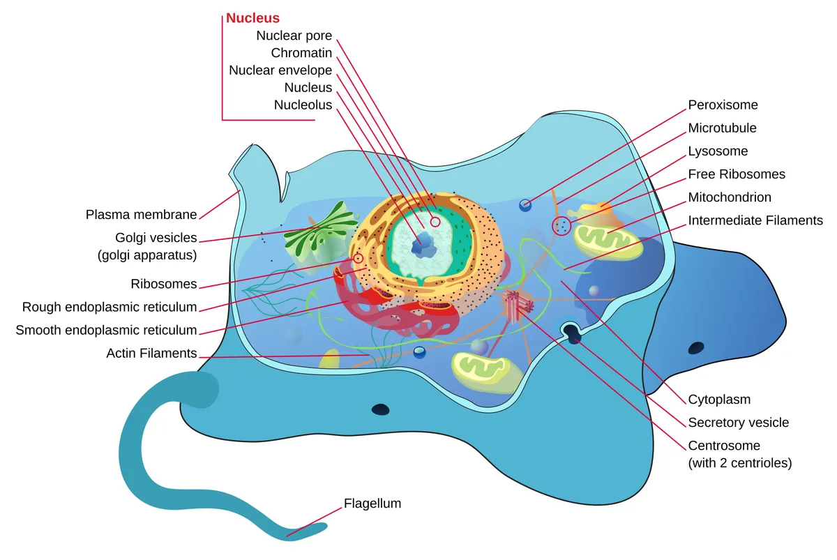
A eukaryotic cell consists of a plasma membrane enclosing the cytoplasm and nucleus. The cytoplasm contains membrane-bound organelles (endoplasmic reticulum, Golgi apparatus, mitochondria, lysosomes, peroxisomes), non-membranous organelles (ribosomes, centrosome) and the cytoskeleton. Each organelle is a specialised compartment, and together they synthesise, transport, generate energy, degrade and divide.
Plasma membrane
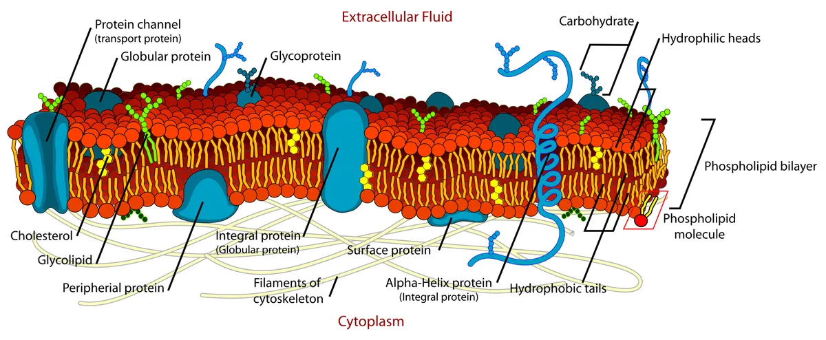
The cell membrane is a phospholipid bilayer about 7–8 nm thick, hydrophobic tails facing inwards, interspersed with cholesterol and proteins. In the fluid-mosaic model, lipids and many proteins diffuse laterally in the plane of the membrane; transmembrane proteins act as channels, pumps, carriers and receptors, while peripheral proteins attach to either face. The two leaflets differ: phosphatidylserine sits in the inner leaflet, and its exposure outside signals macrophages to clear a dying cell.[1]
The membrane is not uniform. Cholesterol and sphingolipids tend to gather into short-lived nanodomains (lipid rafts) that concentrate certain receptors and signalling molecules; the idea was long debated because the domains are too small and transient to see directly. Caveolae are 50–100 nm flask-shaped pits shaped by caveolin and cavin proteins, abundant in endothelium, adipocytes and smooth muscle; they flatten when the membrane is stretched, acting as a reserve that protects it from tearing.[2][3][4]
The outer face is coated by the glycocalyx, the oligosaccharide chains of glycoproteins, glycolipids and proteoglycans. It determines blood groups and mediates cell recognition and adhesion; on vascular endothelium it is several hundred nanometres thick, senses the shear of flowing blood, keeps platelets and leucocytes off the wall, and is rapidly shed in sepsis.[5][6]
Nucleus
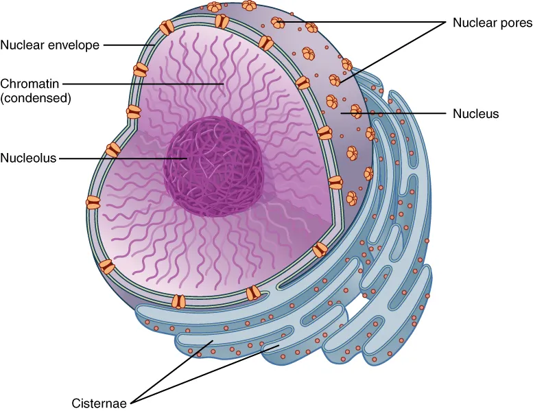
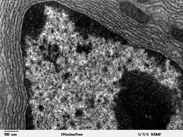
The nucleus (5–10 µm across) is enclosed by a double nuclear envelope; the outer membrane is continuous with rough ER and also carries ribosomes, and the space between the two membranes is continuous with the ER lumen. The inner face is lined by the nuclear lamina of lamins A, B and C, which both shapes the nucleus and anchors chromatin. Mutations of lamin A cause a family of laminopathies such as Emery–Dreifuss muscular dystrophy and Hutchinson–Gilford progeria.[7][8]
All traffic between nucleus and cytoplasm passes through nuclear pores: giant complexes of about 110 MDa built from roughly 1,000 copies of some 30 nucleoporins in eight-fold symmetry. The channel holds a meshwork of phenylalanine–glycine-rich protein chains; small molecules diffuse freely, whereas large proteins and messenger RNAs must be carried by transport receptors (importins, exportins) driven by the Ran-GTP gradient. A nucleus has several thousand pores.[9][10][11]
Some 2 m of DNA is wound around nucleosomes and then folded on several levels: chromatin loops, topologically associating domains (TADs), active and inactive compartments, and finally a separate territory for each chromosome. Dense, transcriptionally quiet heterochromatin tends to lie against the lamina and around the nucleolus, while open, active euchromatin occupies the interior. The nucleolus forms without a membrane around the ribosomal RNA gene clusters; there rRNA is transcribed and ribosomal subunits are assembled before export through the pores.[12][13][14][15]
Endoplasmic reticulum, Golgi and the secretory pathway
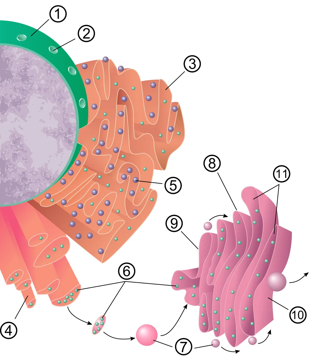
The endoplasmic reticulum is the largest continuous membrane system in the cell, made of flat sheets and tubules continuous with the nuclear envelope. Rough ER consists of stacked flattened cisternae with ribosomes on their outer face: secreted proteins, membrane proteins and lysosomal enzymes are translated straight into the lumen, where chaperones fold them, disulfide bonds form and the first N-linked glycans are attached. Misfolded proteins are retained and degraded; when they accumulate, the cell triggers the unfolded protein response.[16][17]
Smooth ER is a ribosome-free tubular network that makes phospholipids and cholesterol, steroid hormones (adrenal cortex, testis), detoxifies drugs with cytochrome P450 enzymes (hepatocytes) and stores Ca²⁺; in muscle it becomes the sarcoplasmic reticulum. The ER also touches mitochondria, plasma membrane, endosomes and lipid droplets at membrane contact sites 10–30 nm apart, where lipids and Ca²⁺ are handed over without vesicles.[18][19][20]
Proteins leave the ER in COPII-coated vesicles for the Golgi apparatus, a polarised stack of 4–8 flattened cisternae: the cis face receives cargo and the trans face dispatches it. As cargo moves through, its glycans are trimmed and extended in sequence by glycosyltransferases arranged from cis to trans, and lysosomal enzymes receive a mannose-6-phosphate tag. At the trans-Golgi network cargo is sorted into three routes: vesicles to lysosomes, constitutive secretion to the membrane, and regulated secretory granules stored until a signal arrives (insulin, pancreatic enzymes).[21][22][23][24]
Mitochondria
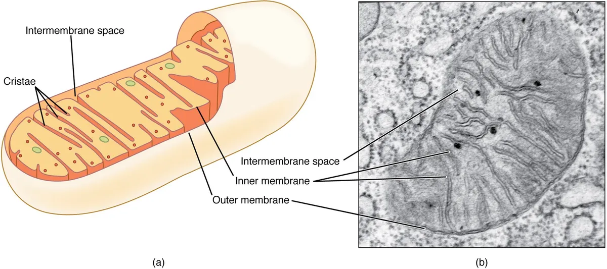
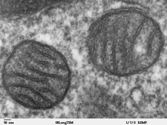
Mitochondria have two membranes: a smooth outer membrane permeable to small molecules and an inner membrane folded into cristae that carries the electron-transport chain and ATP synthase. The proton gradient across the inner membrane drives ATP synthase and supplies most of the cell’s ATP; the matrix holds the Krebs-cycle and fatty-acid oxidation enzymes, circular mitochondrial DNA (37 genes) and its own ribosomes. Their number matches energy demand: they fill about a third of a cardiomyocyte.[25][26][27]
Mitochondria are not fixed individual granules but a dynamic network that constantly fuses and divides. Fusion (mitofusins in the outer membrane, OPA1 in the inner) shares proteins and DNA; fission, by the dynamin DRP1 constricting at contact sites with the ER, separates damaged parts for removal by mitophagy. About 99% of mitochondrial proteins are encoded by nuclear genes and imported through the TOM and TIM complexes.[28][29][30][31]
Beyond energy, mitochondria buffer Ca²⁺, produce reactive oxygen species used as signals and decide programmed cell death: when the outer membrane is permeabilised, cytochrome c escapes and activates caspases. Mitochondrial DNA released into the cytosol is sensed as foreign and drives inflammation. Disturbed mitochondrial shape and dynamics feature in neurodegeneration, heart failure and kidney disease.[32][33][34]
Lysosomes, autophagy and peroxisomes
Lysosomes are single-membrane vesicles whose acidic lumen (pH about 4.5–5, maintained by the V-ATPase proton pump) holds more than 50 hydrolases. Material reaches them by three routes: endocytosis (receptors and fluid from the membrane via early and late endosomes), phagocytosis (bacteria and dead cells in macrophages) and autophagy. Maturation from endosome to lysosome is orchestrated by the GTPases Rab5 and then Rab7. Lack of one lysosomal enzyme causes a storage disease such as Gaucher or Tay–Sachs disease.[35][36]
In autophagy a double membrane engulfs a portion of cytoplasm, protein aggregates or damaged organelles to form an autophagosome, which fuses with a lysosome for digestion and recycling of the products. It is vital during starvation and the main quality-control system of neurons, which cannot dilute waste by dividing; failure of the autophagy–lysosome pathway contributes to Alzheimer, Parkinson and Huntington diseases.[37][38][39]
Peroxisomes are small (0.2–1 µm) single-membrane vesicles containing oxidases that generate H₂O₂ and catalase that destroys it. They β-oxidise very-long-chain fatty acids, α-oxidise phytanic acid and make plasmalogens (key myelin lipids) and bile acids. Their proteins are imported from the cytosol by peroxins (PEX genes), whose defects cause peroxisome biogenesis disorders such as Zellweger syndrome.[40][41][42]
Cytoskeleton
Three filament systems make up the cytoskeleton. Actin microfilaments (7 nm) are densest in the cortex beneath the membrane, build microvilli, lamellipodia and the cytokinetic ring, and generate force with myosin. Microtubules (25 nm) are hollow α/β-tubulin tubes growing from the centrosome with their plus ends outwards; they continuously grow and collapse (“dynamic instability”), serve as tracks for kinesins (towards the periphery) and dyneins (towards the centre) and form the mitotic spindle.[43]
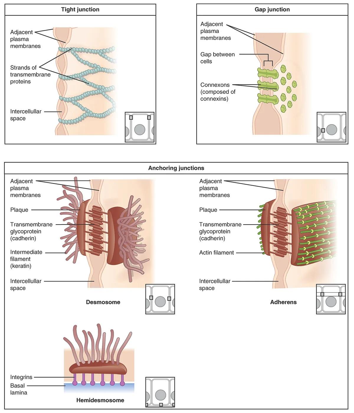
Intermediate filaments (10 nm) are mechanically tough and specific to cell type: keratins in epithelia, vimentin in mesenchymal cells, desmin in muscle, GFAP in astrocytes, neurofilaments in neurons and lamins in the nucleus. They attach to desmosomes and hemidesmosomes to spread tension through the tissue; keratin mutations make skin blister after minor friction. Because they are cell-type specific they serve as tumour markers, and neurofilament in blood is a biomarker of axonal damage.[44][45][46]
The centrosome consists of two orthogonal centrioles, cylinders about 250 nm wide and 500 nm long whose walls are nine microtubule triplets, surrounded by pericentriolar material where microtubules are nucleated. Centrioles also serve as basal bodies of cilia: the axoneme has a 9 + 2 arrangement in which dyneins make the cilium beat. Multiciliated cells of the airway carry hundreds of cilia beating in synchrony to move mucus; ciliary dyskinesia causes recurrent bronchitis, sinusitis and infertility.[47][48][49]
Cell cycle and division
The cell cycle comprises interphase (G1, S for DNA replication, G2) and M phase. Checkpoints controlled by cyclin–CDK complexes let the cell proceed only when DNA is intact and fully copied; the transcription factor MYC drives cells into the cycle and is over-active in many cancers. Highly differentiated cells such as neurons leave the cycle permanently for G0; senescent cells stop dividing for good but keep secreting inflammatory factors.[50][51]
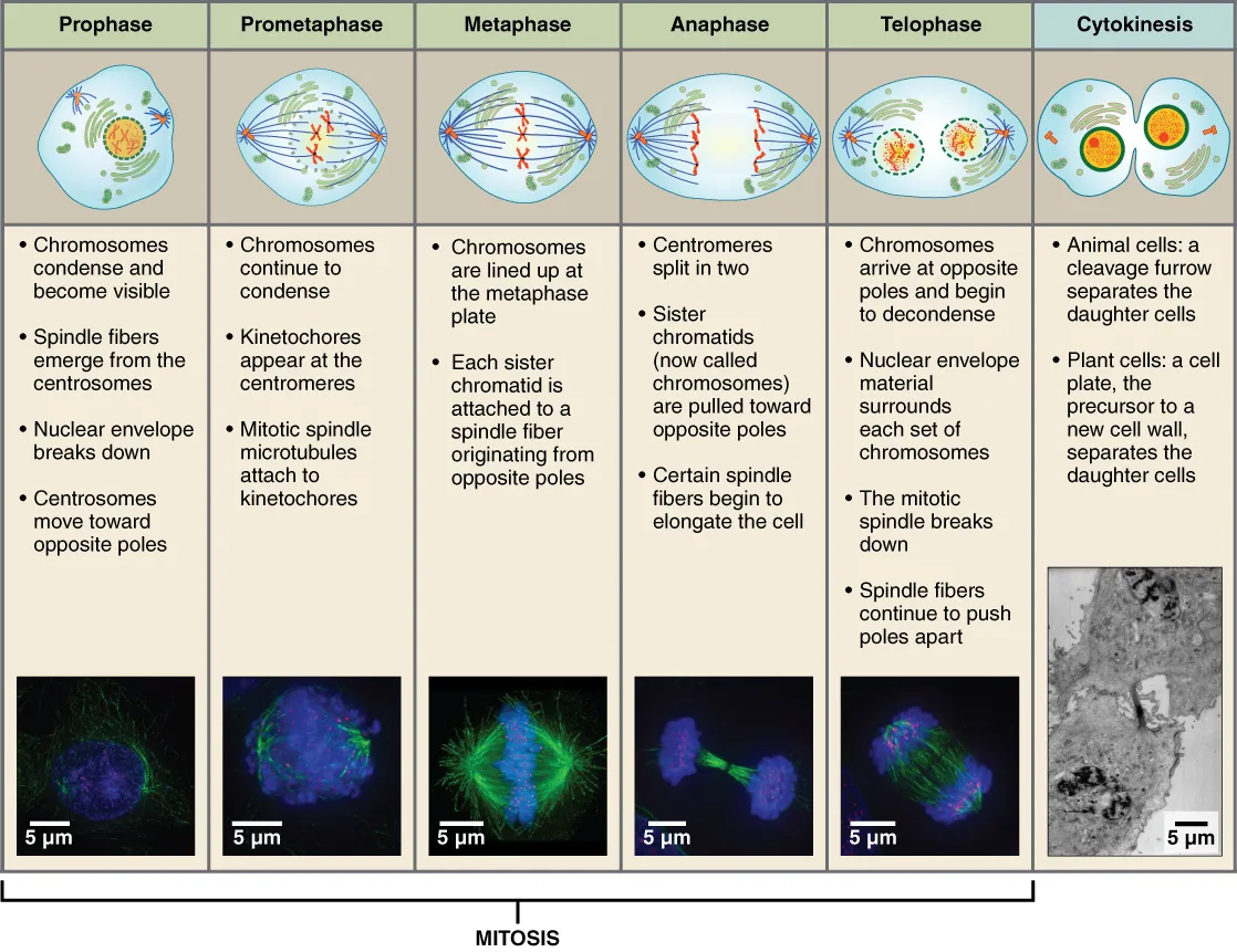
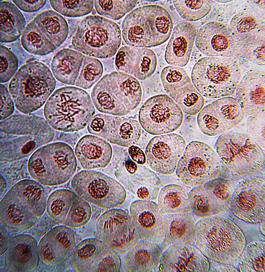
In mitosis the chromosomes condense and the nuclear envelope breaks down (prophase, prometaphase), and spindle microtubules capture the centromeres through kinetochores, protein complexes on the centromere that bind microtubule ends. The spindle checkpoint holds the cell in metaphase until every chromosome is attached to both poles; cohesin is then cleaved, sister chromatids move apart (anaphase), the nuclear envelope reforms (telophase) and an actomyosin ring pinches the cytoplasm in two. Segregation errors produce aneuploidy, a leading cause of miscarriage and a hallmark of many tumours.[52][53][54][55]
Full-text reading (StatPearls)
The StatPearls chapters on this topic, reproduced verbatim. Their CC BY-NC-ND 4.0 licence allows unmodified copies only, so they are not translated or shortened.
Anatomy, Back, Nucleus PulposusNedresky D, Reddy V, Singh G · StatPearls 2023 · NBK535373 · 8k characters
Loading…
Nedresky D, Reddy V, Singh G. Anatomy, Back, Nucleus Pulposus. StatPearls [Internet]. Treasure Island (FL): StatPearls Publishing; 2023. NBK535373 · PMID 30570994 · CC BY-NC-ND 4.0. Reproduced verbatim, unmodified.
Histology, Rough Endoplasmic ReticulumSanvictores T, Davis DD · StatPearls 2023 · NBK563126 · 7k characters
Loading…
Sanvictores T, Davis DD. Histology, Rough Endoplasmic Reticulum. StatPearls [Internet]. Treasure Island (FL): StatPearls Publishing; 2023. NBK563126 · PMID 33085273 · CC BY-NC-ND 4.0. Reproduced verbatim, unmodified.
Organs in this system
References
Sources for the values and descriptions on this page, indexed in PubMed (US National Library of Medicine).
- [1]Essay on Biomembrane StructureGerle C, The Journal of membrane biology 2019PMID 30877332
- [2]Lipid Rafts: Controversies Resolved, Mysteries RemainLevental I et al., Trends in cell biology 2020PMID 32302547
- [3]Lipid rafts as a membrane-organizing principleLingwood D et al., Science 2010PMID 20044567
- [4]CaveolaeParton RG et al., Current biology 2018PMID 29689223
- [5]The endothelial glycocalyx: composition, functions, and visualizationReitsma S et al., Pflugers Archiv 2007PMID 17256154
- [6]The glycocalyx: a novel diagnostic and therapeutic target in sepsisUchimido R et al., Critical care 2019PMID 30654825
- [7]Lamina-associated domains: Tethers and loosenersManzo SG et al., Current opinion in cell biology 2022PMID 35189475
- [8]Shared mechanisms in physiological and pathological nucleoplasmic reticulum formationDrozdz MM et al., Nucleus 2017PMID 27797635
- [9]The Structure of the Nuclear Pore Complex (An Update)Lin DH et al., Annual review of biochemistry 2019PMID 30883195
- [10]Structure, function and assembly of nuclear pore complexesPetrovic S et al., Nature reviews. Molecular cell biology 2026PMID 40926106
- [11]Aggregation, Phase Separation and Spatial Morphologies of the Assemblies of FG NucleoporinsZilman A, Journal of molecular biology 2018PMID 30017917
- [12]Chromosome territories and the global regulation of the genomeFritz AJ et al., Genes, chromosomes & cancer 2019PMID 30664301
- [13]Decoding the organization, dynamics, and function of the 4D genomeAboelnour E et al., Developmental cell 2021PMID 33984271
- [14]The nucleolusPederson T, Cold Spring Harbor perspectives in biology 2011PMID 21106648
- [15]Eukaryotic Ribosome AssemblyVanden Broeck A et al., Annual review of biochemistry 2024PMID 38768392
- [16]Histology, Rough Endoplasmic ReticulumSanvictores T et al., StatPearls 2023PMID 33085273
- [17]The endoplasmic reticulum: structure, function and response to cellular signalingSchwarz DS et al., Cellular and molecular life sciences 2016PMID 26433683
- [18]Structure and function of ER membrane contact sites with other organellesPhillips MJ et al., Nature reviews. Molecular cell biology 2016PMID 26627931
- [19]Endoplasmic reticulum-mitochondria contacts: function of the junctionRowland AA et al., Nature reviews. Molecular cell biology 2012PMID 22992592
- [20]Zooming into lipid droplet biology through the lens of electron microscopyDudka W et al., FEBS letters 2024PMID 38726814
- [21]Making COPII coatsKirchhausen T, Cell 2007PMID 17604713
- [22]The Golgi complex: a hub of the secretory pathwayPark K et al., BMB reports 2021PMID 33612152
- [23]Golgi glycosylationStanley P, Cold Spring Harbor perspectives in biology 2011PMID 21441588
- [24]Mechanistic insights into cargo sorting and export from the Golgi apparatusWatson ET et al., Nature reviews. Molecular cell biology 2025PMID 41214333
- [25]Mitochondrial proteins: from biogenesis to functional networksPfanner N et al., Nature reviews. Molecular cell biology 2019PMID 30626975
- [26]Structure and Function of the Mitochondrial RibosomeGreber BJ et al., Annual review of biochemistry 2016PMID 27023846
- [27]Kinetic Mathematical Modeling of Oxidative Phosphorylation in Cardiomyocyte MitochondriaTseng WW et al., Cells 2022PMID 36552784
- [28]Molecular mechanisms of mitochondrial dynamicsTábara LC et al., Nature reviews. Molecular cell biology 2025PMID 39420231
- [29]The cell biology of mitochondrial membrane dynamicsGiacomello M et al., Nature reviews. Molecular cell biology 2020PMID 32071438
- [30]Mitochondrial Machineries for Protein Import and AssemblyWiedemann N et al., Annual review of biochemistry 2017PMID 28301740
- [31]Mechanisms of mitophagyYoule RJ et al., Nature reviews. Molecular cell biology 2011PMID 21179058
- [32]Mitochondria and cell death-associated inflammationVringer E et al., Cell death and differentiation 2023PMID 36447047
- [33]Mitochondria in disease: changes in shapes and dynamicsJenkins BC et al., Trends in biochemical sciences 2024PMID 38402097
- [34]Mitochondrial dysfunction and the AKI-to-CKD transitionJiang M et al., American journal of physiology. Renal physiology 2020PMID 33073587
- [35]Rab GTPase Function in Endosome and Lysosome BiogenesisLangemeyer L et al., Trends in cell biology 2018PMID 30025982
- [36]Lysosomal quality control ReviewHenn D et al., Autophagy 2025PMID 39968899
- [37]Autophagy pathway: Cellular and molecular mechanismsYu L et al., Autophagy 2018PMID 28933638
- [38]Mechanisms of autophagy-lysosome dysfunction in neurodegenerative diseasesNixon RA et al., Nature reviews. Molecular cell biology 2024PMID 39107446
- [39]Autophagy genes in biology and diseaseYamamoto H et al., Nature reviews. Genetics 2023PMID 36635405
- [40]Peroxisome: Metabolic Functions and BiogenesisOkumoto K et al., Advances in experimental medicine and biology 2020PMID 33417203
- [41]Peroxisomes: a nexus for lipid metabolism and cellular signalingLodhi IJ et al., Cell metabolism 2014PMID 24508507
- [42]Alpha-oxidationJansen GA et al., Biochimica et biophysica acta 2006PMID 16934890
- [43]Microtubules and Microtubule-Associated ProteinsGoodson HV et al., Cold Spring Harbor perspectives in biology 2018PMID 29858272
- [44]Plasticity of cytoplasmic intermediate filament architecture determines cellular functionsSchwarz N et al., Current opinion in cell biology 2023PMID 37918274
- [45]GFAP at 50Messing A et al., ASN neuro 2020PMID 32811163
- [46]Neurofilaments as biomarkers in neurological disorders - towards clinical applicationKhalil M et al., Nature reviews. Neurology 2024PMID 38609644
- [47]Centriole structureWiney M et al., Philosophical transactions of the Royal Society of London. Series B, Biological sciences 2014PMID 25047611
- [48]Multiciliated cellsBrooks ER et al., Current biology 2014PMID 25291643
- [49]Towards an atomic model of a beating ciliary axonemeGrossman-Haham I, Current opinion in structural biology 2023PMID 36586349
- [50]Myc and cell cycle controlBretones G et al., Biochimica et biophysica acta 2015PMID 24704206
- [51]A guide to assessing cellular senescence in vitro and in vivoGonzález-Gualda E et al., The FEBS journal 2021PMID 32961620
- [52]PrometaphaseFerreira LT et al., Seminars in cell & developmental biology 2021PMID 34127384
- [53]The kinetochoreCheeseman IM, Cold Spring Harbor perspectives in biology 2014PMID 24984773
- [54]Mitotic genome foldingHirano T, The Journal of cell biology 2025PMID 40492990
- [55]Human aneuploidy: mechanisms and new insights into an age-old problemNagaoka SI et al., Nature reviews. Genetics 2012PMID 22705668