Nervous tissue
Model
Hover or tap to see names, click to read a description; click a name in the legend to fly to it. Procedurally built to histological proportions; colours follow illustration conventions.
Description
Tissue made of neurons, which conduct electrical impulses, and neuroglia, which support, insulate, nourish and protect them. The human brain holds about 86 billion neurons and a roughly similar number of glial cells.
The neuron
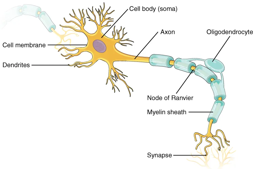
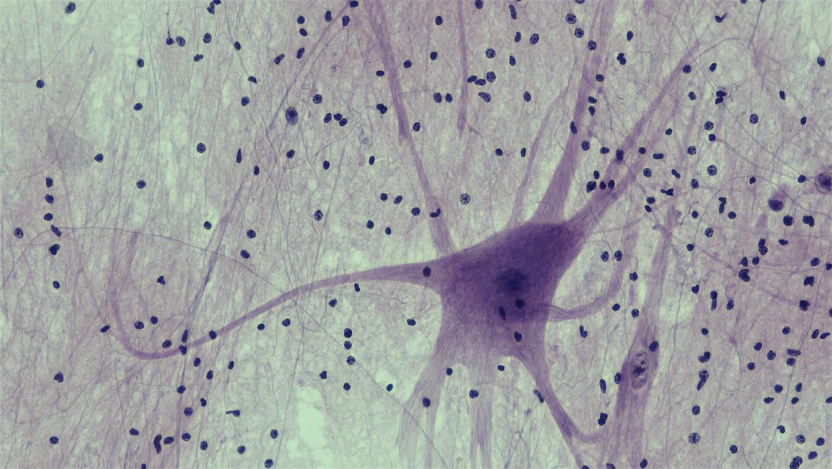
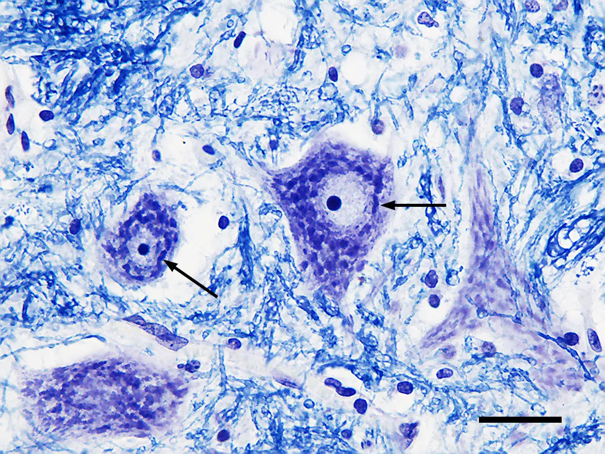
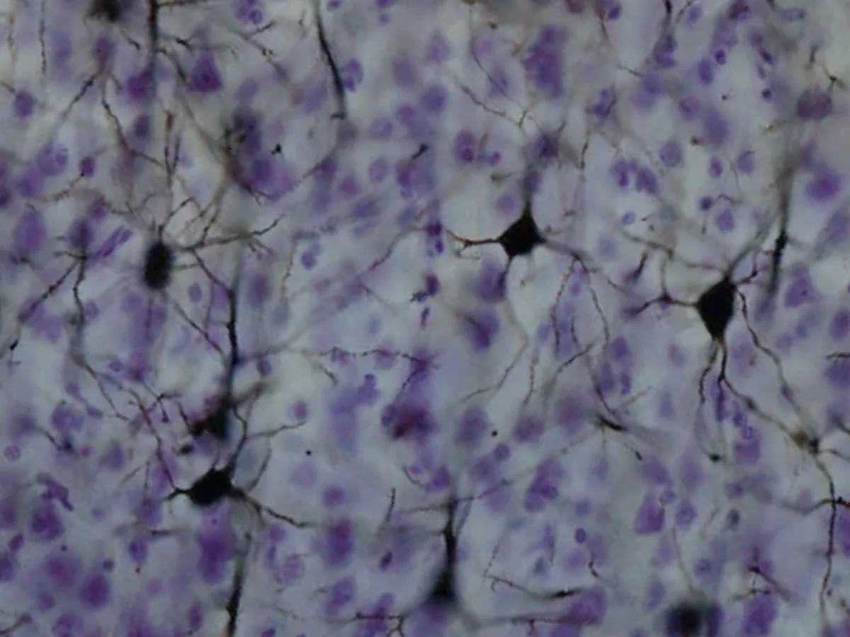
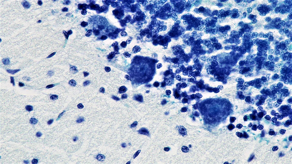
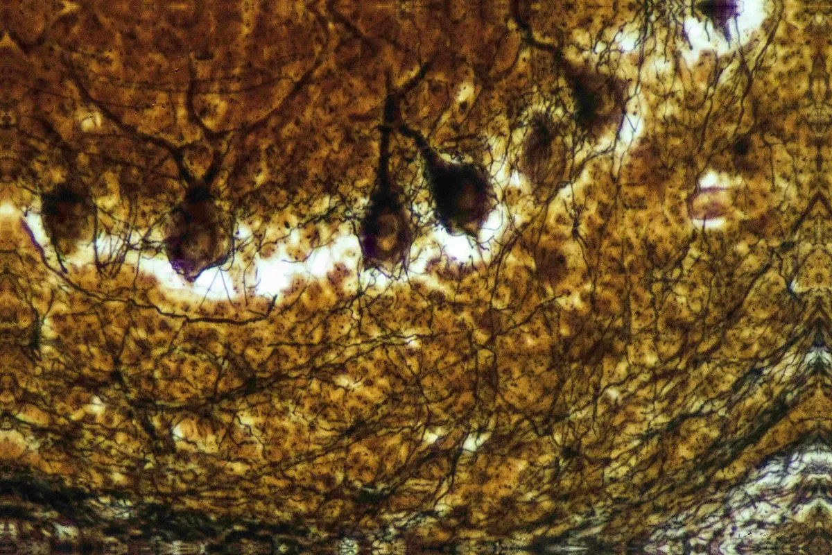
The neuron conducts electrical and chemical signals and has three parts: a cell body with a large nucleus, prominent nucleolus and Nissl bodies (stacks of rough ER making protein), dendrites that receive signals, and a single axon that carries them away. By number of processes neurons are multipolar (most motor and cortical neurons), bipolar (retina, olfactory epithelium) or pseudounipolar (dorsal root ganglia). The cortical pyramidal neuron has a long apical dendrite rising to superficial layers and many basal dendrites; distal and proximal parts of the tree receive different input streams, and ion channels along the dendrites decide which signals summate at the soma.[1][2]
Dendrites bear thousands of spines, small mushroom, thin or stubby protrusions, each usually carrying one excitatory synapse; spines change shape and number with activity and are the structural basis of memory. The axon begins at the axon hillock and the 20–60 µm axon initial segment, where Na⁺ channel density is highest and action potentials are initiated. The initial segment is organised by ankyrin G and spectrin and also acts as a filter that keeps dendritic and axonal proteins in their proper compartments, maintaining neuronal polarity. Axonal transport by kinesin and dynein on microtubules carries organelles to terminals as far as a metre from the soma.[3][4][1]
The axon initial segment has its own molecular architecture: the membrane proteins neurofascin and NrCAM and ion channels are anchored to ankyrin G, which links to βIV-spectrin and actin beneath the membrane and to microtubules through EB proteins. Nav1.2 sodium channels gather proximally and Nav1.6 distally, where action potentials usually start; Kv7.2/7.3 and Kv1.1 potassium channels set threshold and spike duration. The initial segment is not fixed: when input falls it lengthens to raise excitability, and with prolonged depolarisation it moves away from the soma to lower it. Mutations in these channels or in ankyrin G cause epilepsy, and the initial segment is altered in autism, bipolar disorder and Alzheimer disease.[5]
The synapse
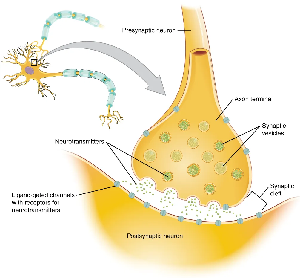
At a chemical synapse the bouton holds hundreds of synaptic vesicles about 40 nm across filled with transmitter, gathered at the active zone, where a scaffold of RIM, Munc13 and ELKS docks vesicles close to voltage-gated Ca²⁺ channels. When an action potential arrives, Ca²⁺ rushes in and binds synaptotagmin, and the SNARE complex zips the vesicle membrane into the plasma membrane in under a millisecond. Transmitter crosses the 20 nm cleft and binds receptors on the postsynaptic membrane, where the postsynaptic density concentrates glutamate receptors and the scaffold protein PSD-95. Vesicles are retrieved by ultrafast endocytosis within tens of milliseconds and refilled.[6][7][8]
A synapse’s effect is set by its receptors: glutamate acting on AMPA and NMDA receptors excites, GABA and glycine open Cl⁻ channels to inhibit, while noradrenaline, dopamine and serotonin act more slowly through G-protein-coupled receptors. At noradrenergic synapses the signal ends mainly when transmitter is pumped back into the bouton, the target of many antidepressants. Trans-synaptic adhesion molecules such as neurexins and neuroligins align pre- and postsynaptic membranes and specify which kind of synapse forms; mutations in these genes occur in autism. Electrical synapses through gap junctions are rarer but transmit instantly, synchronising groups of neurons.[6][9][10]
Neuroglia
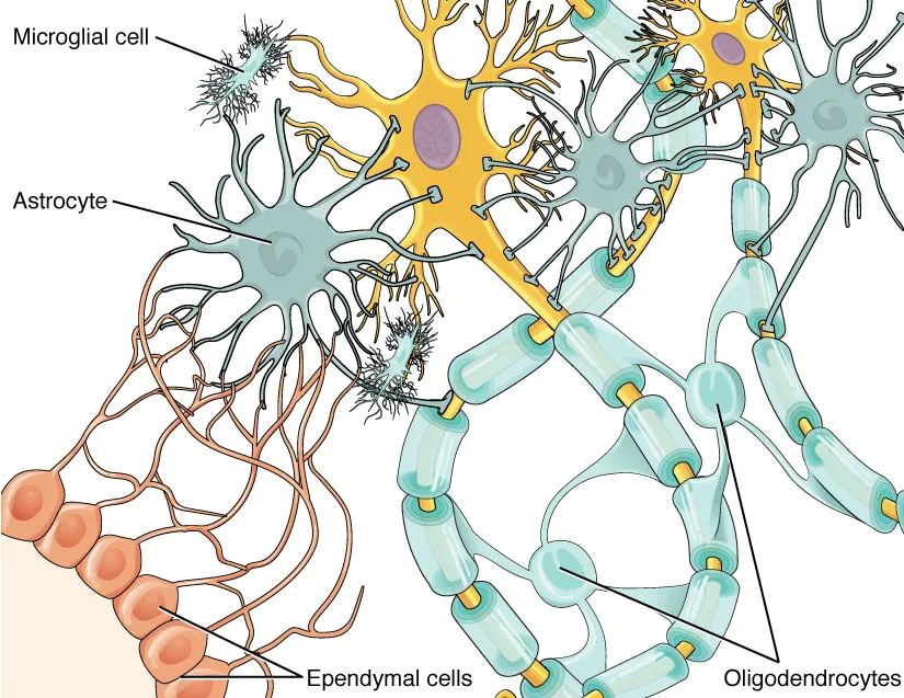
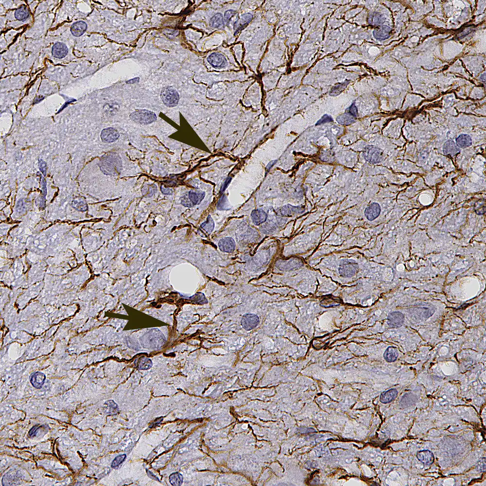
Glial cells are as numerous as neurons and fire no action potentials, yet every neural function depends on them. Astrocytes have hundreds of fine processes wrapping synapses and endfeet sheathing capillaries; they clear excess glutamate and K⁺ from the cleft, supply lactate to neurons, match blood flow to activity and with the endothelium form the blood–brain barrier. After injury they hypertrophy and form a glial scar. Ciliated ependymal cells line the ventricles and central canal and help produce and circulate cerebrospinal fluid.[11][12][13]
Microglia are the resident macrophages of the central nervous system, derived from the yolk sac in the embryo; their small bodies and branching processes constantly survey the surroundings, phagocytose dead cells and prune surplus synapses in development, and become activated in inflammation, neurodegeneration and brain tumours. Oligodendrocytes in the central nervous system and Schwann cells in peripheral nerves make myelin. Oligodendrocyte precursor cells remain scattered throughout the adult brain, continuing to generate new oligodendrocytes and even receiving synaptic input from neurons. In peripheral ganglia, satellite cells envelop neuronal cell bodies.[11][14][15]
Pruning of surplus synapses borrows the immune complement system: C1q and C3 tag weak or less active synapses, and microglia recognise the tag through complement receptor 3 and engulf them; astrocytes also phagocytose synapses through the MEGF10 and MERTK receptors. This refines circuits during development, but overactivity strips synapses in Alzheimer disease and after stroke, so complement inhibition is being tested as a neuroprotective strategy.[16]
Myelin and saltatory conduction
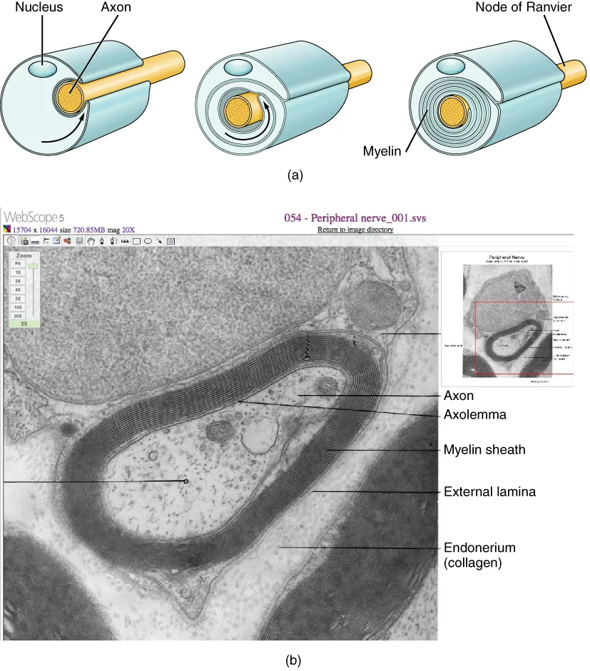
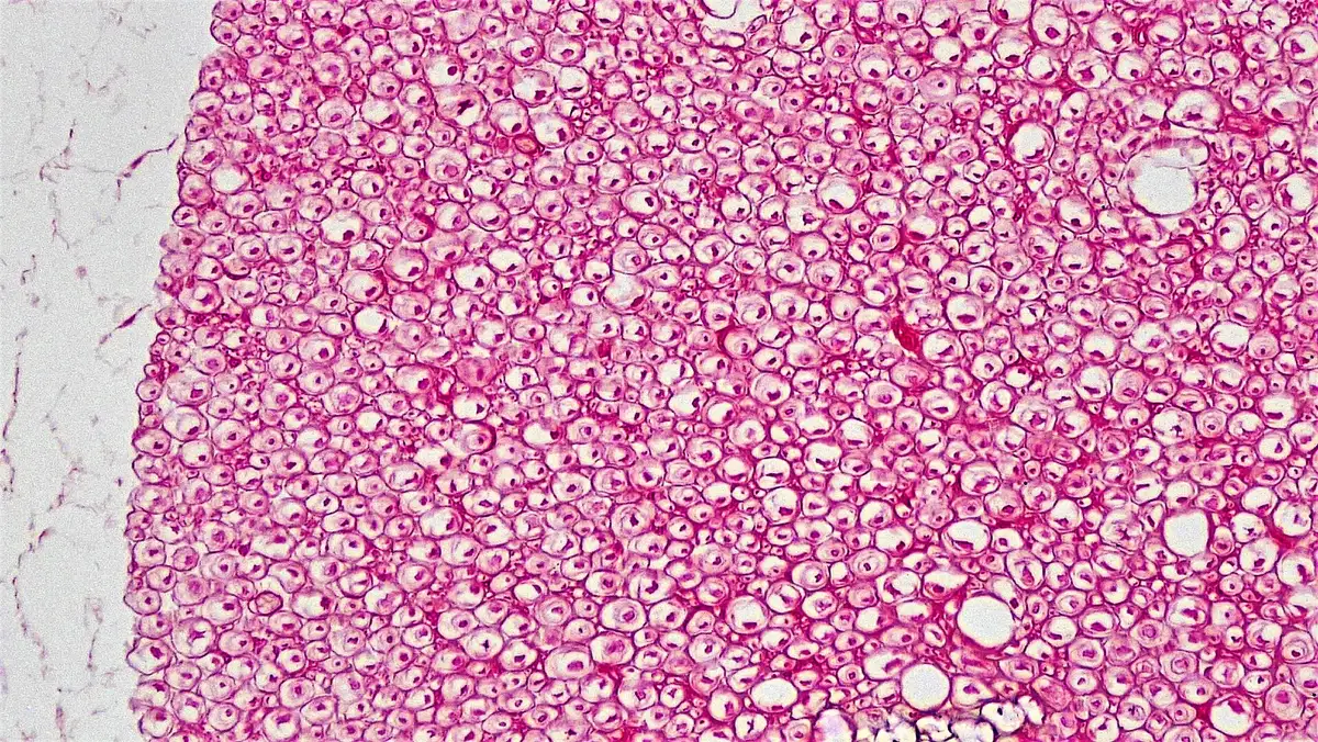
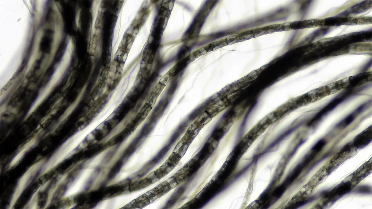
Myelin is glial membrane wrapped many times around the axon and compacted to squeeze out cytoplasm, forming a lipid-rich insulating sheath (about 70% lipid, rich in cholesterol and galactocerebroside). One oligodendrocyte can myelinate segments of dozens of different axons, whereas each Schwann cell wraps a single segment of a single axon. Between segments are nodes of Ranvier about 1 µm long, where Na⁺ channels are densely clustered; the action potential “jumps” from node to node, speeding conduction up to fiftyfold and saving energy. Large myelinated fibres conduct at up to 100 m/s, unmyelinated fibres at about 1 m/s.[17][18][19]
Myelin is more than insulation: oligodendrocytes supply lactate to the axon through channels within the sheath, and losing this support makes axons degenerate even with myelin intact. Myelination is most intense from late gestation to early adulthood, and adults still add myelin in response to activity when learning new skills. In multiple sclerosis the immune system attacks central myelin, slowing or blocking conduction; in Guillain–Barré syndrome and Charcot–Marie–Tooth disease the damage lies in Schwann-cell myelin. Peripheral nerves can regenerate because Schwann cells dedifferentiate into repair cells that guide regrowing axons.[20][21][22][23][24]
Schwann cells arise from neural-crest Schwann cell precursors that migrate along growing nerves, guided by axonal neuregulin-1 acting on ErbB2/3 receptors. The amount of neuregulin decides their fate: large axons are myelinated, whereas many small axons are wrapped together by one non-myelinating Schwann cell in a Remak bundle. Schwann cell precursors are also far more versatile: they give rise to parasympathetic neurons, the chromaffin cells of the adrenal medulla and a substantial share of enteric neurons.[25]
Full-text reading (StatPearls)
The StatPearls chapters on this topic, reproduced verbatim. Their CC BY-NC-ND 4.0 licence allows unmodified copies only, so they are not translated or shortened.
Histology, AstrocytesWei DC, Morrison EH · StatPearls 2023 · NBK545142 · 23k characters
Loading…
Wei DC, Morrison EH. Histology, Astrocytes. StatPearls [Internet]. Treasure Island (FL): StatPearls Publishing; 2023. NBK545142 · PMID 31424726 · CC BY-NC-ND 4.0. Reproduced verbatim, unmodified.
Physiology, SynapseCaire MJ, Reddy V, Varacallo MA · StatPearls 2023 · NBK526047 · 16k characters
Loading…
Caire MJ, Reddy V, Varacallo MA. Physiology, Synapse. StatPearls [Internet]. Treasure Island (FL): StatPearls Publishing; 2023. NBK526047 · PMID 30252303 · CC BY-NC-ND 4.0. Reproduced verbatim, unmodified.
Physiology, Noradrenergic SynapseHussain LS, Reddy V, Maani CV · StatPearls 2023 · NBK540977 · 16k characters
Loading…
Hussain LS, Reddy V, Maani CV. Physiology, Noradrenergic Synapse. StatPearls [Internet]. Treasure Island (FL): StatPearls Publishing; 2023. NBK540977 · PMID 31082021 · CC BY-NC-ND 4.0. Reproduced verbatim, unmodified.
Histology, Schwann CellsFallon M, Tadi P · StatPearls 2023 · NBK544316 · 13k characters
Loading…
Fallon M, Tadi P. Histology, Schwann Cells. StatPearls [Internet]. Treasure Island (FL): StatPearls Publishing; 2023. NBK544316 · PMID 31335036 · CC BY-NC-ND 4.0. Reproduced verbatim, unmodified.
Histology, MyelinJohnson JH, Al Khalili Y · StatPearls 2023 · NBK541009 · 8k characters
Loading…
Johnson JH, Al Khalili Y. Histology, Myelin. StatPearls [Internet]. Treasure Island (FL): StatPearls Publishing; 2023. NBK541009 · PMID 31082053 · CC BY-NC-ND 4.0. Reproduced verbatim, unmodified.
Histology, Glial CellsLudwig PE, Das JM · StatPearls 2023 · NBK441945 · 7k characters
Loading…
Ludwig PE, Das JM. Histology, Glial Cells. StatPearls [Internet]. Treasure Island (FL): StatPearls Publishing; 2023. NBK441945 · PMID 28722974 · CC BY-NC-ND 4.0. Reproduced verbatim, unmodified.
Organs in this system
References
Sources for the values and descriptions on this page, indexed in PubMed (US National Library of Medicine).
- [1]Pyramidal neurons: dendritic structure and synaptic integrationSpruston N, Nature reviews. Neuroscience 2008PMID 18270515
- [2]Architectures of neuronal circuitsLuo L, Science 2021PMID 34516844
- [3]Axon initial segment structure and function in health and diseaseJenkins PM et al., Physiological reviews 2025PMID 39480263
- [4]The function of the axon initial segment in neuronal polarityEichel K et al., Developmental biology 2022PMID 35640681
- [5]Contribution of Axon Initial Segment Structure and Channels to Brain PathologyGarrido JJ, Cells 2023PMID 37190119
- [6]Physiology, SynapseCaire MJ et al., StatPearls 2023PMID 30252303
- [7]The presynaptic active zoneSüdhof TC, Neuron 2012PMID 22794257
- [8]Synaptic Vesicle Recycling Through the Lens of Ultrafast EndocytosisWatanabe S, Annual review of neuroscience 2025PMID 40670291
- [9]Physiology, Noradrenergic SynapseHussain LS et al., StatPearls 2023PMID 31082021
- [10]Synaptic organizing complexesSiddiqui TJ et al., Current opinion in neurobiology 2011PMID 20832286
- [11]Histology, Glial CellsLudwig PE et al., StatPearls 2023PMID 28722974
- [12]Histology, AstrocytesWei DC et al., StatPearls 2023PMID 31424726
- [13]Neuronal regulation of the blood-brain barrier and neurovascular couplingKaplan L et al., Nature reviews. Neuroscience 2020PMID 32636528
- [14]Microglia in GliomaGarofalo S et al., Advances in neurobiology 2024PMID 39207710
- [15]Myelination-independent functions of oligodendrocyte precursor cells in health and diseaseXiao Y et al., Nature neuroscience 2023PMID 37653126
- [16]The Role of the Complement System in Synaptic Pruning after StrokeLiu H et al., Aging and disease 2024PMID 39012667
- [17]Histology, MyelinJohnson JH et al., StatPearls 2023PMID 31082053
- [18]Myelin in the Central Nervous System: Structure, Function, and PathologyStadelmann C et al., Physiological reviews 2019PMID 31066630
- [19]Myelination of the nervous system: mechanisms and functionsNave KA et al., Annual review of cell and developmental biology 2014PMID 25288117
- [20]Oligodendrocytes: Myelination and Axonal SupportSimons M et al., Cold Spring Harbor perspectives in biology 2015PMID 26101081
- [21]Activity-Dependent MyelinationKato D et al., Advances in experimental medicine and biology 2019PMID 31760637
- [22]Myelin BiologyBolino A, Neurotherapeutics 2021PMID 34244924
- [23]Schwann Cell Development and MyelinationSalzer J et al., Cold Spring Harbor perspectives in biology 2024PMID 38503507
- [24]Histology, Schwann CellsFallon M et al., StatPearls 2023PMID 31335036
- [25]Neuronal lineages derived from the nerve-associated Schwann cell precursorsKameneva P et al., Cellular and molecular life sciences 2021PMID 32748156