Quadriceps femoris
Model
Layers
Z-Anatomy anatomical model based on BodyParts3D, licensed CC BY-SA 4.0.
Description
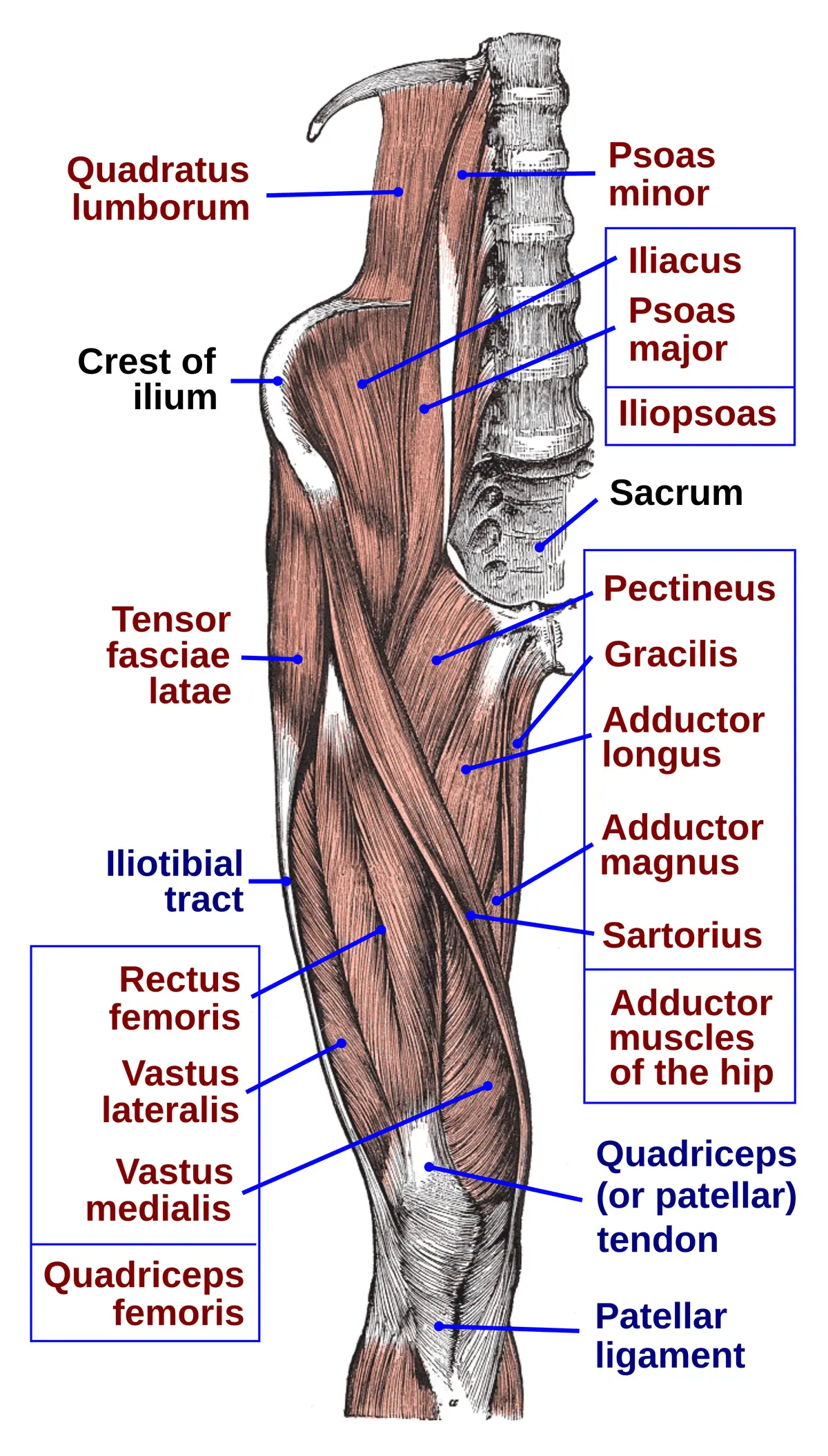
The anterior thigh group made up of rectus femoris, vastus lateralis, vastus medialis and vastus intermedius; inserting on the tibial tuberosity via the patellar ligament, it is the principal extensor of the knee.
Parts
Each part has its own card; press “Show on the model” to fly to it and select it in the 3D model above.
- 01
Rectus femoris
Musculus rectus femoris
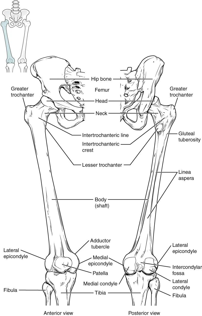
The only head that crosses two joints: it arises from the anterior inferior iliac spine (straight head) and above the acetabulum (reflected head), so it both flexes the hip and extends the knee. It is a bipennate muscle in the middle of the front of the thigh, often strained in kicking.
- 02
Vastus lateralis
Musculus vastus lateralis
The largest head, arising from the greater trochanter and the lateral lip of the linea aspera. It is the safe intramuscular injection site in infants (middle third of the anterolateral thigh) and a common muscle biopsy site.
- 03
Vastus medialis
Musculus vastus medialis
Arises from the intertrochanteric line and the medial lip of the linea aspera; its almost horizontal lower fibres (vastus medialis obliquus) pull the patella medially and resist lateral dislocation. It is the first to waste after knee injury or surgery.
- 04
Vastus intermedius
Musculus vastus intermedius
Lies deep to rectus femoris, arising from the anterior and lateral surfaces of the femoral shaft. A few deepest fibres form articularis genus, which pulls the suprapatellar bursa up during extension so it is not pinched.
- 05
Quadriceps tendon, patella and patellar ligament
Tendo musculi quadricipitis femoris
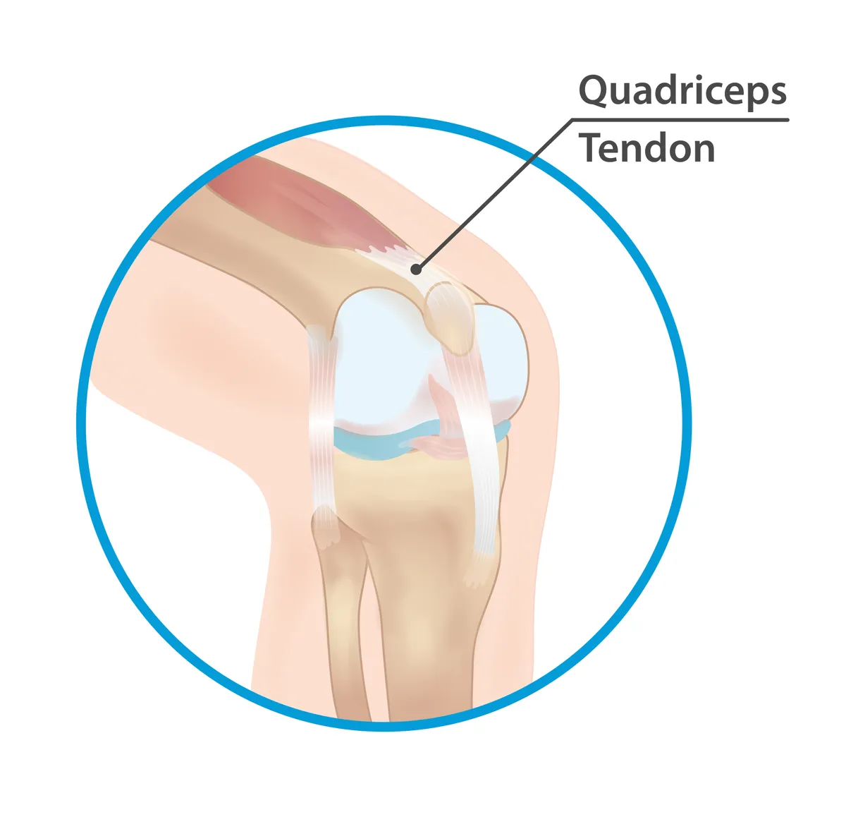
The four heads unite in the quadriceps tendon, which encloses the patella (the largest sesamoid bone) and continues as the patellar ligament to the tibial tuberosity. The patella increases the extensor lever arm; tapping the patellar ligament elicits the knee jerk (L3–L4).
- 06
Nerve and blood supply
Innervatio et vascularisatio
The femoral nerve (L2–L4) emerges below the inguinal ligament into the femoral triangle and branches to all four heads. Blood comes mainly from the lateral circumflex femoral artery and branches of the deep femoral artery. Femoral nerve palsy abolishes knee extension, making slopes and rising from a chair difficult.
Form and structure
The quadriceps femoris has four parts: rectus femoris (from the anterior inferior iliac spine, crossing the hip as well) and vastus lateralis, medialis and intermedius (from the femoral shaft). They form the quadriceps tendon onto the patella, continued by the patellar ligament to the tibial tuberosity.
Vessels and nerves
It is innervated by the femoral nerve (L2–L4); its main action is knee extension, and rectus femoris also flexes the hip. The patellar reflex tests the L3–L4 roots. The oblique part of vastus medialis keeps the patella tracking correctly during extension.
Fibre types and ageing
The quadriceps femoris contains a mix of type I and type II fibres, whose proportions vary markedly between individuals and change with training. It is also one of the muscles most affected by age-related wasting (sarcopenia), which reduces knee-extension strength and the ability to rise from a chair or climb stairs in older adults.[1][2]
Function and arthrogenic inhibition
The quadriceps comprises four muscles of the anterior thigh (rectus femoris, vastus medialis, intermedius and lateralis) and is the main knee extensor; rectus femoris crosses two joints and also flexes the hip. Common sports injuries include strains, tears, avulsions and contusions, mostly of rectus femoris.[3]
Arthritis, surgery or knee injury cause a long-lasting inability to fully activate the quadriceps, termed arthrogenic muscle inhibition. Altered discharge from sensory receptors of the damaged joint (swelling, inflammation, laxity) reduces spinal and cortical motor-neuron excitability; severity depends on damage, time since injury and knee angle. It is a major barrier to rehabilitation after ACL reconstruction.[4][5]
Quadriceps strengthening reduces pain and improves function in knee osteoarthritis, and neuromuscular electrical stimulation increases strength after ACL reconstruction. Prospective studies identify weak knee-extensor (quadriceps) strength as a predictor of patellofemoral pain.[6][7][8]
Full-text reading (StatPearls)
The StatPearls chapters on this topic, reproduced verbatim. Their CC BY-NC-ND 4.0 licence allows unmodified copies only, so they are not translated or shortened.
Anatomy, Bony Pelvis and Lower Limb: Thigh Quadriceps MuscleBordoni B, Varacallo MA · StatPearls 2023 · NBK513334 · 35k characters
Loading…
Bordoni B, Varacallo MA. Anatomy, Bony Pelvis and Lower Limb: Thigh Quadriceps Muscle. StatPearls [Internet]. Treasure Island (FL): StatPearls Publishing; 2023. NBK513334 · PMID 30020706 · CC BY-NC-ND 4.0. Reproduced verbatim, unmodified.
Organs in this system
References
Sources for the values and descriptions on this page, indexed in PubMed (US National Library of Medicine).
- [1]Fiber types in mammalian skeletal musclesSchiaffino S et al., Physiological reviews 2011PMID 22013216
- [2]Muscle aging and sarcopenia: The pathology, etiology, and most promising therapeutic targetsGrima-Terrén M et al., Molecular aspects of medicine 2024PMID 39312874
- [3]Quadriceps muscle injuries in athletes: a narrative reviewAriyaratne SP et al., The British journal of radiology 2025PMID 39989034
- [4]Quadriceps arthrogenic muscle inhibition: neural mechanisms and treatment perspectivesRice DA et al., Seminars in arthritis and rheumatism 2010PMID 19954822
- [5]Arthrogenic muscle inhibition after ACL reconstruction: a scoping review of the efficacy of interventionsSonnery-Cottet B et al., British journal of sports medicine 2019PMID 30194224
- [6]Effectiveness of Quadriceps Strength Training in Adults With Knee Osteoarthritis: A Systematized ReviewHegde DD et al., Musculoskeletal care 2025PMID 40462206
- [7]Neuromuscular electrical stimulation is effective in strengthening the quadriceps muscle after anterior cruciate ligament surgeryHauger AV et al., Knee surgery, sports traumatology, arthroscopy 2018PMID 28819679
- [8]Risk factors for patellofemoral pain: a systematic review and meta-analysisNeal BS et al., British journal of sports medicine 2019PMID 30242107
- [9]Anatomy, Bony Pelvis and Lower Limb, Gluteus Maximus MuscleStatPearls (NCBI Bookshelf)PMID 30855781Supports: Gluteus maximus