Humerus
Model
Hover or tap to see names, click to read a description; click a name in the legend to fly to it. Procedurally built to histological proportions; colours follow illustration conventions.
Description
The long bone of the arm, articulating with the scapula at the shoulder and with the radius and ulna at the elbow. Many important muscles and nerves attach to or run close to it.
Parts
Each part has its own card; press “Show on the model” to fly to it and select it in the 3D model above.
- 01
Head and anatomical neck
Caput et collum anatomicum humeri
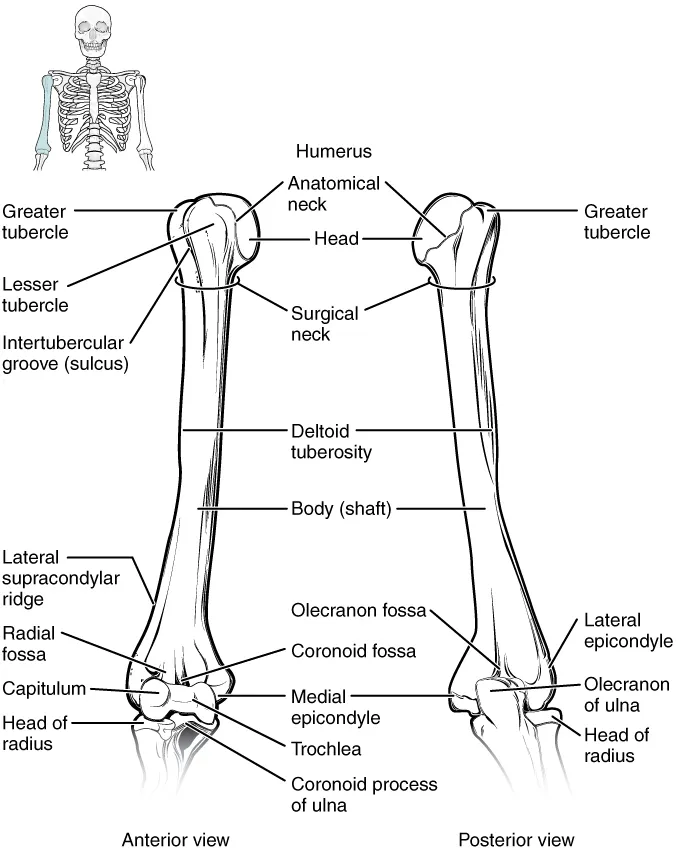
The head, about a third of a sphere, faces medially, upwards and backwards to meet the glenoid cavity of the scapula. The glenoid is only about a third the size of the head, making the shoulder the most mobile and most often dislocated joint, usually anteriorly. The anatomical neck is the narrow groove around the head where the capsule attaches.
- 02
Greater and lesser tubercles
Tuberculum majus et minus
The greater tubercle laterally receives three rotator cuff muscles (supraspinatus, infraspinatus, teres minor); the lesser tubercle in front receives subscapularis. Between them the intertubercular groove carries the tendon of the long head of biceps, held by the transverse humeral ligament.
- 03
Surgical neck
Collum chirurgicum humeri
The constriction just below the tubercles where the upper end joins the shaft, a common fracture site in osteoporotic elderly people. The axillary nerve and posterior circumflex humeral artery wind around it and can be injured, causing deltoid paralysis and loss of sensation over the “regimental badge” area.
- 04
Shaft
Corpus humeri
Cylindrical above and triangular in section below. The lateral surface bears the deltoid tuberosity; the posterior surface has the radial groove running obliquely down and out, carrying the radial nerve and the deep brachial artery. A midshaft fracture can injure the radial nerve and cause wrist drop.
- 05
Distal end
Extremitas distalis humeri
The condyle comprises the capitulum (meeting the radial head) and the trochlea (meeting the trochlear notch of the ulna). Above lie the coronoid fossa in front and the olecranon fossa behind, which take the corresponding ulnar processes in flexion and extension. Supracondylar fracture is the most common elbow fracture in children and endangers the brachial artery.
- 06
Medial and lateral epicondyles
Epicondyli humeri
The lateral epicondyle is the common origin of the forearm extensors; inflammation of this origin is “tennis elbow”. The larger medial epicondyle is the common flexor origin (“golfer’s elbow”); the ulnar nerve runs in a groove just behind it, where a knock sends tingling down to the little finger.
Position and relations
The proximal end has the head (articulating with the glenoid), the anatomical neck, the greater and lesser tubercles (rotator cuff attachments) separated by the intertubercular groove for the long head of biceps, and the surgical neck, a common fracture site in older people. The shaft bears the deltoid tuberosity and the radial groove running obliquely behind. The distal end has the condyle (capitulum for the radius, trochlea for the ulna) and two epicondyles.
Form and structure
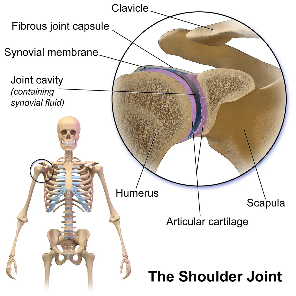
The shoulder is the most mobile joint because a large head meets a shallow glenoid; stability relies mainly on the labrum, capsule and rotator cuff muscles (supraspinatus, infraspinatus, teres minor, subscapularis). Shoulder dislocation (usually anterior) is therefore common.
Vessels and nerves
The bone is supplied by the anterior and posterior circumflex humeral arteries (around the surgical neck) and the profunda brachii. Three nerves lie on it: the axillary at the surgical neck, the radial in the radial groove of the shaft and the ulnar behind the medial epicondyle; each fracture site has its typical palsy.
Landmarks and proximal humerus fractures
The humerus is the largest bone of the upper limb. Proximally its head forms a ball-and-socket joint with the glenoid cavity of the scapula. The anatomical neck, the remnant of the epiphyseal plate, separates the head from the greater and lesser tubercles, with the intertubercular groove between them. Below lies the surgical neck, a frequent fracture site. The cylindrical shaft carries the deltoid tuberosity laterally and the radial (spiral) groove posteriorly.[1]
Distally the bone widens into the medial and lateral epicondyles and ends in the condyle: the capitulum articulates with the radial head and the trochlea with the trochlear notch of the ulna. Above the trochlea the coronoid fossa receives the ulnar coronoid process, and above the capitulum the radial fossa receives the radial head in elbow flexion; posteriorly the olecranon fossa receives the olecranon.[1]
Proximal humerus fractures are common. The ascending branch of the anterior circumflex humeral artery perfuses most of the head. The greater tuberosity tends to displace posterosuperiorly and the lesser tuberosity and shaft medially; the head may be pulled by an attached tuberosity, impacted in valgus or, in severe cases, dislocated, impacted or split. Local osteopenia can compromise fixation, and fixed-angle devices anchored in the strongest bone have improved results.[2]
Full-text reading (StatPearls)
The StatPearls chapters on this topic, reproduced verbatim. Their CC BY-NC-ND 4.0 licence allows unmodified copies only, so they are not translated or shortened.
Anatomy, Shoulder and Upper Limb, HumerusMostafa E, Imonugo O, Varacallo MA · StatPearls 2023 · NBK534821 · 18k characters
Loading…
Mostafa E, Imonugo O, Varacallo MA. Anatomy, Shoulder and Upper Limb, Humerus. StatPearls [Internet]. Treasure Island (FL): StatPearls Publishing; 2023. NBK534821 · PMID 30521242 · CC BY-NC-ND 4.0. Reproduced verbatim, unmodified.
Organs in this system
References
Sources for the values and descriptions on this page, indexed in PubMed (US National Library of Medicine).
- [1]Anatomy, Shoulder and Upper Limb, HumerusMostafa E et al., StatPearls 2023PMID 30521242
- [2]Proximal humerus fracturesSanchez-Sotelo J, Clinical anatomy 2006PMID 16506205
- [3]Anatomy, BonesStatPearls (NCBI Bookshelf)PMID 30725884Supports: Number of bones
- [4]Calcium metabolism in health and diseasePeacock M, 2010PMID 20089499Supports: Skeletal calcium store