Salivary glands
Model
Layers
Z-Anatomy anatomical model based on BodyParts3D, licensed CC BY-SA 4.0.
Description
Three pairs of major salivary glands (parotid, submandibular, sublingual) and hundreds of minor glands secrete about 0.5–1.5 litres of saliva a day, which moistens food, starts starch digestion with amylase and protects the teeth and mucosa.
Parts
Each part has its own card; press “Show on the model” to fly to it and select it in the 3D model above.
- 01
Parotid gland
Glandula parotidea
The largest, purely serous salivary gland, in front of the ear and behind the ramus of the mandible within a tough capsule. The facial nerve and its branches pass through it (dividing it into superficial and deep lobes), so parotid surgery risks facial palsy; secretomotor supply comes from IX via the otic ganglion.
- 02
Parotid duct (Stensen)
Ductus parotideus
About 5 cm long, it crosses masseter a fingerbreadth below the zygomatic arch, pierces buccinator and opens into the mouth opposite the upper second molar. An accessory parotid gland sometimes lies along it.
- 03
Submandibular gland
Glandula submandibularis
A mixed, mainly serous gland in the submandibular triangle, folded round the posterior border of mylohyoid into superficial and deep parts. It produces about 70% of resting saliva; its secretomotor supply is the chorda tympani (VII) via the submandibular ganglion.
- 04
Submandibular duct (Wharton)
Ductus submandibularis
About 5 cm long, it runs forward in the floor of the mouth, crossing the lingual nerve, and opens at the sublingual caruncle beside the frenulum. Its long uphill course and more viscous saliva explain why about 80% of salivary stones form here.
- 05
Sublingual gland
Glandula sublingualis
The smallest of the three major glands, mainly mucous, lying under the mucosa of the floor of the mouth on mylohyoid and opening by 8–20 small ducts (of Rivinus) on the sublingual fold. Duct obstruction causes a mucous cyst of the floor of the mouth (ranula).
- 06
Minor salivary glands
Glandulae salivariae minores
Some 600–1000 small glands scattered beneath the mucosa of lips, cheeks, palate and tongue, secreting continuously to keep the mucosa moist. Lower-lip gland biopsy helps diagnose Sjögren syndrome, and they are the usual source of labial mucoceles.
Position and relations
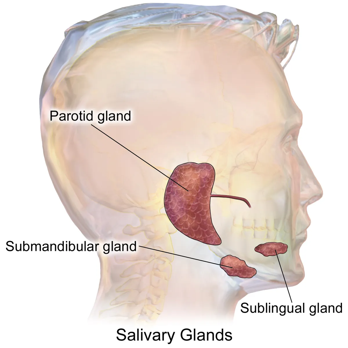
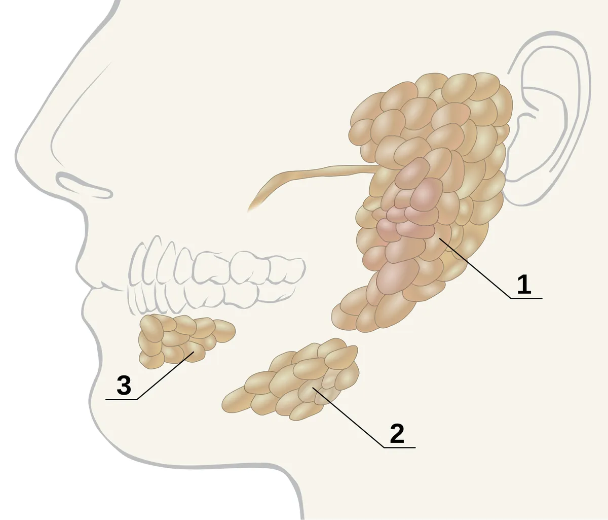
The parotid, the largest, lies in front of the ear behind the mandibular ramus; its duct (Stensen) crosses masseter, pierces buccinator and opens into the oral vestibule opposite the second upper molar. The submandibular gland lies in the submandibular triangle, its duct (Wharton) opening at the sublingual papilla beside the frenulum. The sublingual gland lies under the mucosa of the floor of the mouth and drains by many small ducts.
Form and structure
The parotid is almost purely serous, the submandibular mixed but mostly serous, the sublingual mixed but mostly mucous. Each gland has acini that make primary saliva, intercalated ducts, striated ducts that modify electrolytes, and myoepithelial cells that squeeze the acini. The facial nerve runs through the parotid, dividing it into superficial and deep lobes.
Vessels and nerves
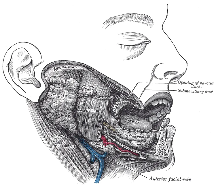
The parotid is supplied by branches of the external carotid (superficial temporal, transverse facial), the submandibular and sublingual glands by the facial and lingual arteries. Secretomotor parasympathetic fibres reach the parotid from IX via the otic ganglion, and the submandibular and sublingual glands from VII via the chorda tympani and submandibular ganglion; sympathetic input thickens saliva.
The three paired major glands
The parotid is the largest salivary gland. It occupies the retromandibular fossa, bounded above by the zygomatic arch, in front by the masseter and behind by the sternocleidomastoid; its superficial lobe extends forward over the mandibular ramus and the back of the masseter. The facial nerve runs through the gland, which demands particular care during parotidectomy.[1][2]
The submandibular gland, second in size, lies in the submandibular triangle; its superficial and deep lobes are separated by the mylohyoid. Wharton’s duct opens at the sublingual caruncle beside the frenulum. At rest the submandibular gland makes about 70% of saliva, whereas the parotid dominates under stimulation. The sublingual gland, the smallest, lies among the floor-of-mouth muscles and drains through the many ducts of Rivinus and Bartholin’s duct, which joins Wharton’s duct near the caruncle; it contributes about 5% of saliva.[3][4]
Salivary secretion is a nerve-mediated reflex whose volume depends on the intensity and type of taste, chewing or tactile stimulus: long periods of low resting flow alternate with short bursts of high flow during meals. Cholinergic parasympathetic nerves (via muscarinic M3 receptors) and adrenergic sympathetic nerves both stimulate acinar cells, which secrete an isotonic Na⁺- and Cl⁻-rich fluid that the ducts render hypotonic. Dry mouth during anxiety shows how higher centres modulate the reflex.[5]
Full-text reading (StatPearls)
The StatPearls chapters on this topic, reproduced verbatim. Their CC BY-NC-ND 4.0 licence allows unmodified copies only, so they are not translated or shortened.
Anatomy, Head and Neck, Sublingual GlandGrewal JS, Bordoni B, Shah J, Ryan J · StatPearls 2023 · NBK535426 · 10k characters
Loading…
Grewal JS, Bordoni B, Shah J, Ryan J. Anatomy, Head and Neck, Sublingual Gland. StatPearls [Internet]. Treasure Island (FL): StatPearls Publishing; 2023. NBK535426 · PMID 30571047 · CC BY-NC-ND 4.0. Reproduced verbatim, unmodified.
Anatomy, Head and Neck, Submandibular GlandGrewal JS, Jamal Z, Ryan J · StatPearls 2022 · NBK542272 · 10k characters
Loading…
Grewal JS, Jamal Z, Ryan J. Anatomy, Head and Neck, Submandibular Gland. StatPearls [Internet]. Treasure Island (FL): StatPearls Publishing; 2022. NBK542272 · PMID 31194412 · CC BY-NC-ND 4.0. Reproduced verbatim, unmodified.
Anatomy, Head and Neck, Parotid GlandChason HM, Downs BW · StatPearls 2024 · NBK534225 · 9k characters
Loading…
Chason HM, Downs BW. Anatomy, Head and Neck, Parotid Gland. StatPearls [Internet]. Treasure Island (FL): StatPearls Publishing; 2024. NBK534225 · PMID 30480958 · CC BY-NC-ND 4.0. Reproduced verbatim, unmodified.
Organs in this system
References
Sources for the values and descriptions on this page, indexed in PubMed (US National Library of Medicine).
- [1]Anatomy, Head and Neck, Parotid GlandChason HM et al., StatPearls 2024PMID 30480958
- [2]Facial Nerve and Parotid Gland AnatomyKochhar A et al., Otolaryngologic clinics of North America 2016PMID 27040583
- [3]Anatomy, Head and Neck, Submandibular GlandGrewal JS et al., StatPearls 2022PMID 31194412
- [4]Anatomy, Head and Neck, Sublingual GlandGrewal JS et al., StatPearls 2023PMID 30571047
- [5]The physiology of salivary secretionProctor GB, Periodontology 2000 2016PMID 26662479
- [6]Gastrointestinal mean transit times in young and middle-aged healthy subjectsPMID 11318834Supports: Gastrointestinal transit time
- [7]Revised Estimates for the Number of Human and Bacteria Cells in the BodySender R, Fuchs S, Milo R, 2016PMID 27541692Supports: Colonic bacterial count