Kidneys
Model
Hover or tap to see names, click to read a description; click a name in the legend to fly to it. Procedurally built to histological proportions; colours follow illustration conventions.
Description
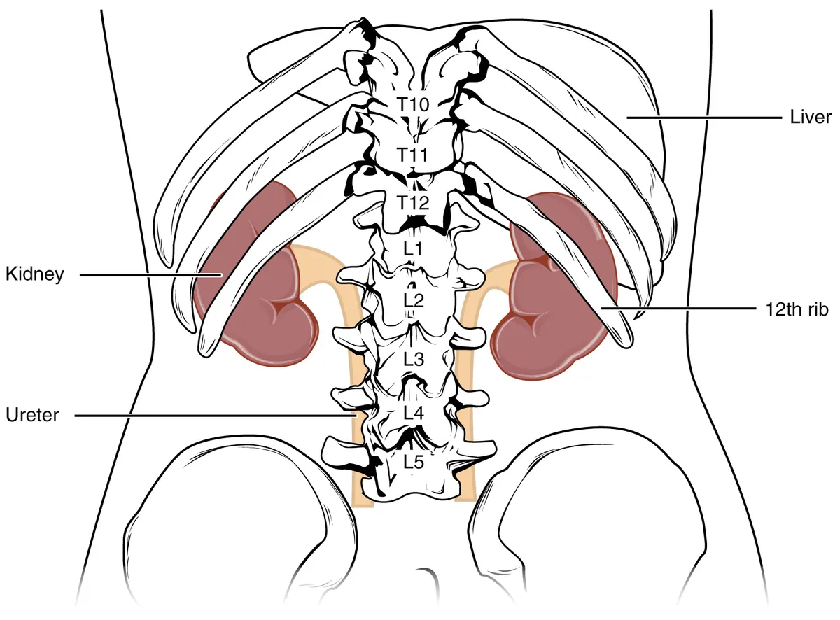
Bean-shaped retroperitoneal organs lying either side of the vertebral column at the T12–L3 level. Each kidney contains about one million nephrons.
Parts
Each part has its own card; press “Show on the model” to fly to it and select it in the 3D model above.
- 01
Capsule, perirenal fat and renal fascia
Capsula fibrosa; Capsula adiposa; Fascia renalis
From inside out: a thin fibrous capsule that strips easily from a healthy kidney, perirenal fat, the renal (Gerota) fascia enclosing kidney and adrenal, and pararenal fat. The fascia contains perirenal haematomas or collections and is open below, so the kidney can descend.
- 02
Renal cortex
Cortex renalis
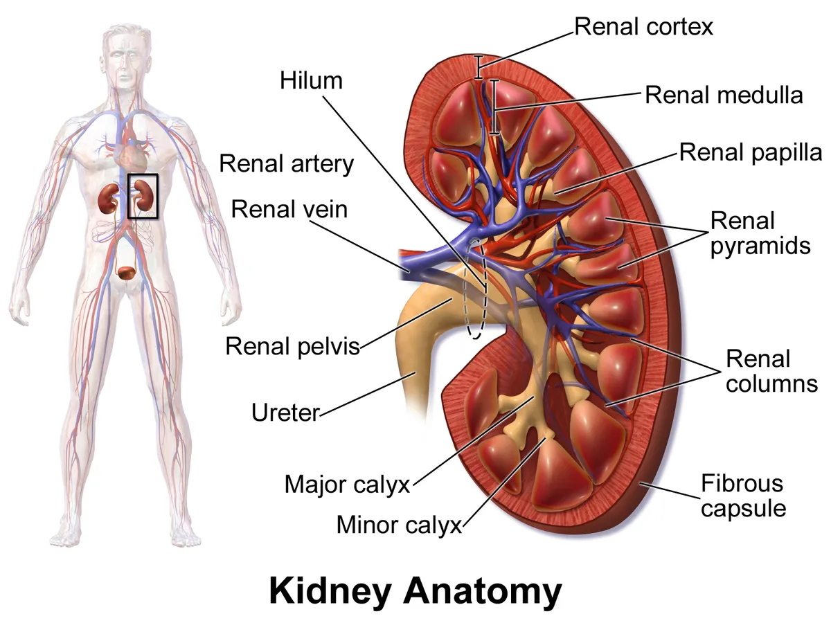
The outer layer, about 1 cm thick and red-brown, holding the renal corpuscles and proximal and distal convoluted tubules; it receives about 90% of renal blood flow. Medullary rays (straight collecting ducts) run up into it, dividing it into lobules.
- 03
Medulla and renal pyramids
Medulla renalis; Pyramides renales
The medulla consists of 8–18 striated pyramids (loops of Henle, collecting ducts, vasa recta) whose bases face the cortex. Its countercurrent arrangement builds an osmotic gradient rising to about 1200 mOsm/kg at the papilla, allowing urine to be concentrated.
- 04
Renal columns (of Bertin)
Columnae renales
Bands of cortical tissue extending between the pyramids and carrying the interlobar vessels. A prominent column can mimic a tumour on ultrasound but is a normal variant.
- 05
Renal papillae
Papillae renales
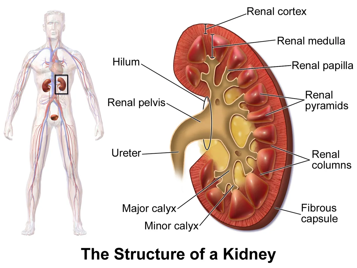
The apex of each pyramid projects into a minor calyx; its area cribrosa carries 10–25 openings of the papillary ducts (of Bellini) that discharge urine. Supplied by the end of the vasa recta, papillae are prone to necrosis (diabetes, analgesic abuse).
- 06
Minor and major calyces
Calices renales minores et majores
Each minor calyx (7–14) cups one or more papillae; they join into 2–3 major calyces (upper, middle, lower) that open into the pelvis. Smooth muscle in their walls propels urine; lower-pole calyceal stones clear least readily.
- 07
Renal pelvis and sinus
Pelvis renalis; Sinus renalis
The renal sinus is the space within the hilum containing fat, calyces, pelvis and vessels. The funnel-shaped pelvis collects urine from the calyces and narrows into the ureter at the pelviureteric junction, the first narrowing, a common site of stone impaction and congenital obstruction.
- 08
Renal hilum
Hilum renale
A slit on the medial border at L1 where structures are arranged from front to back as renal vein, renal artery and pelvis. The left kidney lies about 1–2 cm higher than the right because the liver pushes the right one down.
- 09
Renal vessels
Arteria et vena renalis; Arteriae segmentales
The renal artery gives five segmental (end) arteries → interlobar arteries in the columns → arcuate arteries at the pyramid bases → interlobular arteries → afferent arterioles. The kidneys receive about 20–25% of cardiac output; the relatively avascular plane (of Brödel) behind the lateral border is used for nephrolithotomy.
- 10
Nephron
Nephronum
The functional unit (about a million per kidney): the glomerulus filters some 180 L a day, the proximal tubule reabsorbs about two-thirds, the loop of Henle builds the gradient, and the distal tubule and collecting duct fine-tune under aldosterone and ADH. Juxtamedullary nephrons have long loops reaching the inner medulla.
Position and relations
The two kidneys lie retroperitoneally on either side of the spine, from T12 to L3; the right is 1–2 cm lower than the left, pushed down by the liver. Each is enclosed by a fibrous capsule, perirenal fat and the renal fascia (Gerota’s). The suprarenal gland caps the upper pole.
Form and structure
In coronal section, the kidney shows an outer cortex and an inner medulla of 8–18 renal pyramids whose apices (papillae) drain urine into minor calyces, major calyces and the renal pelvis. Each kidney has about one million nephrons, each made of a glomerulus and a tubule (proximal convoluted tubule, loop of Henle, distal convoluted tubule, collecting duct).[1]
Vessels and nerves
The renal artery arises directly from the abdominal aorta at L1–L2 and divides into segmental arteries that are end arteries (without anastomoses), so occlusion of one causes infarction of a renal segment. The kidneys receive about 20–25% of cardiac output.
The nephron
Each nephron consists of a glomerulus and a tubule. Filtrate crosses fenestrated endothelium, the glomerular basement membrane (laminins, type IV collagen) and the slit diaphragms between podocyte foot processes. Damage to this barrier lets proteins into the tubule, triggering tubulointerstitial inflammation and fibrosis; conversely, tubular injury feeds back to harm the glomerulus.[2][3]
Glomerulus and tubule
The glomerulus is a unique capillary tuft: fenestrated endothelium, a complex protein mesh forming the glomerular basement membrane, and visceral epithelial cells (podocytes) whose interdigitating foot processes create slit diaphragms. This three-layer filter allows continuous filtration of plasma; single-nephron filtration is set by physical forces (hydrostatic and oncotic pressures, filtration coefficient).[4][2]
Nephron number is determined during fetal life: a small pool of nephron progenitors must balance epithelial differentiation with self-renewal to generate large numbers of stereotypically arrayed nephrons. No new nephrons form after birth, so the initial endowment influences later kidney disease risk.[5]
Claudins are tight-junction proteins lining the paracellular pore and set the selective permeability of each tubule segment; mutations in two claudin genes cause familial hypomagnesaemia with hypercalciuria and nephrocalcinosis. The kidney also links acid–base and potassium balance: acidosis reduces K⁺ secretion in the collecting duct, alkalosis causes hypokalaemia, and potassium depletion increases H⁺ secretion and ammoniagenesis.[6][7]
Clinical correlations
Glomeruli and tubules interact as chronic kidney disease progresses: a damaged filtration barrier lets serum proteins, complement and cytokines reach tubular cells and promotes tubulointerstitial fibrosis, while tubular injury in turn affects glomeruli. Acute tubular injury is therefore a driving force toward chronic kidney disease.[3][8]
Glomerular hyperfiltration (each remaining nephron filtering more) is adaptive but damages glomeruli over time and is a therapeutic target in chronic kidney disease; in diabetic kidney disease, podocyte death is a key step towards proteinuria.[9][10]
Full-text reading (StatPearls)
The StatPearls chapters on this topic, reproduced verbatim. Their CC BY-NC-ND 4.0 licence allows unmodified copies only, so they are not translated or shortened.
Anatomy, Abdomen and Pelvis: KidneysGarza FA, Leslie SW · StatPearls 2025 · NBK482385 · 28k characters
Loading…
Garza FA, Leslie SW. Anatomy, Abdomen and Pelvis: Kidneys. StatPearls [Internet]. Treasure Island (FL): StatPearls Publishing; 2025. NBK482385 · PMID 29494007 · CC BY-NC-ND 4.0. Reproduced verbatim, unmodified.
Anatomy, Retroperitoneum: Nerve Supply to the KidneysOfumwengbe O, Leslie SW · StatPearls 2025 · NBK459339 · 12k characters
Loading…
Ofumwengbe O, Leslie SW. Anatomy, Retroperitoneum: Nerve Supply to the Kidneys. StatPearls [Internet]. Treasure Island (FL): StatPearls Publishing; 2025. NBK459339 · PMID 29083631 · CC BY-NC-ND 4.0. Reproduced verbatim, unmodified.
Organs in this system
References
Sources for the values and descriptions on this page, indexed in PubMed (US National Library of Medicine).
- [1]Design-based stereological methods for estimating numbers of glomerular podocytesPuelles VG et al., Annals of anatomy = Anatomischer Anzeiger 2014PMID 23845787
- [2]Complexities of the glomerular basement membraneNaylor RW et al., Nature reviews. Nephrology 2021PMID 32839582
- [3]Crosstalk between glomeruli and tubulesFogo AB et al., Nature reviews. Nephrology 2025PMID 39643696
- [4]The glomerulus: the sphere of influencePollak MR et al., Clinical journal of the American Society of Nephrology 2014PMID 24875196
- [5]Kidney Nephron DeterminationOxburgh L, Annual review of cell and developmental biology 2018PMID 30125139
- [6]Biology of claudinsAngelow S et al., American journal of physiology. Renal physiology 2008PMID 18480174
- [7]Acid-base and potassium homeostasisLee Hamm L et al., Seminars in nephrology 2013PMID 23953803
- [8]Renal tubule injury: a driving force toward chronic kidney diseaseLiu BC et al., Kidney international 2018PMID 29361307
- [9]Glomerular hyperfiltration as a therapeutic target for CKDKanbay M et al., Nephrology, dialysis, transplantation 2024PMID 38308513
- [10]Podocyte Death in Diabetic Kidney Disease: Potential Molecular Mechanisms and Therapeutic TargetsZhong S et al., International journal of molecular sciences 2024PMID 39201721
- [11]Normal reference values for glomerular filtration rate: what do we really know?Delanaye P et al., 2012PMID 22802582Supports: Glomerular filtration rate