Abdomen and pelvis
Model
Layers
Z-Anatomy anatomical model based on BodyParts3D, licensed CC BY-SA 4.0.
Overview
A regional model of the abdomen and pelvis: the anterolateral and posterior abdominal walls, lumbar spine and bony pelvis, liver and biliary tree, stomach, pancreas, small and large intestine, kidneys, ureters and bladder, suprarenal glands, genital organs, the abdominal aorta and its branches, inferior vena cava and portal system, the lumbosacral plexus and lymph nodes.
Boundaries and subdivisions
The abdominal cavity is bounded above by the diaphragm, opens below into the pelvis through the pelvic inlet, has the lumbar spine with psoas and quadratus lumborum behind, and the musculoaponeurotic wall in front and at the sides. Clinically the anterior wall is divided into nine regions by the two midclavicular lines and two horizontal planes (subcostal and intertubercular), or into four quadrants through the umbilicus. The peritoneum lines the inner wall and covers the viscera; intraperitoneal organs (stomach, jejunum and ileum, spleen) hang on mesenteries, whereas retroperitoneal structures (most of the duodenum, pancreas, kidneys, ureters, abdominal aorta, inferior vena cava) lie against the posterior wall.[1][2][3]
Layers from superficial to deep
The anterolateral wall consists of skin, superficial fascia (fatty Camper and membranous Scarpa layers), the three flat muscles external oblique, internal oblique and transversus abdominis, whose aponeuroses blend to form the rectus sheath and the linea alba, then transversalis fascia, extraperitoneal fat and parietal peritoneum. Rectus abdominis lies within its sheath and is crossed by tendinous intersections. The posterior wall is formed by psoas major, iliacus and quadratus lumborum; iliopsoas inserts on the lesser trochanter of the femur. Within the peritoneal cavity the greater omentum hangs from the greater curvature of the stomach in front of the bowel, and the lesser omentum joins the stomach to the liver and bounds the omental bursa.[4][5]
Vessels, nerves and lymphatics
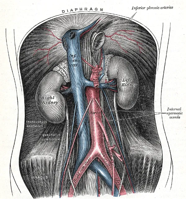
The abdominal aorta passes through the aortic hiatus at T12 and bifurcates into the common iliac arteries at about L4. Its three unpaired visceral branches are the coeliac trunk (foregut: stomach, liver, spleen, upper duodenum), the superior mesenteric artery (midgut) and the inferior mesenteric artery (hindgut); the paired branches are the inferior phrenic, suprarenal, renal, gonadal and lumbar arteries. The inferior vena cava lies to the right of the aorta. Blood from the gut, spleen and pancreas drains into the portal vein (formed by the superior mesenteric and splenic veins behind the neck of the pancreas) and then into the liver. The renal nerves travel in the renal plexus, receiving fibres from the sympathetic trunk and the vagus.[6][7][8][9][10]
Embryology
In weeks 3 and 4 the embryonic disc folds in four directions (cranial, caudal and two lateral folds): the ectoderm rolls into the neural tube while endoderm and mesoderm fold the opposite way into the gut tube and ventral body wall. The lateral folds meet at the umbilical ring by the end of week 4; the muscles and fascia of the abdominal wall come from mesoderm, and the thoracoabdominal nerves keep their segmental order, supplying the wall in bands. The peritoneum is also mesodermal: the parietal layer from somatic and the visceral layer from splanchnic mesoderm; the primitive gut hangs on ventral and dorsal mesenteries, and the ventral mesentery later becomes the lesser omentum and falciform ligament. In the female, the ovaries are the first reproductive organs to develop, from the gonadal ridge on the mesonephros, before descending into the pelvis. Renal innervation appears at about 9 weeks, and renal nerves help guide kidney development.[11][12][13][14][10]
Anatomical variants
The anterolateral abdominal wall shows “classic” anatomy in only just over half of specimens: sensory territories vary, pyramidalis may be present or absent, an accessory internal oblique may occur, and the composition of the anterior and posterior rectus sheath differs. The ilioinguinal nerve arises from L1 alone in only 65% of people; otherwise from T12–L1 (14%), L1–L2 (11%) or L2–L3 (10%), which explains some failed ilioinguinal blocks. The femoral nerve may split in the pelvis around accessory psoas slips. The uterine artery usually arises from the anterior division of the internal iliac in a common trunk with the umbilical artery (about 81% in a series of 218 patients). The renal plexus collects fibres from the aorticorenal ganglia and the lumbar and least splanchnic nerves and follows the main renal artery, segmental branches and accessory renal arteries at variable intervals. Failure of the abdominal wall muscles to form produces prune belly syndrome, with undescended testes and urinary tract malformations.[15][16][14][10][11]
Clinical correlations
Abdominal aortic aneurysms usually lie below the renal arteries and may be felt as a pulsatile mass around the umbilicus. Portal hypertension (as in cirrhosis) opens portosystemic anastomoses, causing oesophageal varices and periumbilical collateral veins. A psoas abscess can track down within the muscle sheath beneath the inguinal ligament and present as a mass in the thigh. Knowing the regions of the abdominal wall helps localise pain: appendicitis pain, for example, starts around the umbilicus and then settles in the right iliac fossa.[7][9][4][17]
Surgical and procedural considerations
Choosing an incision balances exposure against morbidity (cosmesis, wound healing, incisional hernia): an emergency exploratory laparotomy uses a long vertical midline incision, whereas elective surgery may use transverse or paramedian incisions with fewer hernias. The transversus abdominis plane block, between transversus abdominis and internal oblique, anaesthetises the thoracoabdominal, iliohypogastric and ilioinguinal nerves that run in that plane. Lateral approaches to the lumbar spine can injure the lumbar plexus within psoas major; bony landmarks such as the midline of the vertebral bodies, the iliac crests and the anterior superior iliac spines help define safe corridors. At caesarean section the bladder must be identified, especially when previous scarring tethers it to the anterior uterine wall, and any uterine surgery requires identification of the ureter passing under the uterine artery. A transplanted kidney loses its sympathetic supply but still autoregulates blood flow through myogenic and tubuloglomerular mechanisms.[12][18][16][14][10]
Key structures
Vagina musculi recti abdominis
- Name
- Rectus sheath
- Position, role
- Formed by the aponeuroses of the three flat muscles; encloses rectus abdominis
Linea alba
- Name
- Linea alba
- Position, role
- Midline raphe where the aponeuroses interlace
Omentum majus
- Name
- Greater omentum
- Position, role
- Hangs from the greater curvature in front of the bowel
Bursa omentalis
- Name
- Omental bursa
- Position, role
- Behind the stomach; opens through the omental foramen
Truncus coeliacus
- Name
- Coeliac trunk
- Position, role
- At T12; supplies liver, stomach and spleen
Vena portae hepatis
- Name
- Hepatic portal vein
- Position, role
- Formed behind the neck of the pancreas; brings gut blood to the liver
Spatium retroperitoneale
- Name
- Retroperitoneal space
- Position, role
- Kidneys, pancreas, duodenum, aorta, inferior vena cava
| Terminologia Anatomica | Name | Position, role |
|---|---|---|
| Vagina musculi recti abdominis | Rectus sheath | Formed by the aponeuroses of the three flat muscles; encloses rectus abdominis |
| Linea alba | Linea alba | Midline raphe where the aponeuroses interlace |
| Omentum majus | Greater omentum | Hangs from the greater curvature in front of the bowel |
| Bursa omentalis | Omental bursa | Behind the stomach; opens through the omental foramen |
| Truncus coeliacus | Coeliac trunk | At T12; supplies liver, stomach and spleen |
| Vena portae hepatis | Hepatic portal vein | Formed behind the neck of the pancreas; brings gut blood to the liver |
| Spatium retroperitoneale | Retroperitoneal space | Kidneys, pancreas, duodenum, aorta, inferior vena cava |
What each layer contains
SkinCutis12
- Epigastric region
- Gluteal region
- Hypochondriac region
- Hypogastric region
- Inguinal region
- Lateral region of abdomen
- Lumbar region
- Sacral region
- Umbilical region
- Urogenital region
- Femoral triangle
- Umbilicus
MusclesMusculi35
- Tendinous arch of levator ani
- Diaphragm
- Inguinal ligament
- Linea alba
- Interspinales lumborum muscles
- Coccygeus muscle
- Inferior gemellus muscle
- Superior gemellus muscle
- Gluteus maximus muscle
- Gluteus medius muscle
- Gluteus minimus muscle
- Iliacus muscle
- Iliococcygeus muscle
- Iliocostalis lumborum muscle
- Latissimus dorsi muscle
- Multifidus lumborum muscle
- External abdominal oblique muscle
- Internal abdominal oblique muscle
- Pectineus muscle
- Piriformis muscle
- Pubo-analis muscle
- Pubococcygeus muscle
- Pyramidalis muscle
- Quadratus femoris muscle
- Quadratus lumborum muscle
- Rectus abdominis muscle
- Serratus posterior inferior muscle
- Transversus abdominis muscle
- Obturator externus
- Obturator internus
- Dorsal parts of lateral intertransversarii lumborum muscles
- Ventral parts of lateral intertransversarii lumborum muscles
- Psoas major
- External anal sphincter
- Tensor fasciae latae
Bones and cartilageOssa et cartilagines15
- Costal cartilage of tenth rib
- Costal cartilage of eighth rib
- Costal cartilage of ninth rib
- Tenth rib
- Twelfth rib
- Coccyx
- Hip bone
- Sacrum
- Eleventh rib
- Vertebra L1
- Vertebra L2
- Vertebra L3
- Vertebra L4
- Vertebra L5
- Vertebra T12
Joints and ligamentsJuncturae et ligamenta38
- Articular capsule of hip joint
- Triradiate cartilage
- Interpubic disc
- Intervertebral disc L1-L2
- Intervertebral disc L2-L3
- Intervertebral disc L3-L4
- Intervertebral disc L4-L5
- Intervertebral disc L5-S1
- Intervertebral disc T11-T12
- Intervertebral disc T12-L1
- Frenula capsulae
- Acetabular labrum
- Ligament of head of femur
- Iliolumbar ligament
- Intercornual ligament
- Ischiofemoral ligament
- Inferior pubic ligament
- Superior pubic ligament
- Pubofemoral ligament
- Anterior sacro-iliac ligament
- Interosseous sacro-iliac ligament
- Posterior sacro-iliac ligament
- Sacrospinous ligament
- Sacrotuberous ligament
- Transverse acetabular ligament
- Obturator membrane
- Nucleus pulposus L1-L2
- Nucleus pulposus L2-L3
- Nucleus pulposus L3-L4
- Nucleus pulposus L4-L5
- Nucleus pulposus L5-S1
- Nucleus pulposus T11-T12
- Nucleus pulposus T12-L1
- Descending part of iliofemoral ligament
- Transverse part of iliofemoral ligament
- Pubic symphysis
- Sacrococcygeal symphysis
- Zona orbicularis
Digestive organsOrgana digestoria19
- Vermiform appendix
- Ascending colon
- Descending colon
- Sigmoid colon
- Transverse colon
- Bile duct
- Pancreatic duct
- Accessory pancreatic duct
- Duodenum
- Stomach
- Jejunum
- Pancreas
- Anterior lateral segment of liver (VI)
- Anterior medial segment of liver (V)
- Free taenia
- Mesocolic taenia
- Omental taenia
- Mucosa of stomach
- Gallbladder
Urogenital organsOrgana urogenitalia19
- Major calices
- Minor calices
- Fibrous capsule of kidney
- Renal columns
- Corpus cavernosum of penis
- Corpus spongiosum of penis
- Renal cortex
- Ductus deferens
- Ejaculatory duct
- Seminal gland
- Suprarenal gland
- Hilum of kidney
- Renal papillae
- Renal pelvis
- Prostate
- Renal pyramids
- Ureter
- Urethra
- Urinary bladder
ArteriesArteriae58
- Abdominal aorta
- Superior anorectal artery
- Appendicular artery
- Lateral circumflex femoral artery
- Medial circumflex femoral artery
- Right colic artery
- Middle colic artery
- Left colic artery
- Dorsal artery of penis
- Inferior epigastric artery
- Superficial epigastric artery
- Superior epigastric artery
- Left gastric artery
- Gastroduodenal artery
- Inferior gluteal artery
- Superior gluteal artery
- Common hepatic artery
- Proper hepatic artery
- Ileocolic artery
- Common iliac artery
- External iliac artery
- Internal iliac artery
- Iliolumbar artery
- Marginal artery
- Inferior mesenteric artery
- Superior mesenteric artery
- Musculophrenic artery
- Obturator artery
- Inferior pancreaticoduodenal artery
- Anterior inferior pancreaticoduodenal artery
- Deep artery of penis
- Deep external pudendal artery
- Superficial external pudendal artery
- Internal pudendal artery
- Right renal artery
- Left renal artery
- Median sacral artery
- Splenic artery
- Subcostal artery
- Inferior suprarenal artery
- Right testicular artery
- Left testicular artery
- Intrarenal arteries of right kidney
- Intrarenal arteries of left kidney
- Lumbar arteries
- Sigmoid arteries
- Anterior division of internal iliac artery
- Posterior division of internal iliac artery
- Anterior branch of renal artery
- Ascending branch of left colic artery
- Colic branch of ileocolic artery
- Descending branch of left colic artery
- Ileal branch of ileocolic artery
- Iliacus branch of iliolumbar artery
- Lumbar branch of iliolumbar artery
- Posterior branch of renal artery
- Spinal branch of iliolumbar artery
- Coeliac trunk
VeinsVenae35
- Inferior vena cava (abdominal part)
- Right colic vein
- Middle colic vein
- Deep dorsal vein of penis
- Inferior epigastric vein
- Superficial epigastric vein
- Right gastro-omental vein
- Left gastro-omental vein
- Common iliac vein
- External iliac vein
- Internal iliac vein
- Iliolumbar vein
- Right ascending lumbar vein
- Left ascending lumbar vein
- Inferior mesenteric vein
- Superior mesenteric vein
- Internal pudendal vein
- Right renal vein
- Left renal vein
- Splenic vein
- Right subcostal vein
- Left subcostal vein
- Right testicular vein
- Left testicular vein
- Lateral circumflex femoral veins
- Medial circumflex femoral veins
- Superficial dorsal veins of penis
- Inferior gluteal veins
- Superior gluteal veins
- Intrarenal veins of right kidney
- Intrarenal veins of left kidney
- Lumbar veins
- External pudendal veins
- Lateral sacral veins
- Sigmoid veins
NervesNervi18
- Cauda equina
- Lateral femoral cutaneous nerve
- Posterior femoral cutaneous nerve
- Femoral nerve
- Genitofemoral nerve
- Superior gluteal nerve
- Iliohypogastric nerve
- Ilio-inguinal nerve
- Nerve to piriformis muscle
- Nerve to quadratus femoris muscle
- Obturator nerve
- Pudendal nerve
- Anterior root of posterior femoral cutaneous nerve
- Posterior root of posterior femoral cutaneous nerve
- Medial crural cutaneous branches of saphenous nerve
- Femoral branch of genitofemoral nerve
- Genital branch of genitofemoral nerve
- Posterior branch of obturator nerve
LymphaticsSystema lymphoideum53
- (Retropyloric nodes)
- (Subpyloric nodes)
- (Intermediate deep inguinal node)
- (Proximal deep inguinal node)
- (Intermediate lacunar node)
- (Lateral lacunar node)
- (Medial lacunar node)
- (Suprapyloric node)
- Spleen
- Lateral aortic nodes
- Appendicular nodes
- Lateral caval nodes
- Coeliac nodes
- Right colic nodes
- Middle colic nodes
- Left colic nodes
- Inferior epigastric nodes
- Right gastric nodes
- Inferior gluteal nodes
- Superior gluteal nodes
- Ileocolic nodes
- Intermediate common iliac nodes
- Lateral common iliac nodes
- Medial common iliac nodes
- Intermediate external iliac nodes
- Medial external iliac nodes
- Inferior superficial inguinal nodes
- Superolateral superficial inguinal nodes
- Superomedial superficial inguinal nodes
- Juxta-intestinal mesenteric nodes
- Intermediate lumbar nodes
- Central superior mesenteric nodes
- Paracolic superior mesenteric nodes
- Obturator nodes
- Inferior pancreatic nodes
- Superior pancreatic nodes
- Superior pancreaticoduodenal nodes
- Pararectal nodes
- Postvesical nodes
- Pre-aortic nodes
- Precaecal nodes
- Precaval nodes
- Prevesical nodes
- Retro-aortic nodes
- Retrocaecal nodes
- Retrocaval nodes
- Lateral sacral nodes
- Median sacral nodes
- Sigmoid nodes
- Splenic nodes
- Subaortic nodes
- Lateral vesical nodes
- Cystic node
Full-text reading (StatPearls)
The StatPearls chapters on this topic, reproduced verbatim. Their CC BY-NC-ND 4.0 licence allows unmodified copies only, so they are not translated or shortened.
Anatomy, Abdomen and Pelvis: Abdominal WallFlynn W, Vickerton P · StatPearls 2023 · NBK551649 · 18k characters
Loading…
Flynn W, Vickerton P. Anatomy, Abdomen and Pelvis: Abdominal Wall. StatPearls [Internet]. Treasure Island (FL): StatPearls Publishing; 2023. NBK551649 · PMID 31869113 · CC BY-NC-ND 4.0. Reproduced verbatim, unmodified.
Anatomy, Abdomen and Pelvis: Female Pelvic CavityMcEvoy A, Tetrokalashvili M · StatPearls 2023 · NBK538435 · 18k characters
Loading…
McEvoy A, Tetrokalashvili M. Anatomy, Abdomen and Pelvis: Female Pelvic Cavity. StatPearls [Internet]. Treasure Island (FL): StatPearls Publishing; 2023. NBK538435 · PMID 30860705 · CC BY-NC-ND 4.0. Reproduced verbatim, unmodified.
Anatomy, Abdomen and Pelvis, Posterior Abdominal Wall NervesNunn JF, Khan YS · StatPearls 2022 · NBK557605 · 13k characters
Loading…
Nunn JF, Khan YS. Anatomy, Abdomen and Pelvis, Posterior Abdominal Wall Nerves. StatPearls [Internet]. Treasure Island (FL): StatPearls Publishing; 2022. NBK557605 · PMID 32491537 · CC BY-NC-ND 4.0. Reproduced verbatim, unmodified.
Anatomy, Anterolateral Abdominal Wall NervesKudzinskas A, Cunha B · StatPearls 2023 · NBK556034 · 13k characters
Loading…
Kudzinskas A, Cunha B. Anatomy, Anterolateral Abdominal Wall Nerves. StatPearls [Internet]. Treasure Island (FL): StatPearls Publishing; 2023. NBK556034 · PMID 32310494 · CC BY-NC-ND 4.0. Reproduced verbatim, unmodified.
Anatomy, Abdomen and Pelvis: Anterolateral Abdominal Wall FasciaJelinek LA, Scharbach S, Kashyap S, Ferguson T · StatPearls 2022 · NBK459392 · 12k characters
Loading…
Jelinek LA, Scharbach S, Kashyap S, Ferguson T. Anatomy, Abdomen and Pelvis: Anterolateral Abdominal Wall Fascia. StatPearls [Internet]. Treasure Island (FL): StatPearls Publishing; 2022. NBK459392 · PMID 29083814 · CC BY-NC-ND 4.0. Reproduced verbatim, unmodified.
Anatomy, Retroperitoneum: Nerve Supply to the KidneysOfumwengbe O, Leslie SW · StatPearls 2025 · NBK459339 · 12k characters
Loading…
Ofumwengbe O, Leslie SW. Anatomy, Retroperitoneum: Nerve Supply to the Kidneys. StatPearls [Internet]. Treasure Island (FL): StatPearls Publishing; 2025. NBK459339 · PMID 29083631 · CC BY-NC-ND 4.0. Reproduced verbatim, unmodified.
Anatomy, Abdomen and Pelvis, PeritoneumKalra A, Wehrle CJ, Tuma F · StatPearls 2023 · NBK534788 · 10k characters
Loading…
Kalra A, Wehrle CJ, Tuma F. Anatomy, Abdomen and Pelvis, Peritoneum. StatPearls [Internet]. Treasure Island (FL): StatPearls Publishing; 2023. NBK534788 · PMID 30521209 · CC BY-NC-ND 4.0. Reproduced verbatim, unmodified.
Anatomy, Abdomen and Pelvis: Anterolateral Abdominal WallSeeras K, Qasawa RN, Ju R, Prakash S · StatPearls 2023 · NBK525975 · 10k characters
Loading…
Seeras K, Qasawa RN, Ju R, Prakash S. Anatomy, Abdomen and Pelvis: Anterolateral Abdominal Wall. StatPearls [Internet]. Treasure Island (FL): StatPearls Publishing; 2023. NBK525975 · PMID 30247850 · CC BY-NC-ND 4.0. Reproduced verbatim, unmodified.
Anatomy, Abdomen and Pelvis, Rectus SheathSevensma KE, Leavitt L, Pihl KD · StatPearls 2023 · NBK537153 · 9k characters
Loading…
Sevensma KE, Leavitt L, Pihl KD. Anatomy, Abdomen and Pelvis, Rectus Sheath. StatPearls [Internet]. Treasure Island (FL): StatPearls Publishing; 2023. NBK537153 · PMID 30725838 · CC BY-NC-ND 4.0. Reproduced verbatim, unmodified.
Anatomy, Abdomen and Pelvis, RetroperitoneumLambert G, Samra NS · StatPearls 2023 · NBK549857 · 8k characters
Loading…
Lambert G, Samra NS. Anatomy, Abdomen and Pelvis, Retroperitoneum. StatPearls [Internet]. Treasure Island (FL): StatPearls Publishing; 2023. NBK549857 · PMID 31751047 · CC BY-NC-ND 4.0. Reproduced verbatim, unmodified.
Anatomy, Abdomen and Pelvis, Posterior Abdominal Wall ArteriesWright N, Burns B · StatPearls 2022 · NBK532972 · 8k characters
Loading…
Wright N, Burns B. Anatomy, Abdomen and Pelvis, Posterior Abdominal Wall Arteries. StatPearls [Internet]. Treasure Island (FL): StatPearls Publishing; 2022. NBK532972 · PMID 30422567 · CC BY-NC-ND 4.0. Reproduced verbatim, unmodified.
Anatomy, Anterolateral Abdominal Wall MusclesVaracallo MA, Scharbach S, Al-Dhahir MA · StatPearls 2023 · NBK470334 · 7k characters
Loading…
Varacallo MA, Scharbach S, Al-Dhahir MA. Anatomy, Anterolateral Abdominal Wall Muscles. StatPearls [Internet]. Treasure Island (FL): StatPearls Publishing; 2023. NBK470334 · PMID 29262084 · CC BY-NC-ND 4.0. Reproduced verbatim, unmodified.
Anatomy, Anterolateral Abdominal Wall VeinsJoseph A, Scharbach S, Samant H · StatPearls 2023 · NBK507909 · 7k characters
Loading…
Joseph A, Scharbach S, Samant H. Anatomy, Anterolateral Abdominal Wall Veins. StatPearls [Internet]. Treasure Island (FL): StatPearls Publishing; 2023. NBK507909 · PMID 29939686 · CC BY-NC-ND 4.0. Reproduced verbatim, unmodified.
Regional anatomy
References
Sources for the values and descriptions on this page, indexed in PubMed (US National Library of Medicine).
- [1]Anatomy, Abdomen and Pelvis: DuodenumOmole AE et al., StatPearls 2026PMID 29494012
- [2]Anatomy, Abdomen and Pelvis, PancreasTalathi SS et al., StatPearls 2025PMID 30422507
- [3]Anatomy, Abdomen and Pelvis: KidneysGarza FA et al., StatPearls 2025PMID 29494007
- [4]Anatomy, Bony Pelvis and Lower Limb: Iliopsoas MuscleBordoni B et al., StatPearls 2026PMID 30285403
- [5]Anatomy, Abdomen and Pelvis: StomachChaudhry SR et al., StatPearls 2024PMID 29493959
- [6]Anatomy, Abdomen and Pelvis: AortaWhite HJ et al., StatPearls 2023PMID 30726004
- [7]Anatomy, Abdomen and Pelvis: Abdominal AortaTran CT et al., StatPearls 2023PMID 30247839
- [8]Anatomy, Abdomen and Pelvis: Inferior Vena CavaTucker WD et al., StatPearls 2023PMID 29493975
- [9]Anatomy, Abdomen and Pelvis, Portal Venous System (Hepatic Portal System)Harkins JM et al., StatPearls 2023PMID 32119476
- [10]Anatomy, Retroperitoneum: Nerve Supply to the KidneysOfumwengbe O et al., StatPearls 2025PMID 29083631
- [11]Anatomy, Abdomen and Pelvis: Anterolateral Abdominal Wall FasciaJelinek LA et al., StatPearls 2022PMID 29083814
- [12]Anatomy, Abdomen and Pelvis: Abdominal WallFlynn W et al., StatPearls 2023PMID 31869113
- [13]Anatomy, Abdomen and Pelvis, PeritoneumKalra A et al., StatPearls 2023PMID 30521209
- [14]Anatomy, Abdomen and Pelvis: Female Pelvic CavityMcEvoy A et al., StatPearls 2023PMID 30860705
- [15]Anatomy, Abdomen and Pelvis: Anterolateral Abdominal WallSeeras K et al., StatPearls 2023PMID 30247850
- [16]Anatomy, Abdomen and Pelvis, Posterior Abdominal Wall NervesNunn JF et al., StatPearls 2022PMID 32491537
- [17]Anatomy, Abdomen and Pelvis: AppendixHodge BD et al., StatPearls 2023PMID 29083761
- [18]Anatomy, Anterolateral Abdominal Wall NervesKudzinskas A et al., StatPearls 2023PMID 32310494