Larynx
Model
Hover or tap to see names, click to read a description; click a name in the legend to fly to it. Procedurally built to histological proportions; colours follow illustration conventions.
Description
The segment of airway joining the pharynx to the trachea at the level of C3–C6, built on a cartilaginous framework (thyroid, cricoid, epiglottis and the two arytenoids) linked by ligaments and membranes. Inside lie the vestibular and vocal folds; the rima glottidis between the vocal folds is the narrowest part of the adult airway. The larynx protects the airway during swallowing and produces the voice.
Parts
Each part has its own card; press “Show on the model” to fly to it and select it in the 3D model above.
- 01
Thyroid cartilage
Cartilago thyroidea
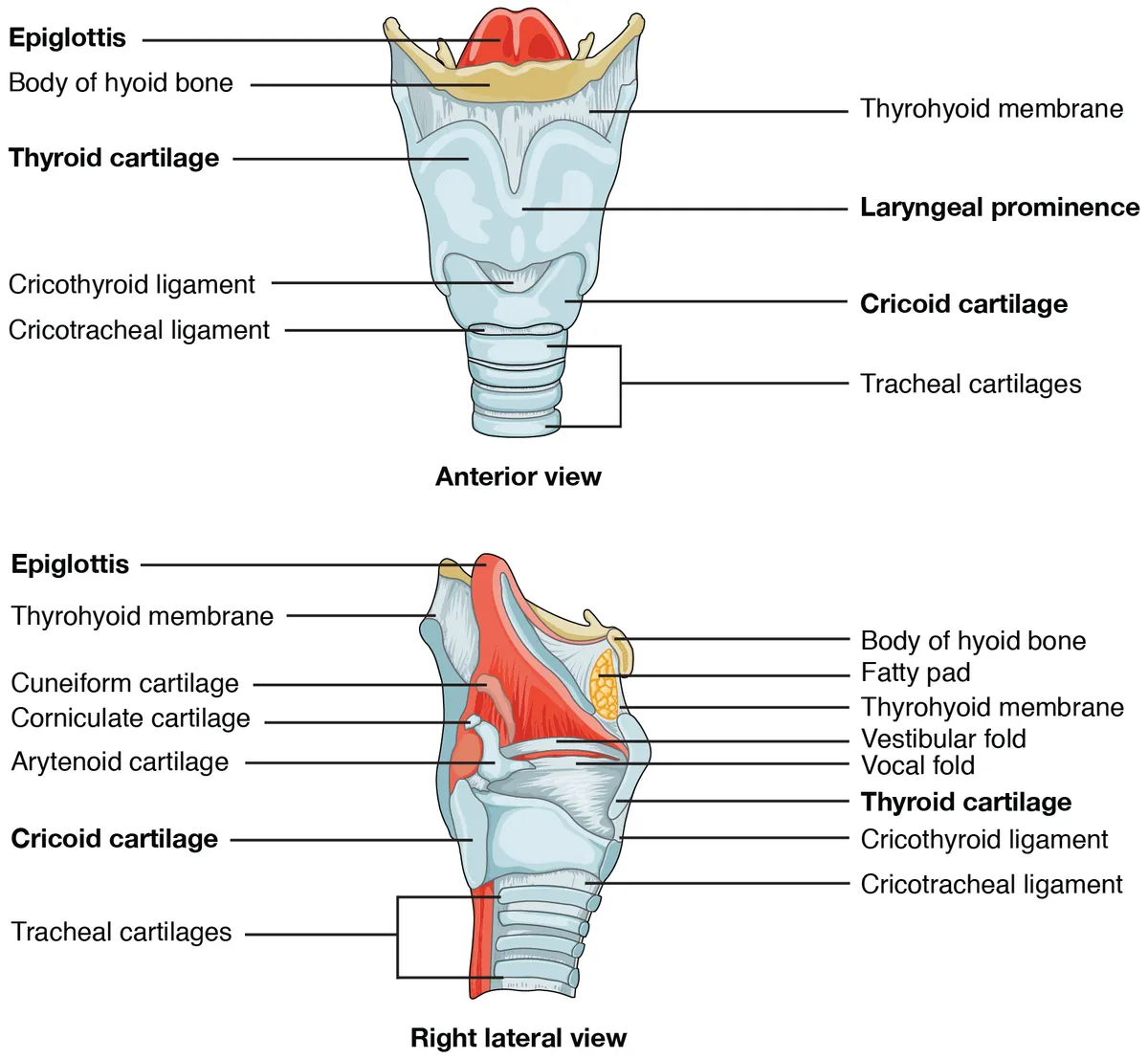
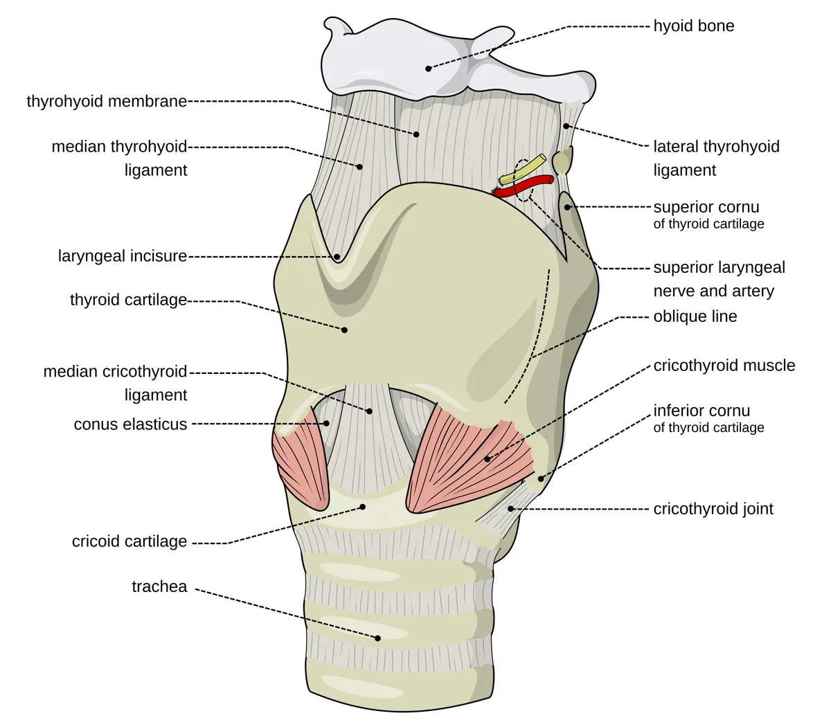
The largest cartilage, two laminae meeting in front at the laryngeal prominence (marked in men, at about 90° versus 120° in women). The superior horns connect to the hyoid and the inferior horns articulate with the cricoid; the vocal ligaments attach to its inner surface in front.
- 02
Cricoid cartilage
Cartilago cricoidea
The only complete cartilage ring of the airway, signet-shaped with its lamina behind, at C6. It is the narrowest point of the airway in young children; the cricothyroid membrane between it and the thyroid cartilage is the site of emergency cricothyroidotomy.
- 03
Arytenoid cartilages
Cartilagines arytenoideae
Two pyramidal cartilages sitting on the upper border of the cricoid lamina, each with a vocal process (for the vocal ligament) and a muscular process (for the crico-arytenoid muscles). The crico-arytenoid joints allow rotation and gliding to open and close the glottis; small corniculate and cuneiform cartilages lie above.
- 04
Epiglottis
Epiglottis
A leaf-shaped elastic cartilage whose stalk attaches inside the thyroid cartilage and whose free upper edge projects behind the tongue base, with the valleculae on either side. During swallowing the larynx rises and the epiglottis tips back over the laryngeal inlet.
- 05
Vocal folds and glottis
Plica vocalis; Glottis
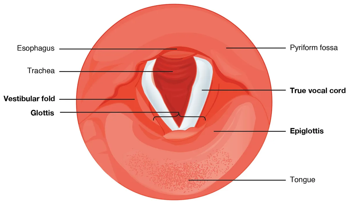
Two folds made of the vocal ligament, vocalis muscle and a covering of non-keratinised stratified squamous epithelium, stretched from the thyroid cartilage to the arytenoid vocal processes. The gap between them, the rima glottidis, is the narrowest part of the adult larynx; their vibration produces voice.
- 06
Vestibular folds and laryngeal ventricle
Plica vestibularis; Ventriculus laryngis
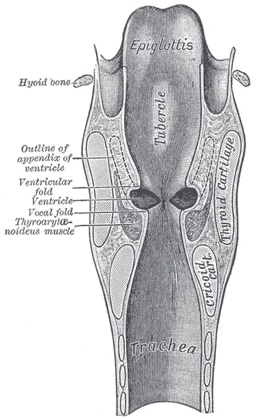
Two thick mucosal folds above the vocal folds (“false cords”), protective during breath-holding and straining. Between them and the vocal folds lies the laryngeal ventricle (of Morgagni), extending up as the saccule whose mucous glands lubricate the vocal folds.
- 07
Laryngeal muscles
Musculi laryngis
The posterior crico-arytenoid is the only abductor of the vocal folds; the lateral crico-arytenoid, interarytenoid and thyro-arytenoid adduct them; cricothyroid tenses them. All are supplied by the recurrent laryngeal nerve except cricothyroid (external branch of the superior laryngeal nerve).
Position and relations
The larynx lies in the front of the neck at C3–C6 in adults (higher in children), anterior to the laryngopharynx and between the carotid sheaths, partly covered by the thyroid lobes and the infrahyoid muscles. The laryngeal prominence (Adam’s apple) is formed by the thyroid cartilage; the cricothyroid membrane between the thyroid and cricoid cartilages is the site for an emergency airway.
Form and structure
The laryngeal cavity has three levels: the vestibule (above the vestibular folds), the glottis (the two vocal folds and the rima glottidis) and the infraglottic cavity (down to the lower border of the cricoid). Each vocal fold contains the vocal ligament and the vocalis muscle under stratified squamous epithelium. Intrinsic muscles open the rima (posterior cricoarytenoid, the only abductor), close it, and adjust vocal-fold tension (cricothyroid).
Vessels and nerves
The superior laryngeal artery (from the superior thyroid) and the inferior laryngeal artery (from the inferior thyroid) supply it. The superior laryngeal nerve (from the vagus) gives an internal branch for supraglottic sensation and an external branch to cricothyroid; the recurrent laryngeal nerve supplies all other intrinsic muscles and infraglottic sensation. The left recurrent nerve loops under the aortic arch, so mediastinal disease can injure it, and both lie close to the thyroid during surgery.
Clinical correlations
The larynx is the gateway between the upper and lower airways and serves phonation, swallowing and airway protection. Knowledge of its cartilages, mucosal spaces, muscles, neurovascular supply and lymphatic drainage underpins imaging-based staging of laryngeal cancer.[1]
Cartilage framework, vocal-fold layers and development
The laryngeal framework has unpaired and paired cartilages. The two laminae of the thyroid cartilage meet anteriorly at the laryngeal prominence (“Adam’s apple”) and stay open behind; the posterior border of each extends into a superior horn, joined to the hyoid by the thyrohyoid membrane and lateral thyrohyoid ligament (which may contain a triticeous cartilage that, when calcified, is mistaken for a foreign body), and an inferior horn that articulates with the cricoid. The thyrohyoid membrane has a foramen for the superior laryngeal vessels and the internal branch of the superior laryngeal nerve. The cricoid, of hyaline cartilage, is the only complete cartilaginous ring, shaped like a signet ring with a broad posterior lamina on which the two arytenoids articulate by ball-and-socket joints. Each arytenoid has a vocal process (for the vocal ligament) and a muscular process (for the cricoarytenoid muscles). The larynx has three levels: supraglottis (epiglottis and vestibular folds), glottis (true vocal folds) and subglottis.[2][3]
The vocal fold has five layers, deep to superficial: thyroarytenoid muscle, deep, intermediate and superficial lamina propria, and stratified squamous epithelium; the deep and intermediate lamina propria form the vocal ligament, while the superficial lamina propria (Reinke space) is loose, gelatinous connective tissue on which the mucosa vibrates during phonation. Being loose, Reinke space readily accumulates fluid under chronic irritation, above all smoking, causing Reinke oedema (polypoid corditis) with a deep, hoarse voice; women often complain that they sound like a man. The laryngeal ventricle is a recess between the vestibular and vocal folds; the piriform sinuses on either side of the aryepiglottic folds are where food commonly lodges. The larynx appears in week 4 as the laryngotracheal groove in the floor of the primitive pharynx; the groove contributes to the oesophagotracheal septum around week 5, separating the anterior airway from the dorsal oesophagus. The lining derives from endoderm, while the cartilages and muscles come from the mesenchyme of pharyngeal arches 3, 4 and 6.[2][4]
Intrinsic muscles and the recurrent laryngeal nerve
Four paired muscles (thyroarytenoid, lateral cricoarytenoid, posterior cricoarytenoid and cricothyroid) and one unpaired muscle (interarytenoid) adduct, abduct and lengthen the vocal folds. The posterior cricoarytenoid is the only abductor; all the others close the glottis. The cricothyroid, with straight and oblique bellies, tilts the thyroid cartilage forwards and down to lengthen and tense the folds for high pitch. The nucleus ambiguus in the brainstem provides motor neurons for the intrinsic muscles. All intrinsic muscles are supplied by the recurrent laryngeal nerve except cricothyroid, supplied by the external branch of the superior laryngeal nerve; the internal branch of the superior laryngeal nerve pierces the thyrohyoid membrane with the superior laryngeal artery, gives sensation to the mucosa above the folds and can be anaesthetised beneath the medial wall of the piriform sinus. The internal and inferior laryngeal nerves communicate through the anastomosis of Galen. The extrinsic muscles comprise the depressors (infrahyoid strap muscles, C1–C3), the elevators (suprahyoid muscles, from C1, V3 and VII) and the pharyngeal constrictors, serving mainly swallowing.[3][2][4]
The left recurrent nerve loops under the aortic arch and the right under the subclavian artery; each then ascends in the tracheo-oesophageal groove, passes deep to the inferior constrictor and enters the larynx behind the cricothyroid joint. Its course makes it vulnerable during thyroid surgery, so surgeons rely on landmarks: the ligament of Berry (joining the thyroid to the first three tracheal rings, the most reliable landmark, superficial to the nerve in about 78% of cases), the tracheo-oesophageal groove (containing the nerve in about 64%), the tubercle of Zuckerkandl and the inferior thyroid artery (the nerve passes behind it in about 61%, in front in 32.5% and between its branches in 6.5%). Extralaryngeal branches occur in 40–60% of people, usually dividing at the ligament of Berry; a large goitre can stretch or envelop the nerve. In about 0.6% of people a “non-recurrent” inferior laryngeal nerve passes straight from the vagus to the larynx in the neck, associated with an aberrant right subclavian artery arising after the left subclavian; it is exceedingly rare on the left.[5][4][2]
Posterior cricoarytenoid
- Action
- Abducts the folds (the only abductor)
- Nerve
- Recurrent laryngeal
Lateral cricoarytenoid
- Action
- Adducts
- Nerve
- Recurrent laryngeal
Interarytenoid
- Action
- Closes the posterior glottis
- Nerve
- Recurrent laryngeal
Thyroarytenoid (vocalis)
- Action
- Adducts, fine-tunes tension
- Nerve
- Recurrent laryngeal
Cricothyroid
- Action
- Lengthens and tenses the folds (high pitch)
- Nerve
- External branch of superior laryngeal
| Muscle | Action | Nerve |
|---|---|---|
| Posterior cricoarytenoid | Abducts the folds (the only abductor) | Recurrent laryngeal |
| Lateral cricoarytenoid | Adducts | Recurrent laryngeal |
| Interarytenoid | Closes the posterior glottis | Recurrent laryngeal |
| Thyroarytenoid (vocalis) | Adducts, fine-tunes tension | Recurrent laryngeal |
| Cricothyroid | Lengthens and tenses the folds (high pitch) | External branch of superior laryngeal |
Full-text reading (StatPearls)
The StatPearls chapters on this topic, reproduced verbatim. Their CC BY-NC-ND 4.0 licence allows unmodified copies only, so they are not translated or shortened.
Anatomy, Head and Neck, Larynx Vocal CordsSaran M, Georgakopoulos B, Bordoni B · StatPearls 2023 · NBK535342 · 28k characters
Loading…
Saran M, Georgakopoulos B, Bordoni B. Anatomy, Head and Neck, Larynx Vocal Cords. StatPearls [Internet]. Treasure Island (FL): StatPearls Publishing; 2023. NBK535342 · PMID 30570963 · CC BY-NC-ND 4.0. Reproduced verbatim, unmodified.
Anatomy, Head and Neck, Larynx Recurrent Laryngeal NerveAllen E, Minutello K, Jozsa F, Murcek BW · StatPearls 2025 · NBK470179 · 21k characters
Loading…
Allen E, Minutello K, Jozsa F, Murcek BW. Anatomy, Head and Neck, Larynx Recurrent Laryngeal Nerve. StatPearls [Internet]. Treasure Island (FL): StatPearls Publishing; 2025. NBK470179 · PMID 29261997 · CC BY-NC-ND 4.0. Reproduced verbatim, unmodified.
Anatomy, Head and Neck: Cervical, Respiratory, Larynx, and CricoarytenoidBenner A, Sharma P, Sharma S · StatPearls 2023 · NBK538307 · 20k characters
Loading…
Benner A, Sharma P, Sharma S. Anatomy, Head and Neck: Cervical, Respiratory, Larynx, and Cricoarytenoid. StatPearls [Internet]. Treasure Island (FL): StatPearls Publishing; 2023. NBK538307 · PMID 30855891 · CC BY-NC-ND 4.0. Reproduced verbatim, unmodified.
Anatomy, Head and Neck: LarynxSuárez-Quintanilla J, Fernández Cabrera A, Sharma S · StatPearls 2023 · NBK538202 · 19k characters
Loading…
Suárez-Quintanilla J, Fernández Cabrera A, Sharma S. Anatomy, Head and Neck: Larynx. StatPearls [Internet]. Treasure Island (FL): StatPearls Publishing; 2023. NBK538202 · PMID 30855790 · CC BY-NC-ND 4.0. Reproduced verbatim, unmodified.
Anatomy, Head and Neck: Larynx Arytenoid CartilageAndaloro C, Sharma P, La Mantia I · StatPearls 2023 · NBK513252 · 13k characters
Loading…
Andaloro C, Sharma P, La Mantia I. Anatomy, Head and Neck: Larynx Arytenoid Cartilage. StatPearls [Internet]. Treasure Island (FL): StatPearls Publishing; 2023. NBK513252 · PMID 30020624 · CC BY-NC-ND 4.0. Reproduced verbatim, unmodified.
Anatomy, Head and Neck: Larynx CartilageFlynn W, Vickerton P · StatPearls 2023 · NBK553185 · 11k characters
Loading…
Flynn W, Vickerton P. Anatomy, Head and Neck: Larynx Cartilage. StatPearls [Internet]. Treasure Island (FL): StatPearls Publishing; 2023. NBK553185 · PMID 31985978 · CC BY-NC-ND 4.0. Reproduced verbatim, unmodified.
Anatomy, Head and Neck: Larynx MusclesJunco K, Chandran SK · StatPearls 2023 · NBK545231 · 10k characters
Loading…
Junco K, Chandran SK. Anatomy, Head and Neck: Larynx Muscles. StatPearls [Internet]. Treasure Island (FL): StatPearls Publishing; 2023. NBK545231 · PMID 31424815 · CC BY-NC-ND 4.0. Reproduced verbatim, unmodified.
Organs in this system
References
Sources for the values and descriptions on this page, indexed in PubMed (US National Library of Medicine).
- [1]Anatomy of the Larynx and Cervical TracheaMcCullagh KL et al., Neuroimaging clinics of North America 2022PMID 36244725
- [2]Anatomy, Head and Neck, Larynx Vocal CordsSaran M et al., StatPearls 2023PMID 30570963
- [3]Anatomy, Head and Neck: Larynx MusclesJunco K et al., StatPearls 2023PMID 31424815
- [4]Anatomy, Head and Neck: LarynxSuárez-Quintanilla J et al., StatPearls 2023PMID 30855790
- [5]Anatomy, Head and Neck, Larynx Recurrent Laryngeal NerveAllen E et al., StatPearls 2025PMID 29261997
- [6]The normal human lung: ultrastructure and morphometric estimation of diffusion capacityGehr P, Bachofen M, Weibel ER, 1978PMID 644146Supports: Alveolar surface area
- [7]Physiology, LungStatPearls (NCBI Bookshelf)PMID 31424761Supports: Respiratory rate