Vermiform appendix
Model
Hover or tap to see names, click to read a description; click a name in the legend to fly to it. Procedurally built to histological proportions; colours follow illustration conventions.
Description
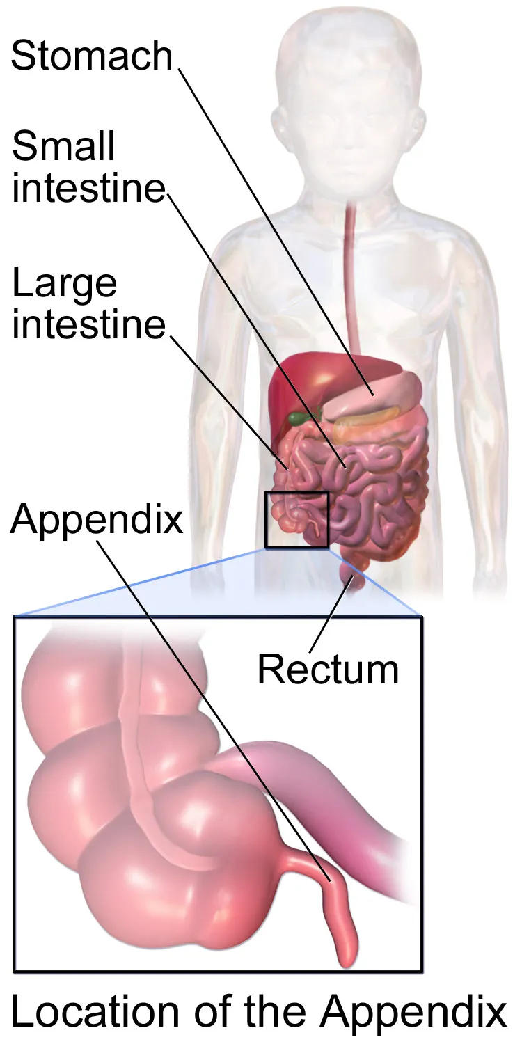
A blind tube averaging 6–10 cm, arising from the posteromedial wall of the caecum where the three taeniae coli converge, about 2 cm below the ileocaecal junction. The position of its tip varies widely (retrocaecal, pelvic and others). Its wall is rich in lymphoid tissue; appendicitis is the commonest abdominal surgical emergency.
Parts
Each part has its own card; press “Show on the model” to fly to it and select it in the 3D model above.
- 01
Base
Basis appendicis
Fixed where the three taeniae coli of the caecum converge, about 2–3 cm below the ileocaecal valve; following the anterior taenia locates the appendix at surgery. It projects to McBurney’s point, one-third of the way from the anterior superior iliac spine to the umbilicus.
- 02
Body, tip and positions
Corpus et apex appendicis; Positiones
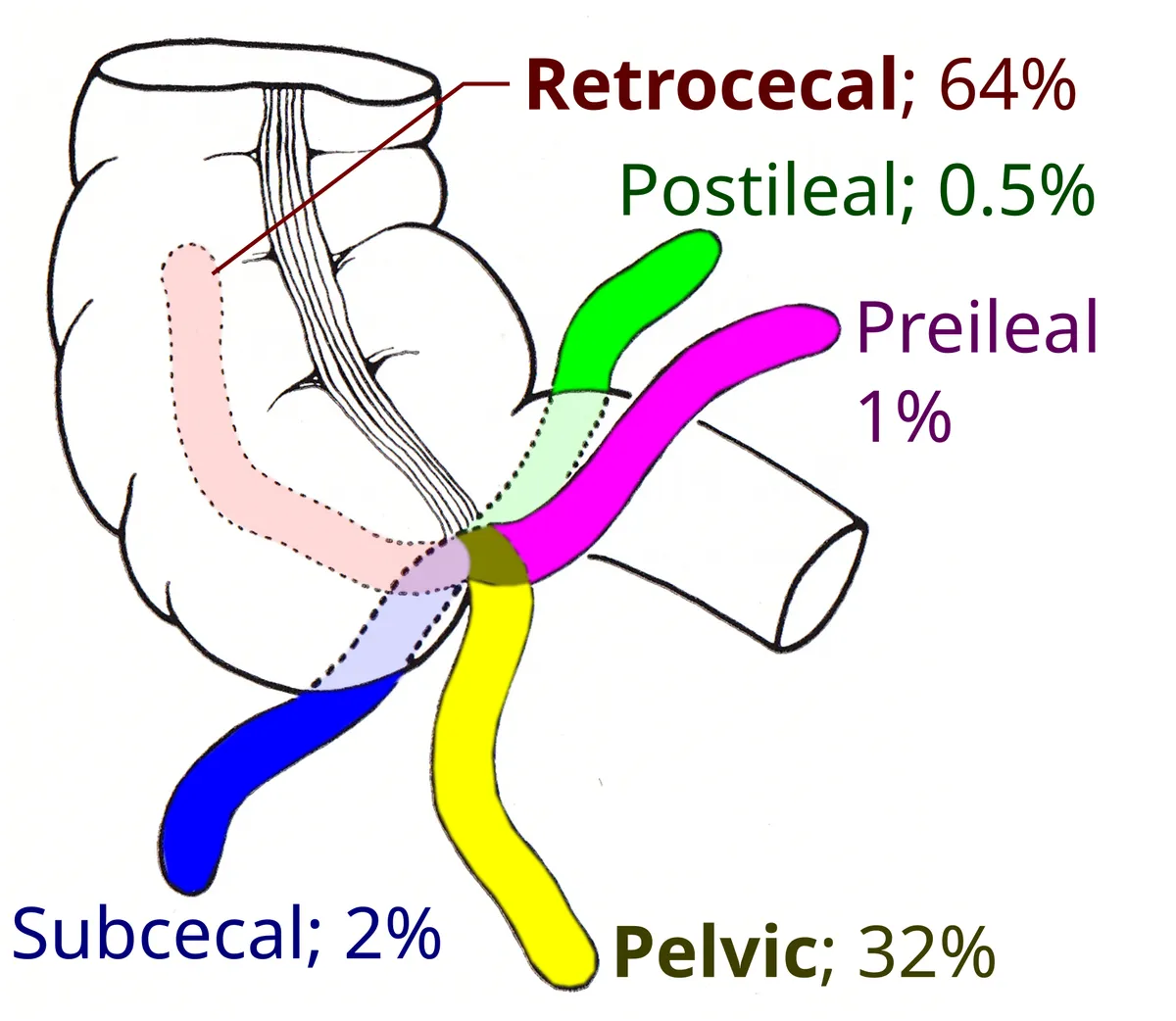
A blind tube averaging 6–10 cm with a narrow lumen. The tip is variable: retrocaecal (the commonest, about 65%), pelvic, subcaecal, pre-ileal or post-ileal; its position shapes the symptoms of appendicitis (flank pain, rectal tenderness, urinary symptoms).
- 03
Mesoappendix and appendicular artery
Mesoappendix; Arteria appendicularis
A triangular peritoneal fold joining the appendix to the back of the ileal mesentery and carrying the appendicular artery (from the ileocolic). It is an end artery, so when the lumen is obstructed and distended the wall readily becomes gangrenous and perforates.
- 04
Lymphoid tissue
Folliculi lymphatici aggregati appendicis
Its wall is rich in submucosal lymphoid follicles, most abundant in adolescence and regressing thereafter, which explains why appendicitis peaks at 10–30 years (lymphoid hyperplasia obstructs the lumen). It may act as a reservoir for the gut microbiota.
Position and relations
The base of the appendix is fairly constant, projecting to McBurney’s point (one-third of the way from the right anterior superior iliac spine to the umbilicus), whereas the tip may be retrocaecal (most often), pelvic, subcaecal, or pre- or post-ileal. The position of the tip shapes the pain and clinical signs of appendicitis.
Form and structure
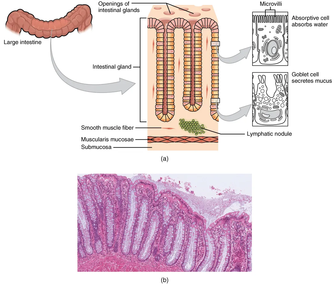
The appendix has a colonic wall structure but a narrow lumen, and its submucosa is packed with lymphoid follicles, most abundant in adolescence and declining thereafter. Obstruction of the lumen (by a faecolith or lymphoid hyperplasia) raises pressure and leads to wall ischaemia and infection, the usual mechanism of acute appendicitis.
Vessels and nerves
The appendicular artery, a branch of the ileocolic artery (from the superior mesenteric), runs in the meso-appendix and is an end artery, so thrombosis readily causes gangrene and perforation. Early pain is periumbilical through visceral fibres at T10, then shifts to the right iliac fossa when the parietal peritoneum becomes irritated.
Anatomical variants
The position and form of the appendix vary widely. Common positions are retrocaecal, pelvic, retro-ileal, pre-ileal, precaecal and paracaecal, the first two being the most frequent. Rarely the appendix is intracaecal, intramural, subhepatic or left-sided, and its base may not match McBurney’s point. Length, diameter, shape and even number (double, triple appendix) vary.[1]
Because its position varies, the appendix can be hard to find on ultrasound, CT and MRI. Knowing its embryology and variants, such as an appendix within an inguinal hernia sac (Amyand’s hernia), helps diagnose appendicitis and appendiceal tumours correctly.[2][3]
Immune function
The appendix is often called an evolutionary remnant, yet phylogenetic data show it has been selected for and is highly conserved in humans, with very rare malformations. Its wall is rich in lymphoid tissue resembling Peyer’s patches and is a primary site of IgA production, which regulates the density and quality of the gut flora.[4]
Thanks to its blind-ended shape and position, the appendix may serve as a reservoir of commensal bacteria: it is rich in biofilms that continuously shed bacteria into the lumen, holds a microbiota as diverse as the colon’s and could replenish healthy flora after diarrhoea. Absence of the appendix predicts worse outcomes of recurrent Clostridioides difficile infection.[4]
In observational studies, appendectomy (especially of an inflamed appendix) is associated with protection against ulcerative colitis; among people who develop the disease, prior appendectomy may protect against colectomy but could raise colorectal cancer risk. Trials of appendectomy as treatment are under way.[5]
How often each position occurs, and size
The appendix continues the caecum caudally in the right iliac fossa, about 2–3 cm below the ileocaecal valve, with a base, body and tip. Its wall has four layers: mucosa, submucosa, muscularis externa and serosa. It is wholly covered by peritoneum and slung by a triangular mesoappendix whose free edge carries the appendicular artery, so an incompletely developed mesoappendix risks poor perfusion and gangrenous or perforated appendicitis.[1]
The retrocaecal position is the commonest variant, in roughly 25.4–71% of people; precaecal occurs in about 4% and paracaecal in 3.1–7.5%. A meta-analysis of 114,080 patients with acute appendicitis found retrocaecal the most frequent on every continent: Africa 44.8%, Asia 32%, South America 36.4%, Europe 27.6% and North America 24.8%, with pelvic second at about 19.5–31.5%. By contrast, a study of 200 cadavers in Iran found pelvic the commonest (55.8%). McBurney’s point is unreliable: in one sample only 1 in 100 subjects had the base of the appendix there.[1]
The appendix measures 0.5 to 23 cm, averaging 5.3–11.7 cm, with a diameter of 3.2–10 mm; mean length is shorter in Western countries (5.3–6.9 cm) and varies within Africa, for example 7.65 cm in Kenya and 11.7 cm in Zambia. Among duplications in the Cave–Wallbridge classification, type B2 is the commonest (about 37%); horseshoe and triplicated appendices are very rare, with 6 and 2 reported cases respectively. Genetics, lifestyle, diet, geography and perhaps sex probably influence its position and size.[1]
Full-text reading (StatPearls)
The StatPearls chapters on this topic, reproduced verbatim. Their CC BY-NC-ND 4.0 licence allows unmodified copies only, so they are not translated or shortened.
Anatomy, Abdomen and Pelvis: AppendixHodge BD, Kashyap S, Khorasani-Zadeh A · StatPearls 2023 · NBK459205 · 8k characters
Loading…
Hodge BD, Kashyap S, Khorasani-Zadeh A. Anatomy, Abdomen and Pelvis: Appendix. StatPearls [Internet]. Treasure Island (FL): StatPearls Publishing; 2023. NBK459205 · PMID 29083761 · CC BY-NC-ND 4.0. Reproduced verbatim, unmodified.
Organs in this system
References
Sources for the values and descriptions on this page, indexed in PubMed (US National Library of Medicine).
- [1]Anatomical Variations of the Vermiform AppendixSakellariadis A et al., Acta medica academica 2024PMID 39720866
- [2]Anatomical variants and pathologies of the vermixDeshmukh S et al., Emergency radiology 2014PMID 24570122
- [3]Amyand's hernia: a reviewIvanschuk G et al., Medical science monitor 2014PMID 24473371
- [4]The immunological functions of the Appendix: An example of redundancy?Girard-Madoux MJH et al., Seminars in immunology 2018PMID 29503124
- [5]The appendix and ulcerative colitis - an unsolved connectionAgrawal M et al., Nature reviews. Gastroenterology & hepatology 2023PMID 37081213
- [6]Gastrointestinal mean transit times in young and middle-aged healthy subjectsPMID 11318834Supports: Gastrointestinal transit time
- [7]Revised Estimates for the Number of Human and Bacteria Cells in the BodySender R, Fuchs S, Milo R, 2016PMID 27541692Supports: Colonic bacterial count