Ankle and foot
Model
Layers
Z-Anatomy anatomical model based on BodyParts3D, licensed CC BY-SA 4.0.
Overview
A regional model of the (right) ankle and foot: the seven tarsal bones, five metatarsals and the phalanges, the talocrural, subtalar and foot joints with their ligaments, the retinacula, the tendons and synovial sheaths of the leg muscles, the plantar aponeurosis and the four layers of plantar muscles, the dorsal foot muscles, the dorsalis pedis and medial and lateral plantar arteries, and the terminal branches of the tibial, deep fibular and superficial fibular nerves.
Boundaries and subdivisions
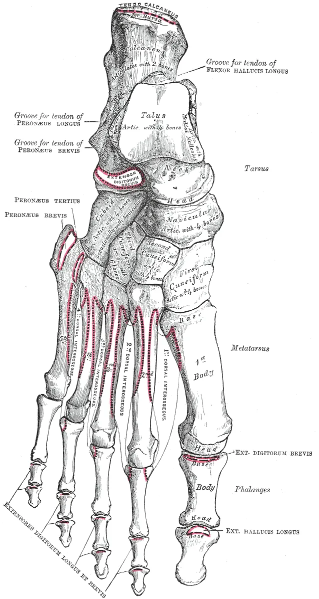
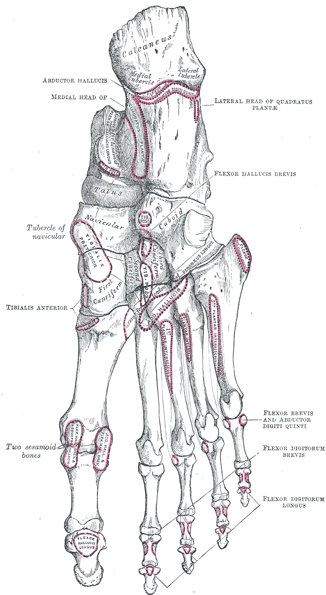
The foot is the part of the lower limb distal to the ankle (talocrural) joint, where the trochlea of the talus fits into the mortise formed by the distal tibia and fibula. It has 26 bones in three parts: the hindfoot (talus and calcaneus, the main weight-bearing part), the midfoot (navicular, cuboid and three cuneiforms) and the forefoot (metatarsals and phalanges). The joint between midfoot and forefoot is the Lisfranc joint, where the base of the second metatarsal keys into the intermediate cuneiform. The bones form medial and lateral longitudinal arches and a transverse arch, held by ligaments, the plantar aponeurosis and tendons.[1][2]
Layers from superficial to deep
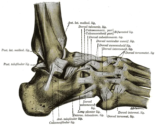
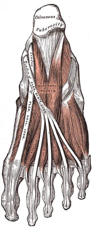
Around the ankle, the retinacula (superior and inferior extensor, flexor, fibular) hold the tendons close to bone, each tendon in its own synovial sheath. Behind the medial malleolus lies the tarsal tunnel, deep to the flexor retinaculum, containing (front to back) tibialis posterior, flexor digitorum longus, the posterior tibial vessels, the tibial nerve and flexor hallucis longus. Laterally, the lateral ligament comprises the anterior talofibular, calcaneofibular and posterior talofibular ligaments; medially lies the strong deltoid ligament. In the sole, the plantar aponeurosis stretches from the calcaneal tuberosity to the toes, with four layers of intrinsic muscles beneath it; the foot has 19 intrinsic muscles. When the toes dorsiflex at push-off the aponeurosis tightens and raises the medial arch (windlass mechanism).[1][3][4][5]
Vessels, nerves and lymphatics
The anterior tibial artery becomes the dorsalis pedis artery after crossing the ankle, giving the medial and lateral tarsal and arcuate arteries; its deep plantar branch dives into the sole to complete the plantar arch formed by the lateral plantar artery. The posterior tibial artery is palpable behind the medial malleolus and divides into the medial and lateral plantar arteries in the tarsal tunnel. The tibial nerve divides into the medial and lateral plantar nerves beneath the flexor retinaculum; the medial plantar nerve runs deep to abductor hallucis. The deep fibular nerve supplies the skin of the first web space, the superficial fibular nerve most of the dorsum, the sural nerve the lateral border and the saphenous nerve the medial border of the foot.[1][6][3][7][8]
Embryology
The lower limb bud grows out in week 4 from lateral plate mesoderm opposite somites L3 to L5; a primitive footplate exists by about 4.5 weeks (stage 15), the cartilaginous skeleton and muscle masses appear within days, followed by the digits (stages 18–19). Proximodistal growth is driven by the apical ectodermal ridge, and most of the limb skeleton is laid down between weeks 4 and 8. The talus and calcaneus appear as cartilage in week 8; the subtalar joint starts to form in week 9, its posterior part faster than the anterior, and the three ossification centres of the talus fuse at about week 10. The nerves of the foot derive from the neural tube and neural crest formed from week 3.[9][1][4][10][11]
Anatomical variants
Common accessory ossicles of the foot and ankle include the os trigonum, accessory navicular, os peroneum, os supranaviculare and os intermetatarseum; many are incidental, but some cause pain. The os trigonum (1–25% of people) is an unfused secondary ossification centre of the posterior talar process and can cause posterior ankle impingement in those who plantarflex repeatedly, such as ballet dancers and footballers. The accessory navicular has three types, of which type II, joined to the navicular by fibrocartilage, is most often symptomatic. Fibularis quartus is an accessory muscle in 6–22% of people, usually arising from fibularis brevis. The superficial fibular nerve may pierce the fascia at different levels or lie in another compartment, and an accessory deep fibular nerve may exist. Arch height ranges from flat foot to high-arched foot, congenital or acquired.[1][10][4][11][9][12]
Clinical correlations
Inversion ankle sprains usually injure the anterior talofibular ligament first, then the calcaneofibular ligament; injury to the distal tibiofibular syndesmosis is called a “high ankle sprain”. Tarsal tunnel syndrome is compression of the tibial nerve beneath the flexor retinaculum, causing burning numbness of the sole. Plantar fasciitis causes heel pain on the first steps in the morning. Lisfranc injuries are easily missed on radiographs but can lead to collapse of the arch. Palpating the posterior tibial and dorsalis pedis pulses is a routine step in assessing lower-limb circulation.[1][3][6]
Surgical and procedural considerations
Abnormal arches disturb gait and may cause pain, deformity and joint degeneration; severe or refractory cases need corrective surgery. In children only rigid flat foot (rare, associated with congenital vertical talus, accessory navicular or tarsal coalition) is operated on; in adults surgery follows failed conservative care, and osteotomies must take talar morphology into account so as not to damage the anterior and middle facets. Foot drop (weak dorsiflexion with a steppage gait) is often iatrogenic, from injury to the deep fibular nerve in leg, ankle or foot surgery, and is treated by neurolysis, nerve repair or tendon transfer. A frequent complication of anterior-approach total ankle arthroplasty is injury to the superficial fibular nerve and its aberrant branches, especially the intermediate dorsal cutaneous nerve. Most fibularis brevis tendon injuries are managed conservatively; active patients may need tendon repair or reconstruction, or retinacular repair when the tendons sublux.[9][12][10][4][11][13]
Key structures
Articulatio talocruralis
- Name
- Ankle (talocrural) joint
- Position, role
- Talar trochlea in the tibiofibular mortise; dorsiflexion and plantarflexion
Articulatio subtalaris
- Name
- Subtalar joint
- Position, role
- Inversion and eversion of the hindfoot
Canalis tarsi (tunnel tarsien)
- Name
- Tarsal tunnel
- Position, role
- Behind the medial malleolus; tibial nerve, posterior tibial vessels, three flexor tendons
Lig. talofibulare anterius
- Name
- Anterior talofibular ligament
- Position, role
- Most often injured in inversion sprains
Lig. deltoideum
- Name
- Deltoid ligament
- Position, role
- Strong medial collateral ligament
Aponeurosis plantaris
- Name
- Plantar aponeurosis
- Position, role
- From the calcaneal tuberosity to the toes; supports the arch
A. dorsalis pedis
- Name
- Dorsalis pedis artery
- Position, role
- Continues the anterior tibial artery; pulse on the dorsum
| Terminologia Anatomica | Name | Position, role |
|---|---|---|
| Articulatio talocruralis | Ankle (talocrural) joint | Talar trochlea in the tibiofibular mortise; dorsiflexion and plantarflexion |
| Articulatio subtalaris | Subtalar joint | Inversion and eversion of the hindfoot |
| Canalis tarsi (tunnel tarsien) | Tarsal tunnel | Behind the medial malleolus; tibial nerve, posterior tibial vessels, three flexor tendons |
| Lig. talofibulare anterius | Anterior talofibular ligament | Most often injured in inversion sprains |
| Lig. deltoideum | Deltoid ligament | Strong medial collateral ligament |
| Aponeurosis plantaris | Plantar aponeurosis | From the calcaneal tuberosity to the toes; supports the arch |
| A. dorsalis pedis | Dorsalis pedis artery | Continues the anterior tibial artery; pulse on the dorsum |
What each layer contains
SkinCutis22
- Distal transverse arch of foot
- Proximal transverse arch of foot
- Dorsum of foot
- Hallucial eminence
- Dorsal surfaces of digits of foot
- Plantar surfaces of digits of foot
- Nail plate (foot)
- Lateral malleolus
- Medial malleolus
- Lateral border of foot
- Medial border of foot
- Lateral part of longitudinal arch of foot
- Medial part of longitudinal arch of foot
- Perionyx (foot)
- Sole
- Anterior region of leg
- Anterior region of ankle
- Heel region
- Metatarsal region
- Posterior region of leg
- Lateral retromalleolar region
- Medial retromalleolar region
Muscles, fasciae and tendon sheathsMusculi, fasciae et vaginae tendinum48
- (Opponens digiti minimi muscle of foot)
- Abductor digiti minimi of foot
- Abductor hallucis
- Plantar aponeurosis
- Subcutaneous calcaneal bursa
- Subcutaneous bursa of medial malleolus
- Subcutaneous bursa of lateral malleolus
- Subtendinous calcaneal bursa
- Subtendinous bursa of tibialis anterior
- Lateral head of flexor hallucis brevis
- Medial head of flexor hallucis brevis
- Oblique head of adductor hallucis
- Transverse head of adductor hallucis
- Extensor digitorum brevis
- Extensor hallucis brevis
- Extensor digitorum longus
- Extensor hallucis longus
- Flexor digitorum brevis
- Flexor digiti minimi of foot
- Flexor hallucis longus
- Superficial transverse metatarsal ligament
- Dorsal interossei muscles of foot
- Plantar interossei muscles
- Lumbrical muscles of foot
- Fibularis brevis muscle
- Fibularis longus muscle
- Fibularis tertius muscle
- Flexor digitorum longus
- Quadratus plantae muscle
- Tibialis anterior muscle
- Tibialis posterior muscle
- Inferior extensor retinaculum of ankle
- Superior extensor retinaculum of ankle
- Inferior fibular retinaculum
- Superior fibular retinaculum
- Flexor retinaculum of ankle
- Anterior intermuscular septum of leg
- Transverse intermuscular septum of leg
- Calcaneal tendon
- Tendon of extensor digitorum longus
- Tendon sheath of tibialis anterior
- Common tendon sheath of fibularis muscles
- Plantar tendon sheath of fibularis longus muscle
- Tendon sheath of extensor hallucis longus
- Tendon sheath of flexor digitorum longus
- Tendon sheath of flexor hallucis longus
- Tendon sheath of tibialis posterior muscle
- Tendon sheath of extensor digitorum longus
Bones and cartilageOssa et cartilagines29
- Calcaneus
- Fibula
- Cuboid bone
- Intermediate cuneiform bone
- Lateral cuneiform bone
- Medial cuneiform bone
- Navicular bone
- First metatarsal bone
- Fourth metatarsal bone
- Fifth metatarsal bone
- Second metatarsal bone
- Talus
- Third metatarsal bone
- Sesamoid bones of foot
- Distal phalanx of first finger of foot
- Distal phalanx of fourth finger of foot
- Distal phalanx of fifth finger of foot
- Distal phalanx of second finger of foot
- Distal phalanx of third finger of foot
- Middle phalanx of fourth finger of foot
- Middle phalanx of fifth finger of foot
- Middle phalanx of second finger of foot
- Middle phalanx of third finger of foot
- Proximal phalanx of first finger of foot
- Proximal phalanx of fourth finger of foot
- Proximal phalanx of fifth finger of foot
- Proximal phalanx of second finger of foot
- Proximal phalanx of third finger of foot
- Tibia
Joints and ligamentsJuncturae et ligamenta48
- Articular capsule of interphalangeal joint of great toe
- Articular capsules of metatarsophalangeal joints
- Articular capsules of proximal interphalangeal joints of foot
- Articular capsules of distal interphalangeal joints of foot
- Cuneometatarsal interosseous ligaments
- Dorsal cuneonavicular ligaments
- Plantar cuneonavicular ligaments
- Dorsal intercuneiform ligaments
- Intercuneiform interosseous ligaments
- Plantar intercuneiform ligaments
- Collateral interphalangeal ligaments of foot
- Plantar interphalangeal ligaments
- Dorsal metatarsal ligaments
- Metatarsal interosseous ligaments
- Plantar metatarsal ligaments
- Collateral metatarsophalangeal ligaments
- Plantar metatarsophalangeal ligaments
- Dorsal tarsometatarsal ligaments
- Plantar tarsometatarsal ligaments
- Calcaneocuboid ligament
- Dorsal calcaneocuboid ligament
- Plantar calcaneocuboid ligament
- Calcaneofibular ligament
- Calcaneonavicular ligament
- Plantar calcaneonavicular ligament
- Tibiocalcaneal ligament
- Dorsal cuboideonavicular ligament
- Plantar cuboideonavicular ligament
- Dorsal cuneocuboid ligament
- Cuneocuboid interosseous ligament
- Plantar cuneocuboid ligament
- Intersesamoid ligament
- Deep transverse metatarsal ligament
- Long plantar ligament
- Anterior talocalcaneal ligament
- Talocalcaneal interosseous ligament
- Lateral talocalcaneal ligament
- Medial talocalcaneal ligament
- Posterior talocalcaneal ligament
- Anterior talofibular ligament
- Posterior talofibular ligament
- Talonavicular ligament
- Posterior tibiotalar ligament
- Anterior tibiofibular ligament
- Posterior tibiofibular ligament
- Transverse tibiofibular ligament
- Tibionavicular ligament
- Interosseous membrane of leg
ArteriesArteriae18
- Plantar arch
- Arcuate artery
- Dorsalis pedis artery
- Lateral plantar artery
- Medial plantar artery
- Deep plantar artery
- Lateral tarsal artery
- Anterior tibial artery
- Posterior tibial artery
- Dorsal digital arteries of foot
- Common plantar digital arteries
- Proper plantar digital arteries
- Dorsal metatarsal arteries
- Plantar metatarsal arteries
- Calcaneal branches of fibular artery
- Calcaneal branches of posterior tibial artery
- Perforating branches of plantar metatarsal arteries
- Superficial branch of medial plantar artery
VeinsVenae12
- Dorsal venous arch of foot
- Plantar venous arch
- Dorsal digital veins of foot
- Plantar digital veins
- Fibular veins
- Intercapitular veins of foot
- Dorsal metatarsal veins
- Plantar metatarsal veins
- Lateral plantar veins
- Medial plantar veins
- Anterior tibial veins
- Posterior tibial veins
NervesNervi14
- Intermediate dorsal cutaneous nerve of foot
- Medial dorsal cutaneous nerve of foot
- Deep fibular nerve
- Superficial fibular nerve
- Lateral plantar nerve
- Medial plantar nerve
- Sural nerve
- Dorsal digital branches of deep fibular nerve
- Dorsal digital branches of superficial fibular nerve
- Common plantar digital branches of lateral plantar nerve
- Common plantar digital branches of medial plantar nerve
- Proper plantar digital branches of lateral plantar nerve
- Proper plantar digital branches of medial plantar nerve
- Muscular branches of deep fibular nerve
Full-text reading (StatPearls)
The StatPearls chapters on this topic, reproduced verbatim. Their CC BY-NC-ND 4.0 licence allows unmodified copies only, so they are not translated or shortened.
Anatomy, Bony Pelvis and Lower Limb: Arches of the FootChauhan HM, Taqi M · StatPearls 2025 · NBK587361 · 36k characters
Loading…
Chauhan HM, Taqi M. Anatomy, Bony Pelvis and Lower Limb: Arches of the Foot. StatPearls [Internet]. Treasure Island (FL): StatPearls Publishing; 2025. NBK587361 · PMID 36508535 · CC BY-NC-ND 4.0. Reproduced verbatim, unmodified.
Anatomy, Bony Pelvis and Lower Limb: Foot and AnkleFicke J, Fakoya AO, Byerly DW · StatPearls 2026 · NBK546698 · 31k characters
Loading…
Ficke J, Fakoya AO, Byerly DW. Anatomy, Bony Pelvis and Lower Limb: Foot and Ankle. StatPearls [Internet]. Treasure Island (FL): StatPearls Publishing; 2026. NBK546698 · PMID 31536304 · CC BY-NC-ND 4.0. Reproduced verbatim, unmodified.
Anatomy, Bony Pelvis and Lower Limb, Foot MusclesCard RK, Bordoni B · StatPearls 2025 · NBK539705 · 27k characters
Loading…
Card RK, Bordoni B. Anatomy, Bony Pelvis and Lower Limb, Foot Muscles. StatPearls [Internet]. Treasure Island (FL): StatPearls Publishing; 2025. NBK539705 · PMID 30969527 · CC BY-NC-ND 4.0. Reproduced verbatim, unmodified.
Anatomy, Bony Pelvis and Lower Limb, Foot NervesTang A, Bordoni B · StatPearls 2023 · NBK537292 · 26k characters
Loading…
Tang A, Bordoni B. Anatomy, Bony Pelvis and Lower Limb, Foot Nerves. StatPearls [Internet]. Treasure Island (FL): StatPearls Publishing; 2023. NBK537292 · PMID 30725977 · CC BY-NC-ND 4.0. Reproduced verbatim, unmodified.
Anatomy, Bony Pelvis and Lower Limb: Medial Longitudinal Arch of the FootBabu D, Bordoni B · StatPearls 2024 · NBK562289 · 19k characters
Loading…
Babu D, Bordoni B. Anatomy, Bony Pelvis and Lower Limb: Medial Longitudinal Arch of the Foot. StatPearls [Internet]. Treasure Island (FL): StatPearls Publishing; 2024. NBK562289 · PMID 32965960 · CC BY-NC-ND 4.0. Reproduced verbatim, unmodified.
Anatomy, Bony Pelvis and Lower Limb, Foot TalusKhan IA, Varacallo MA · StatPearls 2023 · NBK541086 · 18k characters
Loading…
Khan IA, Varacallo MA. Anatomy, Bony Pelvis and Lower Limb, Foot Talus. StatPearls [Internet]. Treasure Island (FL): StatPearls Publishing; 2023. NBK541086 · PMID 31082130 · CC BY-NC-ND 4.0. Reproduced verbatim, unmodified.
Anatomy, Bony Pelvis and Lower Limb, Foot JointsManganaro D, Dollinger B, Nezwek TA, Sadiq NM · StatPearls 2023 · NBK536941 · 15k characters
Loading…
Manganaro D, Dollinger B, Nezwek TA, Sadiq NM. Anatomy, Bony Pelvis and Lower Limb, Foot Joints. StatPearls [Internet]. Treasure Island (FL): StatPearls Publishing; 2023. NBK536941 · PMID 30725626 · CC BY-NC-ND 4.0. Reproduced verbatim, unmodified.
Anatomy, Bony Pelvis and Lower Limb: Foot ArteriesGuillot C, Smith T · StatPearls 2023 · NBK560912 · 12k characters
Loading…
Guillot C, Smith T. Anatomy, Bony Pelvis and Lower Limb: Foot Arteries. StatPearls [Internet]. Treasure Island (FL): StatPearls Publishing; 2023. NBK560912 · PMID 32809747 · CC BY-NC-ND 4.0. Reproduced verbatim, unmodified.
Anatomy, Bony Pelvis and Lower Limb, Foot Peroneus Brevis MuscleBasit H, Shah J, Siccardi MA · StatPearls 2023 · NBK535427 · 12k characters
Loading…
Basit H, Shah J, Siccardi MA. Anatomy, Bony Pelvis and Lower Limb, Foot Peroneus Brevis Muscle. StatPearls [Internet]. Treasure Island (FL): StatPearls Publishing; 2023. NBK535427 · PMID 30571048 · CC BY-NC-ND 4.0. Reproduced verbatim, unmodified.
Anatomy, Bony Pelvis and Lower Limb, Foot FasciaBourne M, Talkad A, Varacallo MA · StatPearls 2023 · NBK526043 · 11k characters
Loading…
Bourne M, Talkad A, Varacallo MA. Anatomy, Bony Pelvis and Lower Limb, Foot Fascia. StatPearls [Internet]. Treasure Island (FL): StatPearls Publishing; 2023. NBK526043 · PMID 30252299 · CC BY-NC-ND 4.0. Reproduced verbatim, unmodified.
Anatomy, Bony Pelvis and Lower Limb: Ankle JointManganaro D, Alsayouri K · StatPearls 2023 · NBK545158 · 10k characters
Loading…
Manganaro D, Alsayouri K. Anatomy, Bony Pelvis and Lower Limb: Ankle Joint. StatPearls [Internet]. Treasure Island (FL): StatPearls Publishing; 2023. NBK545158 · PMID 31424742 · CC BY-NC-ND 4.0. Reproduced verbatim, unmodified.
Anatomy, Bony Pelvis and Lower Limb, Foot VeinsLezak B, Varacallo MA · StatPearls 2022 · NBK542295 · 10k characters
Loading…
Lezak B, Varacallo MA. Anatomy, Bony Pelvis and Lower Limb, Foot Veins. StatPearls [Internet]. Treasure Island (FL): StatPearls Publishing; 2022. NBK542295 · PMID 31194435 · CC BY-NC-ND 4.0. Reproduced verbatim, unmodified.
Regional anatomy
References
Sources for the values and descriptions on this page, indexed in PubMed (US National Library of Medicine).
- [1]Anatomy, Bony Pelvis and Lower Limb: Foot and AnkleFicke J et al., StatPearls 2026PMID 31536304
- [2]Anatomy, Bony Pelvis and Lower Limb: Metatarsal BonesLezak B et al., StatPearls 2026PMID 31751062
- [3]Anatomy, Bony Pelvis and Lower Limb: Tibial NerveDesai SS et al., StatPearls 2026PMID 30725713
- [4]Anatomy, Bony Pelvis and Lower Limb, Foot MusclesCard RK et al., StatPearls 2025PMID 30969527
- [5]Anatomy, Bony Pelvis and Lower Limb: Ankle JointManganaro D et al., StatPearls 2023PMID 31424742
- [6]Anatomy, Bony Pelvis and Lower Limb: Posterior Tibial ArteryLezak B et al., StatPearls 2026PMID 30725666
- [7]Anatomy, Bony Pelvis and Lower Limb: Leg Anterior CompartmentLezak B et al., StatPearls 2026PMID 30969547
- [8]Anatomy, Bony Pelvis and Lower Limb: Foot ArteriesGuillot C et al., StatPearls 2023PMID 32809747
- [9]Anatomy, Bony Pelvis and Lower Limb: Arches of the FootChauhan HM et al., StatPearls 2025PMID 36508535
- [10]Anatomy, Bony Pelvis and Lower Limb, Foot TalusKhan IA et al., StatPearls 2023PMID 31082130
- [11]Anatomy, Bony Pelvis and Lower Limb, Foot NervesTang A et al., StatPearls 2023PMID 30725977
- [12]Anatomy, Bony Pelvis and Lower Limb: Medial Longitudinal Arch of the FootBabu D et al., StatPearls 2024PMID 32965960
- [13]Anatomy, Bony Pelvis and Lower Limb, Foot Peroneus Brevis MuscleBasit H et al., StatPearls 2023PMID 30571048