Urethra
Model
Layers
Z-Anatomy anatomical model based on BodyParts3D, licensed CC BY-SA 4.0.
Description
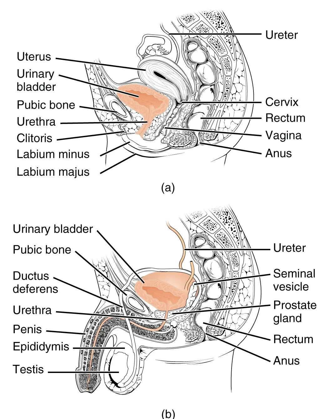
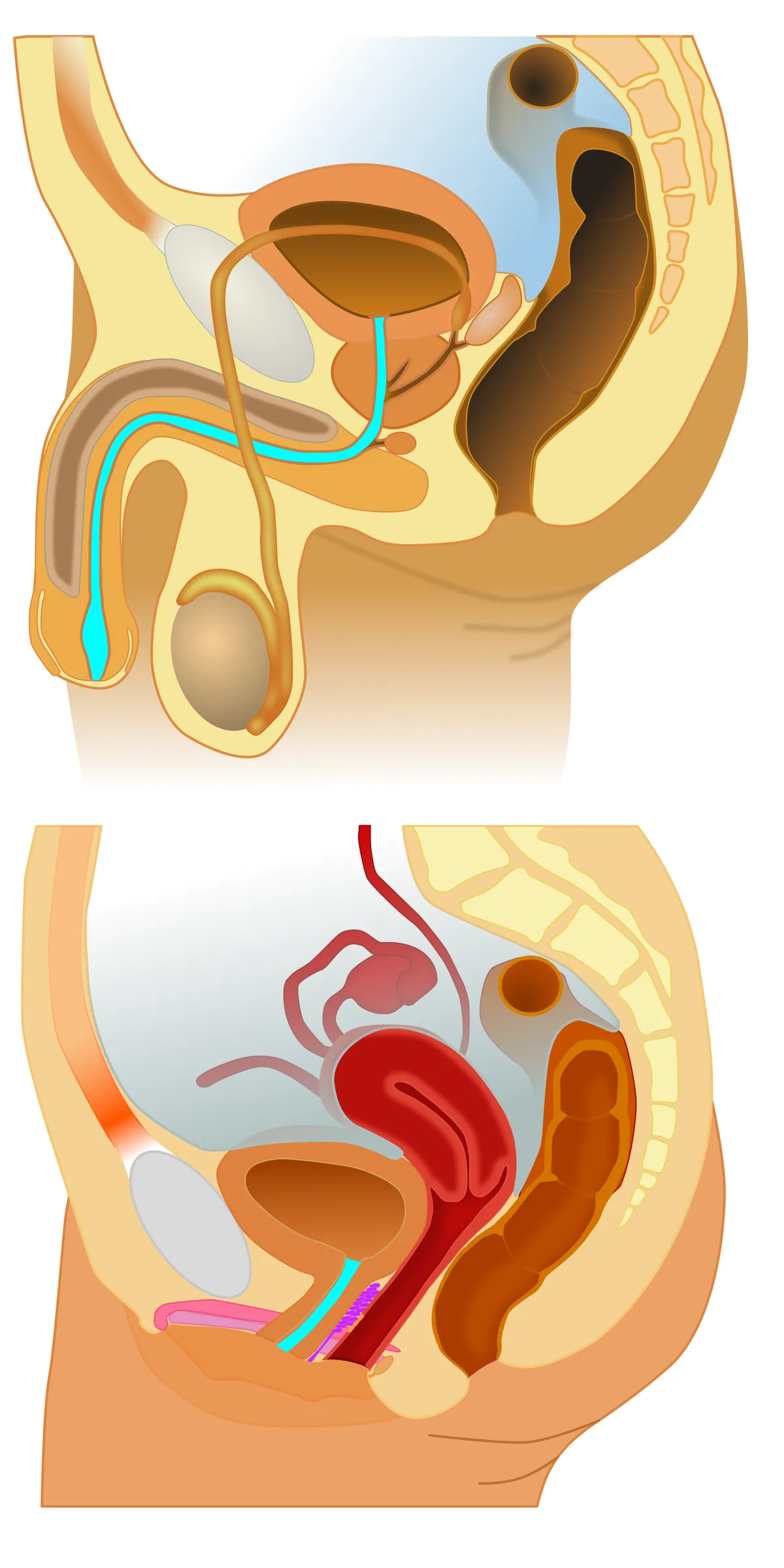
The tube that carries urine from the bladder neck to the exterior. The female urethra is short (about 4 cm); the male urethra is about 18–20 cm long and is a common path for urine and semen.
Parts
Each part has its own card; press “Show on the model” to fly to it and select it in the 3D model above.
- 01
Prostatic urethra
Pars prostatica urethrae
The widest part of the male urethra (about 3–4 cm), passing through the prostate. Its posterior wall carries the urethral crest with the seminal colliculus, on which open the prostatic utricle and the two ejaculatory ducts; the prostatic ducts open into the grooves on either side.
- 02
Membranous urethra
Pars membranacea urethrae
The shortest and narrowest part (about 1–2 cm), traversing the perineum surrounded by the external urethral sphincter (striated, pudendal nerve), which provides voluntary continence. It is torn in pelvic fractures and easily injured by catheters at its junction with the spongy part.
- 03
Spongy urethra
Pars spongiosa urethrae
The longest part (about 15 cm), within the corpus spongiosum, comprising a bulbar part (wide, receiving the bulbo-urethral or Cowper ducts), a penile part and the navicular fossa in the glans before the external meatus, the narrowest point for catheterisation. Urethral (Littré) glands secrete mucus.
- 04
Female urethra
Urethra feminina
Only about 4 cm long, it runs down and forward fused to the anterior vaginal wall and opens in the vestibule between clitoris and vaginal orifice, flanked by the para-urethral (Skene) glands. Its shortness and proximity to the anus explain why urinary infections are commoner in women.
- 05
Urethral sphincters
Musculus sphincter urethrae internus et externus
The internal sphincter is smooth muscle at the bladder neck (well developed in men, involuntary); the external sphincter is striated muscle round the membranous urethra in men and the middle third of the female urethra (voluntary, pudendal nerve S2–S4). Damage after prostatectomy or childbirth causes stress incontinence.
Position and relations
The male urethra has four parts: preprostatic (in the bladder neck), prostatic (through the prostate, with the seminal colliculus and ejaculatory duct openings), membranous (through the urogenital diaphragm, the narrowest and most vulnerable) and spongy (in the corpus spongiosum, with the navicular fossa in the glans). The female urethra runs in front of the vagina and opens into the vestibule between the clitoris and the vaginal orifice.
Form and structure
The internal sphincter (smooth muscle at the bladder neck, distinct in men) prevents retrograde ejaculation; the external sphincter (striated, voluntary) maintains continence. The lining changes from urothelium proximally to pseudostratified columnar and then stratified squamous epithelium at the meatus; small glands (Littré in men, Skene’s paraurethral glands in women) open into the lumen.
Vessels and nerves
Blood comes from branches of the internal pudendal, inferior vesical and (in women) vaginal arteries. The pudendal nerve (S2–S4) supplies the external sphincter and sensation; pudendal or pelvic floor injury after childbirth can cause stress incontinence.
Urethral sphincters and continence
The urethra is a muscular tube leaving the bladder neck. Its inner smooth muscle layer, the internal urethral sphincter, has an inner longitudinal and a thinner outer circular layer; its outer striated layer forms the external urethral sphincter. The internal sphincter is continuous with the detrusor but variably arranged, and the external sphincter blends with fibres of levator ani.[1][2]
Tonic contraction of both smooth and striated muscle creates a urethral closure pressure that exceeds bladder pressure and maintains continence. Smooth muscle probably sustains long-term tone at low energy cost, while striated muscle adds the rise in tone that accompanies sudden increases in abdominal pressure such as coughing. Smooth muscle tone is regulated by noradrenaline, acetylcholine, ATP and nitric oxide, yet it can generate tone without neural input; urethral interstitial cells are candidate pacemakers.[1]
The decision to void relies on communication between several cerebral centres and primitive sacral spinal reflexes. In women the pudendal nerve is most often injured at the sacrospinous ligament. In boys, posterior urethral valves, an obstructing membrane within the urethra, are an important cause of renal damage; severity depends on the membrane’s configuration, and endoscopic valve ablation is the standard treatment.[2][3]
Full-text reading (StatPearls)
The StatPearls chapters on this topic, reproduced verbatim. Their CC BY-NC-ND 4.0 licence allows unmodified copies only, so they are not translated or shortened.
Histology, Male UrethraStoddard N, Leslie SW · StatPearls 2023 · NBK542238 · 10k characters
Loading…
Stoddard N, Leslie SW. Histology, Male Urethra. StatPearls [Internet]. Treasure Island (FL): StatPearls Publishing; 2023. NBK542238 · PMID 31194395 · CC BY-NC-ND 4.0. Reproduced verbatim, unmodified.
Organs in this system
References
Sources for the values and descriptions on this page, indexed in PubMed (US National Library of Medicine).
- [1]Spontaneous Activity in Urethral Smooth MuscleSergeant GP et al., Advances in experimental medicine and biology 2019PMID 31183826
- [2]Continence and micturition: an anatomical basisShah AP et al., Clinical anatomy 2014PMID 24615792
- [3]Posterior urethral valveNasir AA et al., World journal of pediatrics 2011PMID 21822988
- [4]Normal reference values for glomerular filtration rate: what do we really know?Delanaye P et al., 2012PMID 22802582Supports: Glomerular filtration rate