Wrist and hand
Model
Layers
Z-Anatomy anatomical model based on BodyParts3D, licensed CC BY-SA 4.0.
Overview
A regional model of the (right) wrist and hand: the eight carpal bones, five metacarpals and fourteen phalanges, the joints and their ligaments, the flexor and extensor retinacula, the tendons and synovial sheaths of the forearm muscles, the intrinsic muscles (thenar, hypothenar, lumbricals, interossei), the superficial and deep palmar arches and the terminal branches of the median, ulnar and radial nerves.
Boundaries and subdivisions
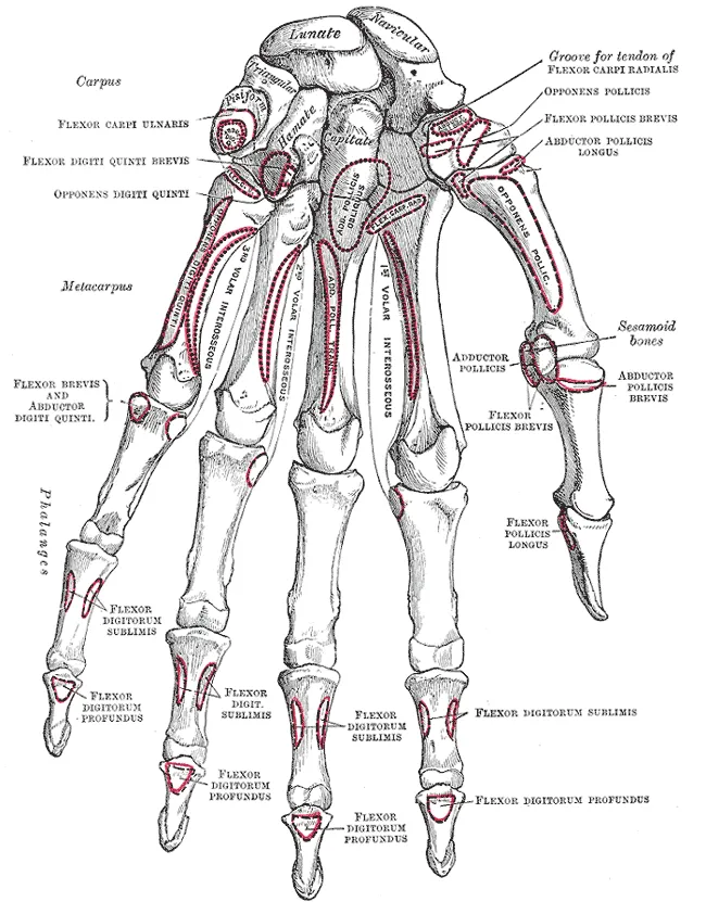
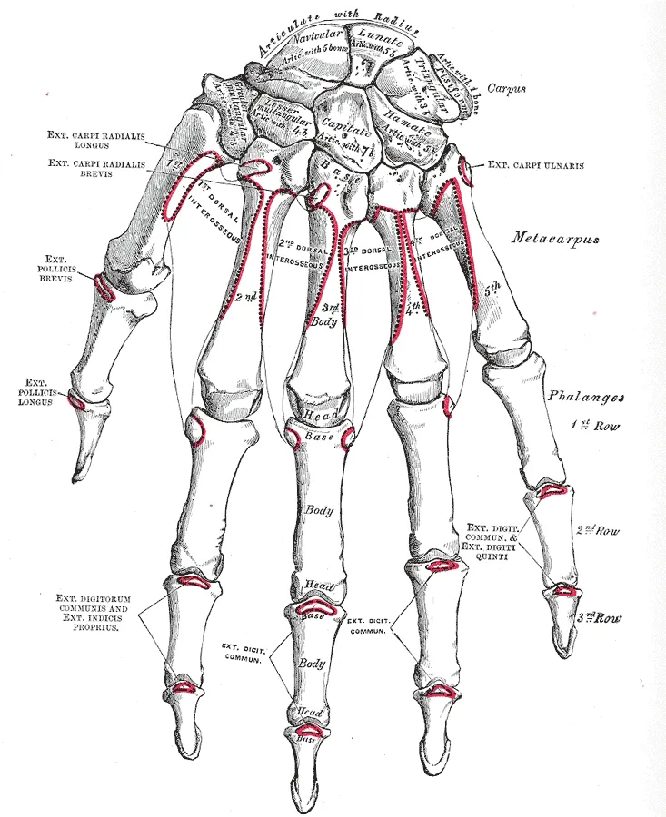
The wrist contains eight carpal bones in two rows: the proximal row scaphoid, lunate, triquetrum and pisiform; the distal row trapezium, trapezoid, capitate and hamate. The hand has five metacarpals and fourteen phalanges (two in the thumb). The palm is divided into the thenar and hypothenar eminences and the central palm; the triangular palmar aponeurosis lies under the palmar skin and continues the tendon of palmaris longus. On the dorsolateral wrist the anatomical snuffbox is bounded behind by the extensor pollicis longus tendon and in front by the tendons of abductor pollicis longus and extensor pollicis brevis; its floor is formed by the scaphoid and trapezium.[1][2]
Layers from superficial to deep
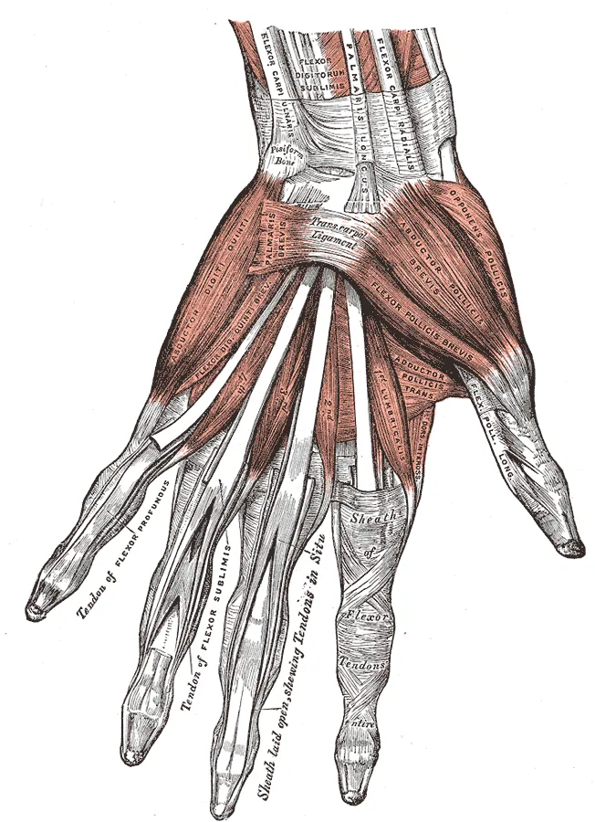
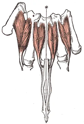
The flexor retinaculum stretches from the scaphoid and trapezium laterally to the pisiform and hamate medially, converting the carpal groove into the carpal tunnel; through it run the median nerve and nine flexor tendons (four superficial, four deep and flexor pollicis longus) in their synovial sheaths. The ulnar nerve and artery pass more superficially through Guyon’s canal between the pisiform and the hook of the hamate. On the dorsum the extensor retinaculum sorts the extensor tendons into six synovial compartments. The intrinsic muscles comprise the thenar group (abductor and flexor pollicis brevis, opponens pollicis, with adductor pollicis), the hypothenar group, four lumbricals arising from the deep flexor tendons, and the palmar (adducting) and dorsal (abducting) interossei.[3][4][5]
Vessels, nerves and lymphatics
The ulnar artery forms most of the superficial palmar arch (just beneath the palmar aponeurosis, level with the distal border of the fully abducted thumb), and the radial artery most of the deep palmar arch at the level of the metacarpal bases; the arches anastomose so the hand stays perfused if one artery is occluded. The median nerve supplies the thenar muscles and the two lateral lumbricals and the palmar skin of the lateral three and a half digits; the ulnar nerve supplies most of the other intrinsic muscles and the medial one and a half digits; the radial nerve gives only sensation to the lateral dorsum. All three come from the brachial plexus.[6][7]
Embryology
The limb bud has three HOX-controlled growth zones: the stylopod (humerus), zeugopod (radius and ulna) and autopod (hand). The ectoderm forms the apical ectodermal ridge, which drives proximodistal growth, and the zone of polarising activity, whose SHH sets the thumb-to-little-finger axis; disrupted signalling causes congenital anomalies such as polydactyly and syndactyly. The hand develops between weeks 6 and 14 in three phases: the hand shape is evident in weeks 6–10, creases appear in weeks 10–13, and the dermal ridges form from week 13. The precursor extensor mass of the forearm splits into three: a radial part (extensor carpi radialis longus and brevis, brachioradialis), a superficial part (extensor digitorum, extensor carpi ulnaris, extensor digiti minimi) and a deep part (abductor pollicis longus, extensor pollicis brevis and longus, extensor indicis). The ulnar and radial arteries branch from the brachial artery, which continues the subclavian, itself derived from the fourth aortic arch.[8][5][9][10][11]
Anatomical variants
In Guyon’s canal the ulnar nerve bifurcates into superficial and deep branches distal to the midpoint of the pisiform in 66–86% of cases and trifurcates in about 10%, which is why ulnar nerve compression at the wrist presents differently from patient to patient. The palmar arches also vary: the ulnar artery is usually larger than the radial but may be equal or smaller, some people have duplicated arches, and in others the deep arch does not connect with the superficial arch, increasing the risk of ischaemia if one artery is injured. The extensors of the posterior forearm and the dorsal wrist compartments often show accessory bellies, duplicated or absent tendons, anomalous insertions and septa within a compartment; anomalies of extensor pollicis longus (duplication, aberrant course) are grouped by the Türker classification. Anomalous muscles and nerves around the intrinsic muscles can distort landmarks or cause compressive neuropathy. The index-to-ring finger length ratio (2D:4D) averages 0.98 and varies with sex and ancestry.[4][11][12][10][5][8]
Clinical correlations
Carpal tunnel syndrome is compression of the median nerve in the tunnel: numbness of the lateral three and a half digits, worse at night, and eventually thenar wasting; the central palm is spared because the palmar branch of the median nerve passes above the retinaculum. Snuffbox tenderness after a fall on the outstretched hand suggests a scaphoid fracture, prone to avascular necrosis of the proximal pole because its blood supply enters distally. Ulnar nerve injury at the wrist produces an ulnar claw hand. The Allen test checks flow through the palmar arches before radial arterial puncture.[4][1]
Surgical and procedural considerations
Hand surgery must preserve collateral flow: in most people the arterial anastomoses keep the hand perfused, but palmar arch variants can put some at risk of ischaemic injury, and digital replantation requires detailed knowledge of the blood supply of the fingers. Metacarpal fractures need fixation when there is any malrotation, so that the patient can make a full fist, or when angulation exceeds accepted limits, using lag screws, plates or percutaneous pins. Because the hand muscles are supplied by the median, ulnar and radial nerves in different patterns, tendon transfers are widely used to restore function after nerve palsy; some intrinsic muscles, such as the first and second lumbricals, have been used as muscle flaps in carpal tunnel syndrome. Stenosing tenosynovitis of extensor pollicis longus is rare but almost always requires complete surgical release to prevent attritional rupture.[11][8][9][5][10]
Key structures
Canalis carpi
- Name
- Carpal tunnel
- Position, role
- Median nerve and nine flexor tendons under the flexor retinaculum
Canalis ulnaris
- Name
- Ulnar canal (Guyon)
- Position, role
- Between the pisiform and hook of hamate; ulnar nerve and artery
Fovea radialis
- Name
- Anatomical snuffbox
- Position, role
- Floor formed by the scaphoid; crossed by the radial artery
Aponeurosis palmaris
- Name
- Palmar aponeurosis
- Position, role
- Protects palmar tendons and vessels; thickens in Dupuytren disease
Arcus palmaris superficialis
- Name
- Superficial palmar arch
- Position, role
- Mainly from the ulnar artery; gives common palmar digital arteries
Mm. lumbricales
- Name
- Lumbricals
- Position, role
- From the deep flexor tendons; flex MCP and extend IP joints
Eminentia thenaris
- Name
- Thenar eminence
- Position, role
- Abductor and flexor pollicis brevis, opponens; recurrent branch of median nerve
| Terminologia Anatomica | Name | Position, role |
|---|---|---|
| Canalis carpi | Carpal tunnel | Median nerve and nine flexor tendons under the flexor retinaculum |
| Canalis ulnaris | Ulnar canal (Guyon) | Between the pisiform and hook of hamate; ulnar nerve and artery |
| Fovea radialis | Anatomical snuffbox | Floor formed by the scaphoid; crossed by the radial artery |
| Aponeurosis palmaris | Palmar aponeurosis | Protects palmar tendons and vessels; thickens in Dupuytren disease |
| Arcus palmaris superficialis | Superficial palmar arch | Mainly from the ulnar artery; gives common palmar digital arteries |
| Mm. lumbricales | Lumbricals | From the deep flexor tendons; flex MCP and extend IP joints |
| Eminentia thenaris | Thenar eminence | Abductor and flexor pollicis brevis, opponens; recurrent branch of median nerve |
What each layer contains
SkinCutis9
- Dorsum of hand
- Dorsal surfaces of digits of hand
- Palmar surfaces of digits of hand
- Radial foveola
- Nail plate
- Palm
- Perionyx
- Anterior region of wrist
- Posterior region of wrist
Muscles, fasciae and tendon sheathsMusculi, fasciae et vaginae tendinum41
- Abductor pollicis brevis
- Abductor digiti minimi of hand
- Palmar aponeurosis
- Humeral head of flexor carpi ulnaris
- Humero-ulnar head of flexor digitorum superficialis
- Oblique head of adductor pollicis
- Deep head of flexor pollicis brevis
- Superficial head of flexor pollicis brevis
- Transverse head of adductor pollicis
- Cruciform part of fibrous sheath of digit of hand
- Extensor pollicis brevis
- Extensor digiti minimi
- Extensor digitorum
- Extensor indicis
- Extensor pollicis longus
- Extensor carpi radialis brevis
- Extensor carpi radialis longus
- Flexor digiti minimi of hand
- Flexor pollicis longus
- Flexor digitorum profundus
- Flexor carpi radialis
- Superficial transverse metacarpal ligament
- Dorsal interossei muscles of hand
- Palmar interossei muscles
- Lumbrical muscles of hand
- Opponens digiti minimi muscle of hand
- Opponens pollicis muscle
- Palmaris longus muscle
- Pronator quadratus
- Extensor retinaculum of wrist
- Flexor retinaculum of wrist
- Tendon sheath of extensor digiti minimi manus
- Tendon sheath of extensors carpi radialis
- Common flexor tendon sheath
- Tendon sheath of extensor carpi ulnaris
- Tendon sheath of flexor pollicis longus
- Tendon sheath of flexor carpi radialis
- Tendon sheath of extensor pollicis longus
- Tendon sheath - abd. pollicis longus - ext. pollicis brevis
- Tendon sheath of extensor digitorum and extensor indicis
- Synovial sheaths of digits of hand
Bones and cartilageOssa et cartilagines27
- Capitate bone
- Hamate bone
- Lunate bone
- Pisiform bone
- First metacarpal bone
- Fourth metacarpal bone
- Fifth metacarpal bone
- Scaphoid bone
- Second metacarpal bone
- Third metacarpal bone
- Trapezium bone
- Trapezoid bone
- Triquetrum bone
- Distal phalanx of first finger of hand
- Distal phalanx of fourth finger of hand
- Distal phalanx of fifth finger of hand
- Distal phalanx of second finger of hand
- Distal phalanx of third finger of hand
- Middle phalanx of fourth finger of hand
- Middle phalanx of fifth finger of hand
- Middle phalanx of second finger of hand
- Middle phalanx of third finger of hand
- Proximal phalanx of first finger of hand
- Proximal phalanx of fourth finger of hand
- Proximal phalanx of fifth finger of hand
- Proximal phalanx of second finger of hand
- Proximal phalanx of third finger of hand
Joints and ligamentsJuncturae et ligamenta45
- Articular capsule of radiocarpal joint
- Articular capsules of proximal interphalangeal joints
- Articular capsules of distal interphalangeal joints
- Articular capsules of metacarpophalangeal joints
- Articular disc of distal radio-ulnar joint
- Dorsal carpometacarpal ligaments
- Palmar carpometacarpal ligaments
- Dorsal intercarpal ligaments
- Collateral interphalangeal ligaments of hand
- Palmar interphalangeal ligaments
- Dorsal metacarpal ligaments
- Interosseous metacarpal ligaments
- Palmar metacarpal ligaments
- Collateral metacarpophalangeal ligaments
- Capitohamate interosseous ligament
- Palmar capitohamate ligament
- Radial collateral ligament of wrist joint
- Ulnar collateral ligament of wrist joint
- Lunotriquetral interosseous ligament
- Palmar lunotriquetral ligament
- Deep transverse metacarpal ligament
- Pisohamate ligament
- Pisometacarpal ligament
- Pisotriquetral ligament
- Radiate carpal ligament
- Radiocapitate ligament
- Dorsal radiocarpal ligament
- Radioscaphocapitate ligament
- Dorsal radio-ulnar ligament
- Palmar radio-ulnar ligament
- Scaphocapitate ligament
- Scapholunate interosseous ligament
- Scaphotrapeziotrapezoidal ligament
- Dorsal scaphotriquetral ligament
- Palmar scaphotriquetral ligament
- Trapeziotrapezoidal interosseous ligament
- Trapezoideocapitate interosseous ligament
- Palmar trapezoideocapitate ligament
- Triquetrocapitate ligament
- Triquetrohamate ligament
- Ulnocapitate ligament
- Dorsal ulnocarpal ligament
- Ulnolunate ligament
- Ulnopisiform ligament
- Ulnotriquetral ligament
ArteriesArteriae10
- Deep palmar arch
- Superficial palmar arch
- Dorsal digital arteries of hand
- Common palmar digital arteries
- Proper palmar digital arteries
- Dorsal metacarpal arteries
- Palmar metacarpal arteries
- Dorsal carpal branch of ulnar artery
- Palmar carpal branch of radial artery
- Dorsal carpal anastomosis
VeinsVenae5
- Deep venous palmar arch
- Superficial venous palmar arch
- Dorsal venous network of hand
- Dorsal digital veins of hand
- Palmar digital veins
NervesNervi13
- (Communicating branch of median nerve with ulnar nerve)
- Dorsal digital branches of radial nerve
- Dorsal digital branches of ulnar nerve
- Common palmar digital branches of median nerve
- Common palmar digital branches of ulnar nerve
- Proper palmar digital branches of median nerve
- Proper palmar digital branches of ulnar nerve
- Dorsal branch of ulnar nerve
- Palmar branch of median nerve
- Palmar branch of ulnar nerve
- Deep branch of ulnar nerve
- Superficial branch of radial nerve
- Superficial branch of ulnar nerve
Full-text reading (StatPearls)
The StatPearls chapters on this topic, reproduced verbatim. Their CC BY-NC-ND 4.0 licence allows unmodified copies only, so they are not translated or shortened.
Anatomy, Shoulder and Upper Limb, Wrist Extensor MusclesReeves N, Crow T, Varacallo MA · StatPearls 2026 · NBK534805 · 29k characters
Loading…
Reeves N, Crow T, Varacallo MA. Anatomy, Shoulder and Upper Limb, Wrist Extensor Muscles. StatPearls [Internet]. Treasure Island (FL): StatPearls Publishing; 2026. NBK534805 · PMID 30521226 · CC BY-NC-ND 4.0. Reproduced verbatim, unmodified.
Anatomy, Shoulder and Upper Limb, Hand Extensor Pollicis Longus MuscleKhan IA, Launico MV · StatPearls 2026 · NBK544294 · 23k characters
Loading…
Khan IA, Launico MV. Anatomy, Shoulder and Upper Limb, Hand Extensor Pollicis Longus Muscle. StatPearls [Internet]. Treasure Island (FL): StatPearls Publishing; 2026. NBK544294 · PMID 31335014 · CC BY-NC-ND 4.0. Reproduced verbatim, unmodified.
Anatomy, Shoulder and Upper Limb, Hand Intrinsic MusclesLaunico MV, Varacallo MA · StatPearls 2025 · NBK539810 · 22k characters
Loading…
Launico MV, Varacallo MA. Anatomy, Shoulder and Upper Limb, Hand Intrinsic Muscles. StatPearls [Internet]. Treasure Island (FL): StatPearls Publishing; 2025. NBK539810 · PMID 30969632 · CC BY-NC-ND 4.0. Reproduced verbatim, unmodified.
Anatomy, Shoulder and Upper Limb, Hand BonesArias DG, Black AC, Varacallo MA · StatPearls 2023 · NBK547684 · 22k characters
Loading…
Arias DG, Black AC, Varacallo MA. Anatomy, Shoulder and Upper Limb, Hand Bones. StatPearls [Internet]. Treasure Island (FL): StatPearls Publishing; 2023. NBK547684 · PMID 31613464 · CC BY-NC-ND 4.0. Reproduced verbatim, unmodified.
Anatomy, Shoulder and Upper Limb, Hand MusclesOkwumabua E, Sinkler MA, Bordoni B · StatPearls 2023 · NBK537229 · 17k characters
Loading…
Okwumabua E, Sinkler MA, Bordoni B. Anatomy, Shoulder and Upper Limb, Hand Muscles. StatPearls [Internet]. Treasure Island (FL): StatPearls Publishing; 2023. NBK537229 · PMID 30725914 · CC BY-NC-ND 4.0. Reproduced verbatim, unmodified.
Anatomy, Shoulder and Upper Limb, Hand ArteriesNguyen JD, Black AC, Duong H · StatPearls 2023 · NBK546583 · 15k characters
Loading…
Nguyen JD, Black AC, Duong H. Anatomy, Shoulder and Upper Limb, Hand Arteries. StatPearls [Internet]. Treasure Island (FL): StatPearls Publishing; 2023. NBK546583 · PMID 31536192 · CC BY-NC-ND 4.0. Reproduced verbatim, unmodified.
Anatomy, Shoulder and Upper Limb, Hand Guyon CanalGarg DR, La Rose J · StatPearls 2026 · NBK534814 · 15k characters
Loading…
Garg DR, La Rose J. Anatomy, Shoulder and Upper Limb, Hand Guyon Canal. StatPearls [Internet]. Treasure Island (FL): StatPearls Publishing; 2026. NBK534814 · PMID 30521235 · CC BY-NC-ND 4.0. Reproduced verbatim, unmodified.
Anatomy, Shoulder and Upper Limb, Hand Carpal BonesTang A, Varacallo MA · StatPearls 2022 · NBK535382 · 13k characters
Loading…
Tang A, Varacallo MA. Anatomy, Shoulder and Upper Limb, Hand Carpal Bones. StatPearls [Internet]. Treasure Island (FL): StatPearls Publishing; 2022. NBK535382 · PMID 30571003 · CC BY-NC-ND 4.0. Reproduced verbatim, unmodified.
Anatomy, Shoulder and Upper Limb: Hand Anatomical Snuff BoxHallett Reid S, Jozsa F, Ashurst JV · StatPearls 2025 · NBK482228 · 12k characters
Loading…
Hallett Reid S, Jozsa F, Ashurst JV. Anatomy, Shoulder and Upper Limb: Hand Anatomical Snuff Box. StatPearls [Internet]. Treasure Island (FL): StatPearls Publishing; 2025. NBK482228 · PMID 29489241 · CC BY-NC-ND 4.0. Reproduced verbatim, unmodified.
Anatomy, Shoulder and Upper Limb, Wrist JointErwin J, Varacallo MA · StatPearls 2023 · NBK534779 · 11k characters
Loading…
Erwin J, Varacallo MA. Anatomy, Shoulder and Upper Limb, Wrist Joint. StatPearls [Internet]. Treasure Island (FL): StatPearls Publishing; 2023. NBK534779 · PMID 30521200 · CC BY-NC-ND 4.0. Reproduced verbatim, unmodified.
Anatomy, Shoulder and Upper Limb, Hand Interossei MusclesValenzuela M, Varacallo MA · StatPearls 2025 · NBK534772 · 11k characters
Loading…
Valenzuela M, Varacallo MA. Anatomy, Shoulder and Upper Limb, Hand Interossei Muscles. StatPearls [Internet]. Treasure Island (FL): StatPearls Publishing; 2025. NBK534772 · PMID 30521193 · CC BY-NC-ND 4.0. Reproduced verbatim, unmodified.
Anatomy, Shoulder and Upper Limb, Hand Volar Arch ArteriesJozsa F, Varacallo MA · StatPearls 2025 · NBK545377 · 10k characters
Loading…
Jozsa F, Varacallo MA. Anatomy, Shoulder and Upper Limb, Hand Volar Arch Arteries. StatPearls [Internet]. Treasure Island (FL): StatPearls Publishing; 2025. NBK545377 · PMID 31430092 · CC BY-NC-ND 4.0. Reproduced verbatim, unmodified.
Anatomy, Shoulder and Upper Limb, Hand Ulnar BursaSawyer E, Black AC, Launico MV, Varacallo MA · StatPearls 2023 · NBK541025 · 10k characters
Loading…
Sawyer E, Black AC, Launico MV, Varacallo MA. Anatomy, Shoulder and Upper Limb, Hand Ulnar Bursa. StatPearls [Internet]. Treasure Island (FL): StatPearls Publishing; 2023. NBK541025 · PMID 31082069 · CC BY-NC-ND 4.0. Reproduced verbatim, unmodified.
Anatomy, Shoulder and Upper Limb, Wrist Flexor RetinaculumDeak N, Black AC, Bordoni B · StatPearls 2023 · NBK545198 · 9k characters
Loading…
Deak N, Black AC, Bordoni B. Anatomy, Shoulder and Upper Limb, Wrist Flexor Retinaculum. StatPearls [Internet]. Treasure Island (FL): StatPearls Publishing; 2023. NBK545198 · PMID 31424782 · CC BY-NC-ND 4.0. Reproduced verbatim, unmodified.
Anatomy, Shoulder and Upper Limb, Hand Palmar Interosseous MuscleValenzuela M, Bordoni B · StatPearls 2025 · NBK537165 · 8k characters
Loading…
Valenzuela M, Bordoni B. Anatomy, Shoulder and Upper Limb, Hand Palmar Interosseous Muscle. StatPearls [Internet]. Treasure Island (FL): StatPearls Publishing; 2025. NBK537165 · PMID 30725850 · CC BY-NC-ND 4.0. Reproduced verbatim, unmodified.
Anatomy, Shoulder and Upper Limb, Hand Dorsal Interossei MuscleValenzuela M, Bordoni B · StatPearls 2025 · NBK536922 · 7k characters
Loading…
Valenzuela M, Bordoni B. Anatomy, Shoulder and Upper Limb, Hand Dorsal Interossei Muscle. StatPearls [Internet]. Treasure Island (FL): StatPearls Publishing; 2025. NBK536922 · PMID 30725607 · CC BY-NC-ND 4.0. Reproduced verbatim, unmodified.
Anatomy, Hand PositioningParekh PS, Gupta V · StatPearls 2023 · NBK557701 · 4k characters
Loading…
Parekh PS, Gupta V. Anatomy, Hand Positioning. StatPearls [Internet]. Treasure Island (FL): StatPearls Publishing; 2023. NBK557701 · PMID 32491633 · CC BY-NC-ND 4.0. Reproduced verbatim, unmodified.
Regional anatomy
References
Sources for the values and descriptions on this page, indexed in PubMed (US National Library of Medicine).
- [1]Anatomy, Shoulder and Upper Limb: Hand Anatomical Snuff BoxHallett Reid S et al., StatPearls 2025PMID 29489241
- [2]Anatomy, Shoulder and Upper Limb, Wrist JointErwin J et al., StatPearls 2023PMID 30521200
- [3]Anatomy, Shoulder and Upper Limb, Wrist Flexor RetinaculumDeak N et al., StatPearls 2023PMID 31424782
- [4]Anatomy, Shoulder and Upper Limb, Hand Guyon CanalGarg DR et al., StatPearls 2026PMID 30521235
- [5]Anatomy, Shoulder and Upper Limb, Hand Intrinsic MusclesLaunico MV et al., StatPearls 2025PMID 30969632
- [6]Anatomy, Shoulder and Upper Limb, Brachial PlexusBayot ML et al., StatPearls 2023PMID 29763192
- [7]Anatomy, Shoulder and Upper Limb, Hand Volar Arch ArteriesJozsa F et al., StatPearls 2025PMID 31430092
- [8]Anatomy, Shoulder and Upper Limb, Hand BonesArias DG et al., StatPearls 2023PMID 31613464
- [9]Anatomy, Shoulder and Upper Limb, Hand MusclesOkwumabua E et al., StatPearls 2023PMID 30725914
- [10]Anatomy, Shoulder and Upper Limb, Hand Extensor Pollicis Longus MuscleKhan IA et al., StatPearls 2026PMID 31335014
- [11]Anatomy, Shoulder and Upper Limb, Hand ArteriesNguyen JD et al., StatPearls 2023PMID 31536192
- [12]Anatomy, Shoulder and Upper Limb, Wrist Extensor MusclesReeves N et al., StatPearls 2026PMID 30521226