Ear
Model
Hover or tap to see names, click to read a description; click a name in the legend to fly to it. Procedurally built to histological proportions; colours follow illustration conventions.
Description
The organ of hearing and balance: the external ear (auricle, external acoustic meatus), the middle ear (tympanic cavity with the tympanic membrane and three ossicles) and the internal ear in the petrous temporal bone (the cochlea for hearing; the vestibule and three semicircular canals for balance).
Parts
Each part has its own card; press “Show on the model” to fly to it and select it in the 3D model above.
- 01
Auricle and external acoustic meatus
Auricula; Meatus acusticus externus
The auricle is a skin-covered frame of elastic cartilage (except the lobule) that gathers and localises sound. The external acoustic meatus is about 2.5 cm long, its outer third cartilaginous with ceruminous glands and inner two-thirds bony; in adults the auricle is pulled up and back to straighten it for otoscopy.
- 02
Tympanic membrane
Membrana tympanica
A tilted cone-shaped membrane about 9–10 mm across with three layers (skin, fibrous, mucosa). The handle of the malleus is fixed to its inner surface at the umbo; a small pars flaccida lies above the large pars tensa, and the cone of light points anteroinferiorly on otoscopy.
- 03
Auditory ossicles
Ossicula auditus
The malleus, incus and stapes, linked by synovial joints, transmit vibrations from the tympanic membrane to the oval window. Lever action and the area ratio between eardrum and stapes footplate amplify pressure about twentyfold; tensor tympani (V3) and stapedius (VII) damp loud sounds.
- 04
Tympanic cavity and auditory tube
Cavitas tympani; Tuba auditiva
The tympanic cavity is an air space in the temporal bone, opening back into the mastoid antrum and air cells and forward into the auditory (Eustachian) tube, about 3.5 cm long, which runs to the nasopharynx and equalises pressure. In young children the tube is shorter and more horizontal, favouring otitis media.
- 05
Cochlea
Cochlea
A bony canal coiled two and a half turns round the modiolus, divided into the scala vestibuli, the cochlear duct (containing endolymph) and the scala tympani. The organ of Corti on the basilar membrane bears hair cells arranged tonotopically: high frequencies at the base, low frequencies at the apex.
- 06
Vestibule (utricle and saccule)
Vestibulum; Utriculus et sacculus
The central cavity of the bony labyrinth, with the oval window in its lateral wall. It houses the utricle and saccule, each with a macula whose otoliths sense linear acceleration and head tilt relative to gravity (utricle mainly horizontal, saccule vertical).
- 07
Semicircular canals
Canales semicirculares
The anterior, posterior and lateral canals lie roughly at right angles; each has an ampulla with a crista and cupula that sense rotational acceleration of the head. The anterior and posterior canals share a common limb; displaced otoconia in the posterior canal cause benign paroxysmal positional vertigo.
External and middle ear
The external ear comprises the auricle (an elastic cartilage frame) and the external acoustic meatus, about 2.5 cm long, ending at the tympanic membrane. The middle ear is an air-filled tympanic cavity in the temporal bone, opening to the nasopharynx through the pharyngotympanic (Eustachian) tube, which equalises pressure, and to the mastoid air cells behind. Three ossicles, malleus, incus and stapes, link the tympanic membrane to the oval window and amplify sound pressure about twentyfold; tensor tympani (V) and stapedius (VII) damp them against loud sounds.
Internal ear
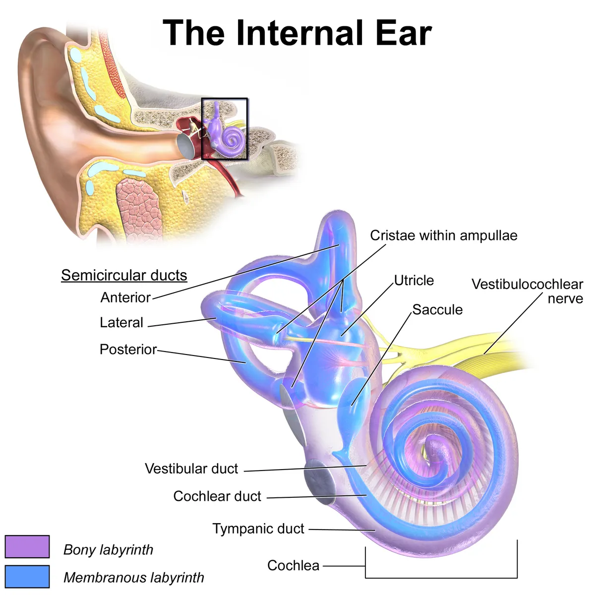
The internal ear is a bony labyrinth in the petrous temporal bone housing a membranous labyrinth filled with endolymph. The cochlea makes about two and a half turns; the perilymph-filled scala vestibuli and scala tympani sandwich the cochlear duct with the organ of Corti, whose hair cells respond to high frequencies at the base and low frequencies at the apex. The vestibular apparatus comprises the utricle and saccule (linear acceleration and gravity) and three mutually perpendicular semicircular canals (rotational acceleration).
Cochlear and vestibular signals travel in nerve VIII through the internal acoustic meatus to the pontomedullary junction and the cochlear and vestibular nuclei of the brainstem. The ascending auditory pathway crosses partially and relays in the inferior colliculus and the medial geniculate body before reaching the auditory cortex in Heschl’s gyri; a one-sided cortical lesion therefore rarely deafens one ear.
Clinical correlations
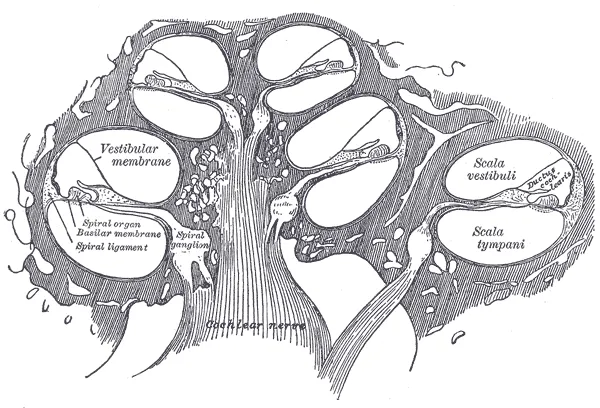
Otitis media is common in young children because their pharyngotympanic tube is shorter and more horizontal. Conductive hearing loss arises in the external or middle ear (wax, perforated tympanic membrane, otosclerosis of the stapes); sensorineural loss arises in the cochlea or nerve VIII. The Rinne and Weber tests distinguish the two. Benign paroxysmal positional vertigo is caused by otoconia displaced into a semicircular canal, usually the posterior one.
Cochlear physiology
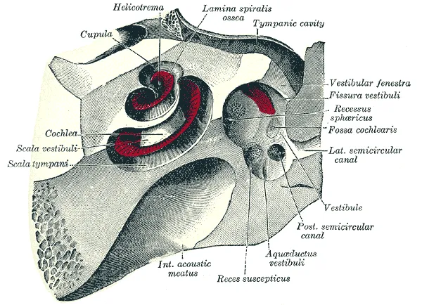
Unlike other senses, hearing is not passive: cochlear hair cells run an active process that amplifies sound several hundred-fold, sharpens frequency selectivity and widens dynamic range. In mammals it combines hair-bundle motility with prestin-based somatic electromotility of the outer hair cells.[1]
Hearing begins when vibrations deflect the stereocilia on the hair-cell apex. Each cell carries dozens of stereocilia in rows of increasing height, a staircase essential for mechanosensitivity that must be maintained for life because mammalian auditory hair cells do not regenerate. Current through the mechanotransduction channels at the stereocilia tips appears to control their dimensions.[2]
The stria vascularis generates the endocochlear potential, the “battery” for outer hair cell amplification; it contains numerous capillary pericytes that form the intrastrial blood–fluid barrier and regulate microcirculation.[3][4]
Clinical correlations
Age-related hearing loss affects the stria vascularis, spiral ligament, hair cells and auditory neurons. Strial dysfunction lowers the endocochlear potential so outer hair cell amplification fails, causing high-frequency loss; neuronal degeneration explains difficulty understanding speech in noise. Noise exposure is a major cause.[3]
Before thresholds rise, noise, ototoxic drugs and ageing can permanently disconnect inner hair cells from subsets of cochlear nerve fibres (“cochlear synaptopathy”, hidden hearing loss). It contributes to speech-in-noise difficulty, tinnitus and hyperacusis even with a normal audiogram.[5]
Semicircular canal dehiscence (most often of the superior canal) creates a “third window” between the inner ear and surrounding structures, disrupting inner-ear mechanics and causing vertigo and oscillopsia with loud sounds or pressure changes, sometimes with hearing loss; surgical repair effectively relieves vestibular symptoms.[6]
Full-text reading (StatPearls)
The StatPearls chapters on this topic, reproduced verbatim. Their CC BY-NC-ND 4.0 licence allows unmodified copies only, so they are not translated or shortened.
External Ear AnatomySutton AE, Geiger Z · StatPearls 2026 · NBK470359 · 40k characters
Loading…
Sutton AE, Geiger Z. External Ear Anatomy. StatPearls [Internet]. Treasure Island (FL): StatPearls Publishing; 2026. NBK470359 · PMID 29262017 · CC BY-NC-ND 4.0. Reproduced verbatim, unmodified.
Middle Ear Anatomy and PhysiologyPeterson DC, Sutton AE · StatPearls 2026 · NBK482338 · 40k characters
Loading…
Peterson DC, Sutton AE. Middle Ear Anatomy and Physiology. StatPearls [Internet]. Treasure Island (FL): StatPearls Publishing; 2026. NBK482338 · PMID 29493963 · CC BY-NC-ND 4.0. Reproduced verbatim, unmodified.
Inner Ear Anatomy and Cochlear PhysiologySutton AE, Peterson DC · StatPearls 2026 · NBK538335 · 40k characters
Loading…
Sutton AE, Peterson DC. Inner Ear Anatomy and Cochlear Physiology. StatPearls [Internet]. Treasure Island (FL): StatPearls Publishing; 2026. NBK538335 · PMID 30855919 · CC BY-NC-ND 4.0. Reproduced verbatim, unmodified.
Histology, EarAnsari A, Tariq MA, Sadiq NM · StatPearls 2023 · NBK545170 · 12k characters
Loading…
Ansari A, Tariq MA, Sadiq NM. Histology, Ear. StatPearls [Internet]. Treasure Island (FL): StatPearls Publishing; 2023. NBK545170 · PMID 31424754 · CC BY-NC-ND 4.0. Reproduced verbatim, unmodified.
Organs in this system
References
Sources for the values and descriptions on this page, indexed in PubMed (US National Library of Medicine).
- [1]Integrating the active process of hair cells with cochlear functionHudspeth AJ, Nature reviews. Neuroscience 2014PMID 25096182
- [2]Lifelong dynamic maintenance of stereocilia bundles in mammalian auditory hair cellsVélez-Ortega AC et al., Current topics in developmental biology 2025PMID 40973236
- [3]Pathology and mechanisms of cochlear agingKeithley EM, Journal of neuroscience research 2020PMID 31066107
- [4]Cochlear Capillary PericytesCanis M et al., Advances in experimental medicine and biology 2019PMID 30937866
- [5]Cochlear synaptopathy in acquired sensorineural hearing loss: Manifestations and mechanismsLiberman MC et al., Hearing research 2017PMID 28087419
- [6]Canal dehiscenceChien WW et al., Current opinion in neurology 2011PMID 21124219
- [7]The human brain in numbers: a linearly scaled-up primate brainHerculano-Houzel S, 2009PMID 19915731Supports: Neuron count
- [8]Determinants of conduction velocity in myelinated nerve fibersWaxman SG, 1980PMID 6245357Supports: Conduction velocity
- [9]Quantitative imaging of energy expenditure in human brainPMID 22487547Supports: Cerebral energy expenditure