Palatine tonsils
Model
Layers
Z-Anatomy anatomical model based on BodyParts3D, licensed CC BY-SA 4.0.
Description
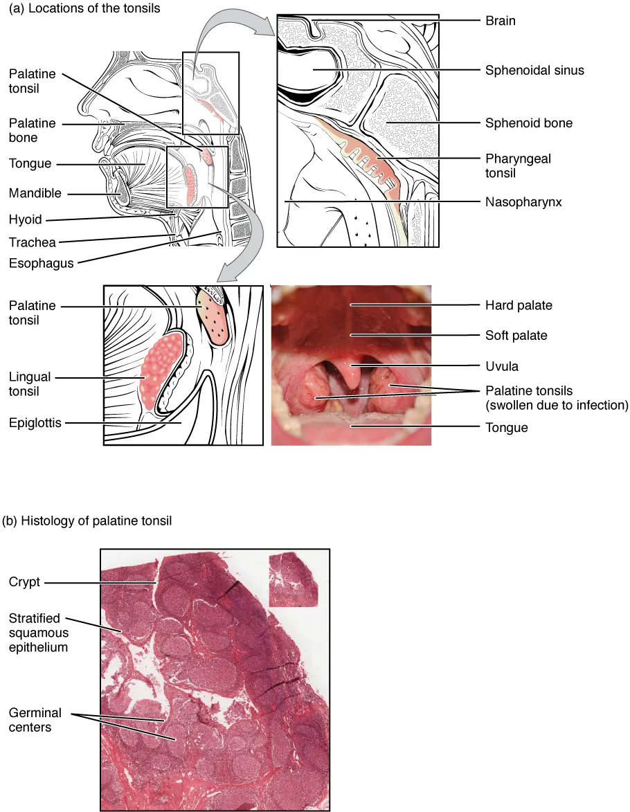
Paired masses of lymphoid tissue in the tonsillar fossa between the palatoglossal and palatopharyngeal arches, forming part of the pharyngeal lymphoid ring (Waldeyer’s ring) with the pharyngeal, tubal and lingual tonsils. Surface crypts enlarge the area exposed to antigen.
Parts
Each part has its own card; press “Show on the model” to fly to it and select it in the 3D model above.
- 01
Palatine tonsil
Tonsilla palatina
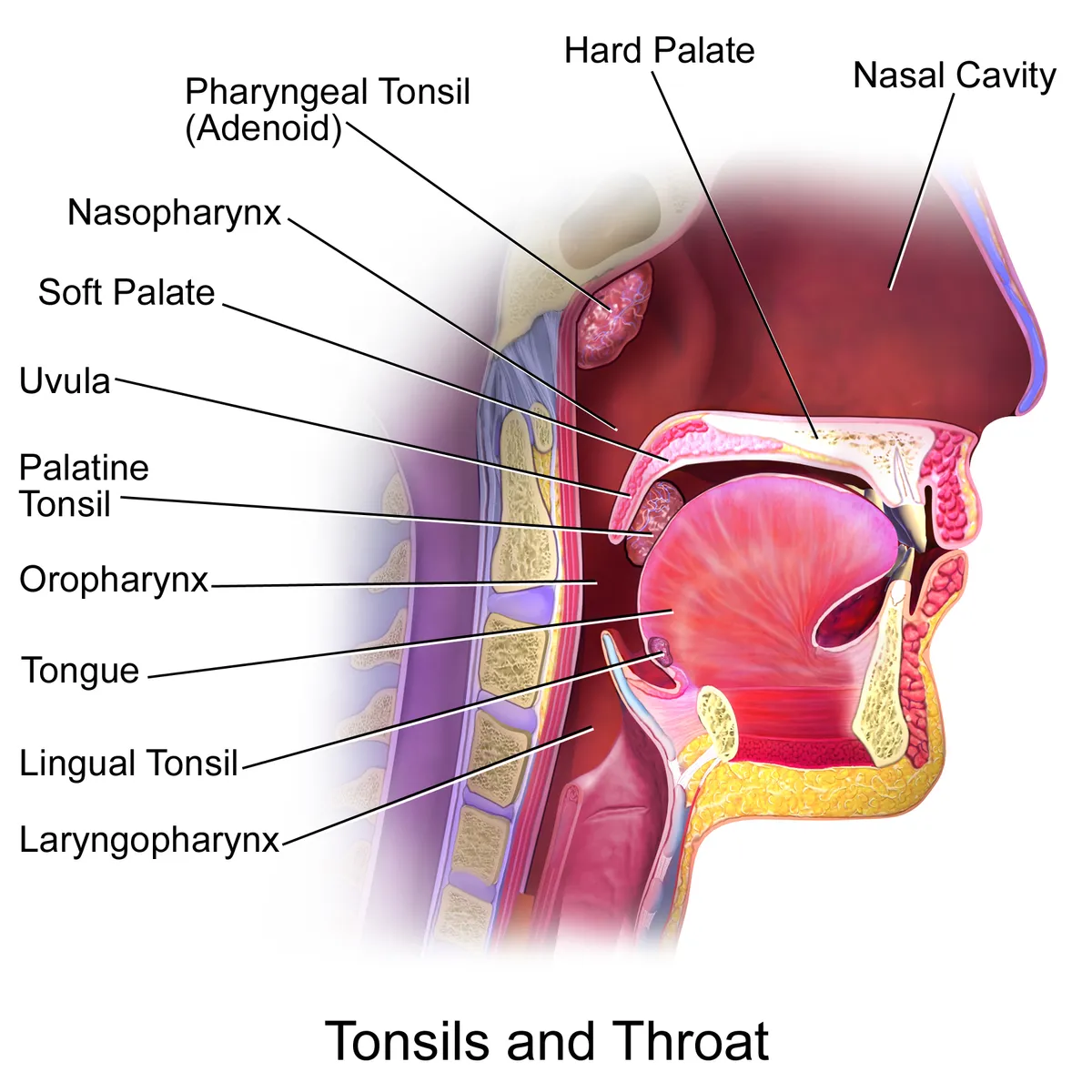
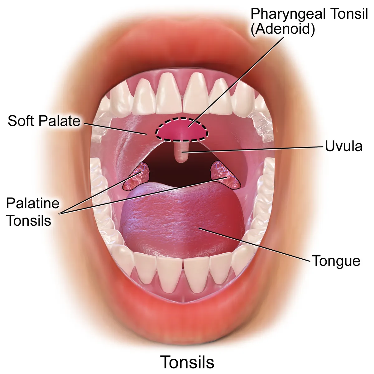
Two almond-shaped masses of lymphoid tissue in the tonsillar fossa, between the palatoglossal arch in front and the palatopharyngeal arch behind. These are “the tonsils” of everyday speech; largest at 4 to 10 years, they then regress. The surface has 10–20 deep crypts where bacteria and debris collect.
- 02
Pharyngeal tonsil (adenoids)
Tonsilla pharyngea
A single mass on the roof and posterior wall of the nasopharynx, covered by ciliated pseudostratified columnar epithelium. When enlarged in young children (adenoids) it causes snoring and mouth breathing and can block the pharyngeal openings of the auditory tubes, leading to recurrent otitis media.
- 03
Tubal tonsil
Tonsilla tubaria
A small aggregate of lymphoid tissue around the pharyngeal opening of the auditory tube on the lateral wall of the nasopharynx, continuous with the pharyngeal tonsil. Its inflammation contributes to tubal obstruction.
- 04
Lingual tonsil
Tonsilla lingualis
Scattered lymphoid follicles on the base of the tongue behind the terminal sulcus, each with a single crypt. Together with the palatine, pharyngeal and tubal tonsils it completes Waldeyer’s ring.
- 05
Tonsillar fossa and bed
Fossa tonsillaris
The palatine tonsil has a fibrous capsule on its lateral surface, separated from the superior pharyngeal constrictor by loose tissue: the cleavage plane used in tonsillectomy and the space where pus collects in a peritonsillar abscess. The glossopharyngeal nerve runs just outside the bed, which explains post-operative referred ear pain.
- 06
Blood supply
Vasa tonsillae palatinae
The tonsillar branch of the facial artery is the main supply, entering the lower pole; further twigs come from the lingual, ascending palatine, descending palatine and ascending pharyngeal arteries. The external palatine (paratonsillar) vein runs on the outer capsule and is a common source of post-tonsillectomy bleeding.
Position and relations
The palatine tonsil lies on the lateral wall of the oropharynx in the tonsillar fossa, bounded by the palatoglossal arch in front and the palatopharyngeal arch behind; the floor of the fossa is the superior pharyngeal constrictor. With the pharyngeal tonsil (adenoid) in the nasopharynx, the tubal tonsils around the auditory tube openings and the lingual tonsil at the base of the tongue, it forms the pharyngeal lymphoid ring guarding the entrance to the airway and gut.
Form and structure
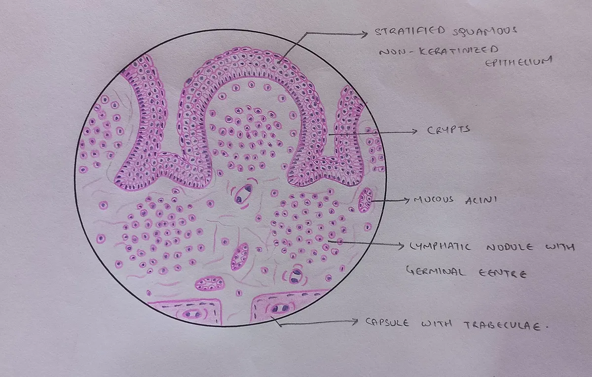
The free surface is covered by stratified squamous epithelium that dips into 10–20 deep crypts; beneath it lie lymphoid follicles with germinal centres. The deep surface has a fibrous capsule separating it from the constrictor; the loose space between capsule and muscle is where a peritonsillar abscess forms. The tonsils are largest in childhood and shrink after puberty.
Vessels and nerves
The main artery is the tonsillar branch of the facial artery, with small branches from the ascending pharyngeal, ascending palatine, lingual and descending palatine arteries. The external palatine vein runs close to the capsule and is a common source of bleeding after tonsillectomy. Lymph drains to the jugulodigastric node below the angle of the mandible, which often swells in pharyngitis.
Clinical correlations
The tonsils are part of the mucosa-associated lymphoid tissue. In IgA nephropathy, studies link tonsillar immunity, tonsil-associated bacteria and serum galactose-deficient IgA1, which is why tonsillectomy is used as a treatment option in some settings.[1]
Crypts, M cells and development
Lying on the lateral wall of the oropharynx at the entrance to the airway and gut, the palatine tonsils are constantly exposed to pathogens and foreign material entering through the mouth. As mucosa-associated lymphoid tissue they are the main lymphoid tissue of the oropharynx: each has about 15 crypts that greatly enlarge its internal surface, lined by non-keratinised stratified squamous epithelium interspersed with reticular epithelium. The follicular germinal centres, mantle zones, extrafollicular area and reticular crypt epithelium together regulate the immune response. Specialised antigen-capturing M cells on the crypt surface take up pathogen-derived antigen and set off the immune cascade; the B cells here mature and can produce every antibody isotype (IgA, IgD, IgE, IgG and IgM). Recent work has also found T-cell developmental intermediates in the tonsils resembling those of the thymus and bone marrow.[2]
The palatine tonsils begin to develop in week 14 of gestation from the second pharyngeal pouch: wandering mononuclear cells migrate into the mesenchyme, crypts grow down into the connective tissue and are infiltrated by T lymphocytes from week 16; they are regarded as the first B-cell regions of fetal lymphoid tissue. Sensory nerves come from the lesser palatine nerve (maxillary division of V) and the tonsillar branches of the glossopharyngeal nerve (IX), which is why a sore throat can be felt in the ear. Veins drain to the peritonsillar plexus, then the pharyngeal and lingual veins and finally the internal jugular vein. Peritonsillar abscess is the leading cause of deep neck infection in adults, mainly in young people, with fever, sore throat, trismus, dysphagia, a peritonsillar bulge, uvular deviation and a “hot potato” voice; it is treated by incision and drainage, antibiotics and support, and if delayed can lead to aspiration, airway obstruction or spread into the retropharyngeal space.[2]
Full-text reading (StatPearls)
The StatPearls chapters on this topic, reproduced verbatim. Their CC BY-NC-ND 4.0 licence allows unmodified copies only, so they are not translated or shortened.
Anatomy, Head and Neck: TonsilsMasters KG, Lasrado S · StatPearls 2025 · NBK539792 · 10k characters
Loading…
Masters KG, Lasrado S. Anatomy, Head and Neck: Tonsils. StatPearls [Internet]. Treasure Island (FL): StatPearls Publishing; 2025. NBK539792 · PMID 30969614 · CC BY-NC-ND 4.0. Reproduced verbatim, unmodified.
Anatomy, Head and Neck, Palatine Tonsil (Faucial Tonsils)Meegalla N, Downs BW · StatPearls 2023 · NBK538296 · 7k characters
Loading…
Meegalla N, Downs BW. Anatomy, Head and Neck, Palatine Tonsil (Faucial Tonsils). StatPearls [Internet]. Treasure Island (FL): StatPearls Publishing; 2023. NBK538296 · PMID 30855880 · CC BY-NC-ND 4.0. Reproduced verbatim, unmodified.
Organs in this system
References
Sources for the values and descriptions on this page, indexed in PubMed (US National Library of Medicine).