Biceps brachii
Model
Layers
Z-Anatomy anatomical model based on BodyParts3D, licensed CC BY-SA 4.0.
Description
A muscle of the anterior compartment of the arm with two heads (long and short) arising from the scapula; its main actions are elbow flexion and forearm supination.
Parts
Each part has its own card; press “Show on the model” to fly to it and select it in the 3D model above.
- 01
Long head
Caput longum musculi bicipitis brachii
Arises from the supraglenoid tubercle of the scapula; its tendon crosses the shoulder joint inside the capsule (but outside the synovium) and descends through the intertubercular groove. It helps stabilise the humeral head; rupture lets the belly bunch up low in the arm (the “Popeye” sign).
- 02
Short head
Caput breve musculi bicipitis brachii
Arises from the tip of the coracoid process together with coracobrachialis, medial to the long head. The two heads unite into a single belly in the middle of the arm.
- 03
Distal tendon and bicipital aponeurosis
Tendo distalis et aponeurosis bicipitalis
The tendon inserts on the radial tuberosity; from it the bicipital aponeurosis fans medially into the forearm fascia, covering the brachial artery and median nerve in the cubital fossa. The tendon is easily felt in the fossa and is tapped to test the biceps reflex (C5–C6).
- 04
Nerve supply
Innervatio
The musculocutaneous nerve (C5–C6, from the lateral cord of the brachial plexus) pierces coracobrachialis, runs between biceps and brachialis, supplies all three anterior arm muscles and continues as the lateral cutaneous nerve of the forearm.
- 05
Actions
Functio
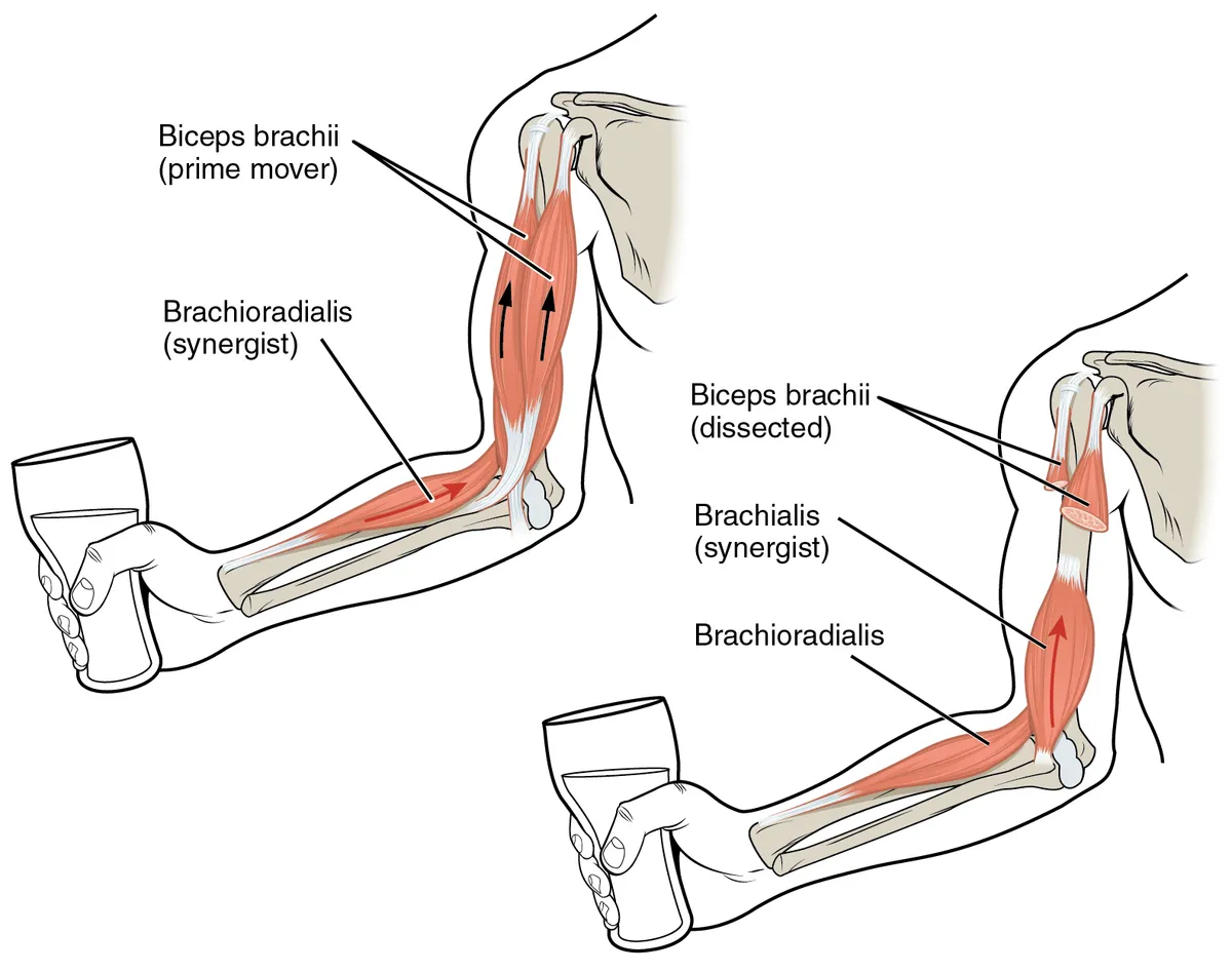
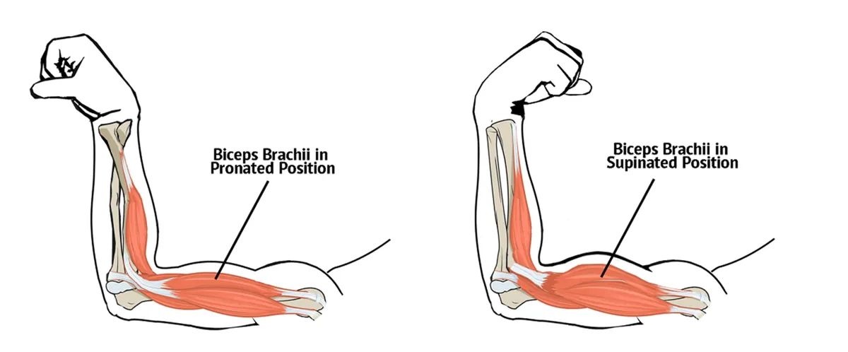
The most powerful supinator of the forearm (with the elbow flexed); it also flexes the elbow and assists shoulder flexion. This is why screws are driven clockwise by supination; brachialis beneath it is the main elbow flexor when the forearm is pronated.
Form and structure
The biceps brachii has two heads: the long head arises from the supraglenoid tubercle, its tendon running through the shoulder joint and the intertubercular groove; the short head arises from the coracoid process. They unite in a single belly that inserts on the radial tuberosity and, via the bicipital aponeurosis, into the forearm fascia.
Vessels and nerves
It is innervated by the musculocutaneous nerve (C5–C6). Its main actions are flexion of the elbow and supination of the forearm (strongest with the elbow at 90°), with a minor role in shoulder flexion. The biceps reflex tests the C5–C6 roots.
Contraction
Like every skeletal muscle, the biceps brachii contracts when the action potential spreads along the fibre membrane and T-tubules, releasing Ca²⁺ from the sarcoplasmic reticulum so that actin and myosin slide past each other. Its force and power depend on cross-sectional area, fibre-type composition and the cytoskeletal architecture of each fibre.[1]
Elbow and related nerves
The biceps tendon inserts on the radial tuberosity, so the muscle both flexes the elbow and is the strongest supinator of the forearm. Almost all daily and athletic upper-limb movements require a stable, painless elbow; its complex anatomy makes MRI a key tool for lesions ranging from acute injury to chronic overuse.[2]
The radial nerve is the largest branch of the posterior cord of the brachial plexus. Its entrapment at the elbow is the third most common compressive neuropathy of the upper limb after carpal and cubital tunnel syndromes; posterior interosseous nerve syndrome is the commonest form, and many compressing agents relate to normal or variant anatomy.[3]
Lateral epicondylitis (“tennis elbow”) is a degenerative disorder of the extensor origin at the lateral humeral epicondyle linked to repetitive wrist extension and supination; most cases improve with conservative care. Elbow dislocation with fractures of the radial head, coronoid or olecranon (complex instability) is far more demanding to manage than simple dislocation.[4][5]
Full-text reading (StatPearls)
The StatPearls chapters on this topic, reproduced verbatim. Their CC BY-NC-ND 4.0 licence allows unmodified copies only, so they are not translated or shortened.
Anatomy, Shoulder and Upper Limb, Biceps MuscleTiwana MS, Charlick M, Varacallo MA · StatPearls 2024 · NBK519538 · 9k characters
Loading…
Tiwana MS, Charlick M, Varacallo MA. Anatomy, Shoulder and Upper Limb, Biceps Muscle. StatPearls [Internet]. Treasure Island (FL): StatPearls Publishing; 2024. NBK519538 · PMID 30137823 · CC BY-NC-ND 4.0. Reproduced verbatim, unmodified.
Organs in this system
References
Sources for the values and descriptions on this page, indexed in PubMed (US National Library of Medicine).
- [1]Skeletal muscle: a brief review of structure and functionFrontera WR et al., Calcified tissue international 2015PMID 25294644
- [2]MR Imaging of the ElbowBinaghi D, Magnetic resonance imaging clinics of North America 2015PMID 26216773
- [3]Radial nerve compression: anatomical perspective and clinical consequencesWęgiel A et al., Neurosurgical review 2023PMID 36781706
- [4]Lateral epicondylitis of the elbowTosti R et al., The American journal of medicine 2013PMID 23398951
- [5]Complex elbow instabilityTashjian RZ et al., The Journal of the American Academy of Orthopaedic Surgeons 2006PMID 16675621
- [6]Anatomy, Bony Pelvis and Lower Limb, Gluteus Maximus MuscleStatPearls (NCBI Bookshelf)PMID 30855781Supports: Gluteus maximus