Intestines
Model
Hover or tap to see names, click to read a description; click a name in the legend to fly to it. Procedurally built to histological proportions; colours follow illustration conventions.
Description
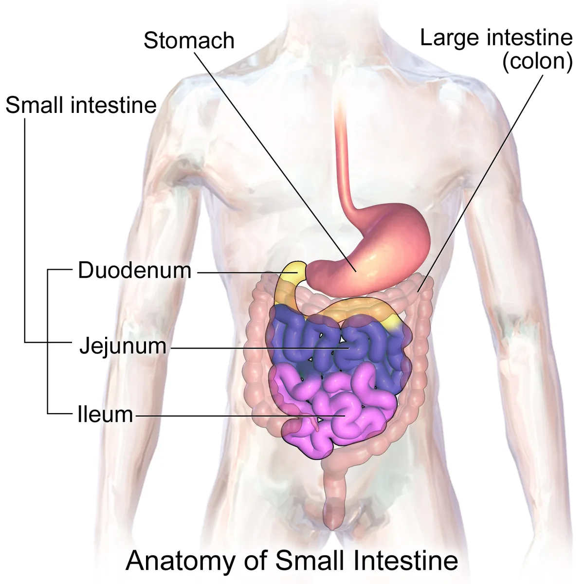
The small intestine (duodenum, jejunum, ileum), about 6–7 m long, absorbs most nutrients; the large intestine (caecum, colon, rectum), about 1.5 m long, absorbs water and electrolytes.
Parts
Each part has its own card; press “Show on the model” to fly to it and select it in the 3D model above.
- 01
Jejunum
Jejunum
The proximal two-fifths of the mesenteric small intestine (about 2.5 m), mainly in the upper left abdomen. It has a thick wall, wide lumen, tall closely packed circular folds, long vasa recta with few arcades and little mesenteric fat; it is the main site of nutrient absorption.
- 02
Ileum
Ileum
The remaining three-fifths (about 3.5 m), mainly in the lower right abdomen and pelvis. It has a thinner wall, low sparse folds, several tiers of arcades, short vasa recta and Peyer’s patches; the terminal ileum absorbs vitamin B12 and bile salts. A Meckel diverticulum lies on its antimesenteric border about 60 cm from the ileocaecal valve.
- 03
Mesentery
Mesenterium
A fan-shaped peritoneal fold whose root, about 15 cm long, runs obliquely from the duodenojejunal flexure (left of L2) to the right sacro-iliac joint, while its intestinal border is some 6 m long. It carries the superior mesenteric vessels and their arcades, lacteals and lymph nodes.
- 04
Caecum and ileocaecal valve
Caecum; Valva ileocaecalis
The caecum is the widest, blind-ended pouch of the colon in the right iliac fossa, below the entry of the ileum; the ileocaecal valve has two lips that limit reflux. Having the largest diameter, it is the most likely to perforate in a closed-loop colonic obstruction (Laplace’s law).
- 05
Ascending colon
Colon ascendens
About 15 cm long, it ascends retroperitoneally on the right to the right colic (hepatic) flexure below the liver. It is supplied by the ileocolic and right colic arteries; its posterior surface is fused to the posterior wall, and right colectomy mobilises it along Toldt’s fascia.
- 06
Transverse colon
Colon transversum
The longest and most mobile segment (about 45 cm), hung on the transverse mesocolon and sagging between the hepatic and splenic flexures. Its right two-thirds are supplied by the middle colic (superior mesenteric) and its left third by the left colic (inferior mesenteric) artery.
- 07
Descending and sigmoid colon
Colon descendens; Colon sigmoideum
The descending colon (about 25 cm) runs retroperitoneally on the left from the splenic flexure; the sigmoid colon (about 40 cm) has a V-shaped mesentery, is mobile and joins the rectum at S3. The sigmoid is the usual site of diverticula, volvulus and cancer; the splenic flexure is a watershed zone prone to ischaemia.
- 08
Taeniae, haustra and omental appendices
Taeniae coli; Haustra coli; Appendices omentales
The longitudinal muscle of the colon is gathered into three bands (mesocolic, omental, free), shorter than the gut so that it puckers into haustra separated by semilunar folds; fat-filled peritoneal tags (omental appendices) hang along it. These features distinguish colon from small bowel and disappear at the rectum.
- 09
Rectum and anal canal
Rectum; Canalis analis
The rectum, about 12–15 cm from S3 to puborectalis, lacks taeniae and appendices and has three transverse folds (of Houston); its ampulla stores faeces before defecation. The anal canal is about 4 cm long; the pectinate line separates an upper part (mucosa, visceral nerves) from a lower part (skin, pudendal nerve, very sensitive); internal (smooth) and external (striated) sphincters provide continence.
Position and relations
The small intestine is about 6–7 m long: the duodenum (fixed, C-shaped around the head of the pancreas), then the jejunum and ileum (mobile, suspended by the mesentery). The large intestine, about 1.5 m long, comprises the caecum and appendix, ascending, transverse, descending and sigmoid colon, rectum and anal canal, framing the small-bowel loops.
Form and structure
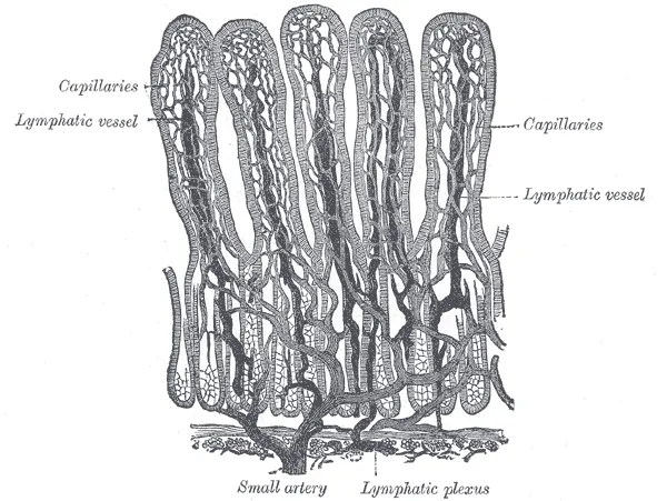
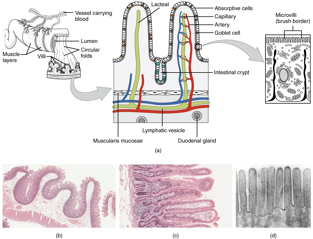
The small-intestinal mucosa bears circular folds, villi and microvilli, multiplying the absorptive surface hundreds of times. The large intestine has no villi; its longitudinal muscle gathers into three taeniae coli that create haustra, and its mucosa, rich in goblet cells, mainly absorbs water and electrolytes.
Vessels and nerves
The small intestine and right colon are supplied by the superior mesenteric artery; the left colon and upper rectum by the inferior mesenteric artery. Venous blood drains to the portal vein and passes through the liver before reaching the heart, making the liver the first filter for everything absorbed.[1]
Physiology
Colonocyte metabolism acts as a switch for microbial homeostasis. Normally colonocytes rely on oxidative phosphorylation (burning bacterial butyrate), consume much oxygen and keep the epithelial surface hypoxic; this favours obligate anaerobes that ferment fibre into beneficial products. When epithelial metabolism shifts (inflammation, antibiotics), oxygenation rises and facultative anaerobes expand, the hallmark of dysbiosis.[2]
The rectosigmoid junction is defined differently by anatomists, endoscopists, physiologists and surgeons, but all agree it is a high-pressure zone where muscle organisation, innervation and vascular anastomoses change; locating it correctly matters in cancer resection.[3]
Clinical correlations
Hirschsprung disease results from absent enteric ganglion cells in the distal bowel and usually presents in neonates with severe constipation or obstruction. Contrast enema and anorectal manometry are screening tools; diagnosis rests on rectal biopsy, aided by acetylcholinesterase histochemistry or calretinin immunohistochemistry.[4]
Ulcerative colitis is a chronic mucosal inflammation extending continuously from the rectum; newer views stress involvement beyond the mucosa and the microbiota. Microscopic colitis causes chronic watery diarrhoea with a normal-looking colon and is diagnosed only on biopsy.[5][6][7]
Full-text reading (StatPearls)
The StatPearls chapters on this topic, reproduced verbatim. Their CC BY-NC-ND 4.0 licence allows unmodified copies only, so they are not translated or shortened.
Anatomy, Abdomen and Pelvis, Small IntestineCollins JT, Nguyen A, Omole AE, Badireddy M · StatPearls 2025 · NBK459366 · 17k characters
Loading…
Collins JT, Nguyen A, Omole AE, Badireddy M. Anatomy, Abdomen and Pelvis, Small Intestine. StatPearls [Internet]. Treasure Island (FL): StatPearls Publishing; 2025. NBK459366 · PMID 29083773 · CC BY-NC-ND 4.0. Reproduced verbatim, unmodified.
Anatomy, Abdomen and Pelvis: Large IntestineOmole AE, Mandiga P, Kahai P, Lobo S · StatPearls 2025 · NBK470577 · 10k characters
Loading…
Omole AE, Mandiga P, Kahai P, Lobo S. Anatomy, Abdomen and Pelvis: Large Intestine. StatPearls [Internet]. Treasure Island (FL): StatPearls Publishing; 2025. NBK470577 · PMID 29261962 · CC BY-NC-ND 4.0. Reproduced verbatim, unmodified.
Organs in this system
References
Sources for the values and descriptions on this page, indexed in PubMed (US National Library of Medicine).
- [1]Liver macrophages: development, dynamics, and functionsNusse Y et al., Cellular & molecular immunology 2025PMID 40461700
- [2]Colonocyte metabolism shapes the gut microbiotaLitvak Y et al., Science 2018PMID 30498100
- [3]Is it possible to give a single definition of the rectosigmoid junction?Massalou D et al., Surgical and radiologic anatomy 2018PMID 29218384
- [4]Diagnosis of Hirschsprung DiseaseAmbartsumyan L et al., Pediatric and developmental pathology 2020PMID 31791203
- [5]Ulcerative ColitisFeuerstein JD et al., Mayo Clinic proceedings 2019PMID 31272578
- [6]Ulcerative colitis: moving beyond the mucosal dogmaEggermont E et al., The lancet. Gastroenterology & hepatology 2026PMID 41173014
- [7]Microscopic colitis: pathophysiology and clinical managementMiehlke S et al., The lancet. Gastroenterology & hepatology 2019PMID 30860066
- [8]Gastrointestinal mean transit times in young and middle-aged healthy subjectsPMID 11318834Supports: Gastrointestinal transit time
- [9]Revised Estimates for the Number of Human and Bacteria Cells in the BodySender R, Fuchs S, Milo R, 2016PMID 27541692Supports: Colonic bacterial count