Aorta
Model
Layers
Z-Anatomy anatomical model based on BodyParts3D, licensed CC BY-SA 4.0.
Description
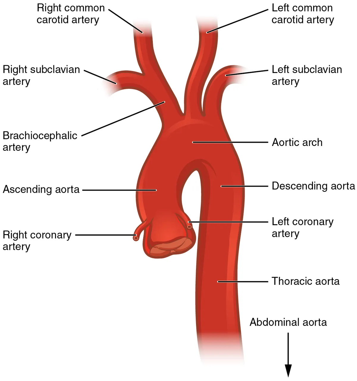
The largest artery, arising from the left ventricle; it comprises the ascending aorta, the aortic arch and the descending (thoracic and abdominal) aorta.
Parts
Each part has its own card; press “Show on the model” to fly to it and select it in the 3D model above.
- 01
Ascending aorta
Aorta ascendens
About 5 cm long, it begins at the aortic valve with a root bulging into the three sinuses of Valsalva, from which the coronary arteries arise. It lies inside the pericardium and runs up and to the right to the level of the second sternocostal joint; it is the usual site of aneurysm and type A dissection.
- 02
Aortic arch
Arcus aortae
Arches backwards and to the left over the left lung root, from the sternal angle to the left side of T4. Its three branches are the brachiocephalic trunk, left common carotid and left subclavian arteries; the ligamentum arteriosum joins its underside to the left pulmonary artery, and the left recurrent laryngeal nerve hooks beneath it.
- 03
Thoracic (descending) aorta
Aorta thoracica
Descends from T4 in the posterior mediastinum, at first left of the midline and then midline, passing through the aortic hiatus of the diaphragm at T12. It gives posterior intercostal (from the third space down), subcostal, bronchial, oesophageal and superior phrenic arteries; some feed the spinal cord (artery of Adamkiewicz).
- 04
Abdominal aorta
Aorta abdominalis
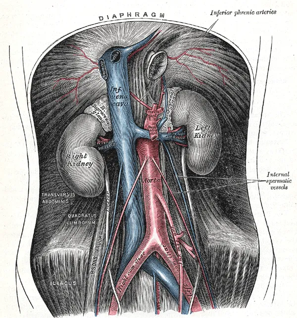
Runs from the diaphragmatic hiatus at T12 to its bifurcation at L4 (umbilical level), in front of the vertebral bodies and left of the inferior vena cava. Unpaired visceral branches: coeliac trunk, superior and inferior mesenteric; paired visceral: middle suprarenal, renal, gonadal; parietal: inferior phrenic and lumbar. Abdominal aortic aneurysms are usually infrarenal.
- 05
Coeliac trunk
Truncus coeliacus
A short trunk about 1 cm long arising at T12 just below the hiatus and dividing into the left gastric, splenic and common hepatic arteries (the last giving the proper hepatic and gastroduodenal). It supplies the foregut organs: abdominal oesophagus, stomach, upper duodenum, liver, biliary tract, pancreas and spleen.
- 06
Superior and inferior mesenteric arteries
Arteria mesenterica superior et inferior
The superior mesenteric artery (L1) crosses in front of the third part of the duodenum into the root of the mesentery and supplies the midgut from the second part of the duodenum to the distal transverse colon. The inferior mesenteric (L3) supplies the hindgut down to the upper anal canal; the two connect through the marginal artery (of Drummond).
- 07
Renal arteries
Arteriae renales
Arise at L1–L2 just below the superior mesenteric; the right is longer and passes behind the inferior vena cava. Each divides into segmental (end) arteries without anastomoses; about a quarter of people have accessory renal arteries.
- 08
Aortic bifurcation and common iliac arteries
Bifurcatio aortae; Arteria iliaca communis
At L4 the aorta divides into the two common iliac arteries, and the small median sacral artery continues downwards. Each common iliac divides in front of the sacro-iliac joint into the internal iliac (pelvic viscera, buttock, perineum) and the external iliac (becoming the femoral artery below the inguinal ligament).
Position and relations
The aorta leaves the left ventricle as the ascending aorta, arches backwards and to the left as the aortic arch, then descends in the posterior mediastinum (thoracic aorta), passes through the aortic hiatus of the diaphragm at T12 to become the abdominal aorta, and bifurcates into the common iliac arteries at L4.
Form and structure
The aortic wall is a typical elastic artery: its tunica media contains many concentric elastic lamellae, so it distends in systole and recoils in diastole, turning the intermittent cardiac output into continuous flow (the Windkessel effect). The arch gives three branches: the brachiocephalic trunk, the left common carotid and the left subclavian arteries.
The aorta: structure and function
The aorta is the “greatest artery”, delivering oxygenated blood from the left ventricle to the organs with every beat (about 200 million litres in a lifetime). Its elastin-rich media distends in systole and recoils in diastole, smoothing flow. Acute factors (cocaine, weight lifting, trauma) and chronic ones (hypertension, phaeochromocytoma) raise wall stress, while medial abnormalities (Marfan syndrome, bicuspid valve, vasculitis, atherosclerosis) weaken the wall.[1]
Aortic stiffness, measured as pulse wave velocity (PWV), is a surrogate marker of cardiovascular disease. A meta-analysis of 17 longitudinal studies (15,877 people, mean follow-up 7.7 years) found that the highest PWV tertile had about 2.3 times the risk of cardiovascular events, 2.0 times cardiovascular mortality and 1.9 times all-cause mortality compared with the lowest.[2]
Development and arch anomalies
Aortic arch variants are best understood on the theoretical embryonic double-arch model: depending on which segment regresses, the result is a typical left arch, a right arch, a double arch or an aberrant right subclavian artery. Some variants form vascular rings around the trachea and oesophagus; an aberrant right subclavian artery may compress the oesophagus (dysphagia lusoria). These anomalies may accompany congenital heart disease and chromosomal abnormalities.[3]
Coarctation is a discrete narrowing of the thoracic aorta but is better regarded as an aortopathy with stiff, poorly relaxing walls. Even after surgical or transcatheter relief, patients remain at risk of hypertension, aortic complications and left ventricular dysfunction and need lifelong imaging surveillance.[4]
Full-text reading (StatPearls)
The StatPearls chapters on this topic, reproduced verbatim. Their CC BY-NC-ND 4.0 licence allows unmodified copies only, so they are not translated or shortened.
Anatomy, Abdomen and Pelvis: Abdominal AortaTran CT, Wu CY, Bordes SJ, Lui F · StatPearls 2023 · NBK525964 · 16k characters
Loading…
Tran CT, Wu CY, Bordes SJ, Lui F. Anatomy, Abdomen and Pelvis: Abdominal Aorta. StatPearls [Internet]. Treasure Island (FL): StatPearls Publishing; 2023. NBK525964 · PMID 30247839 · CC BY-NC-ND 4.0. Reproduced verbatim, unmodified.
Anatomy, Abdomen and Pelvis: AortaWhite HJ, Bordes SJ, Borger J · StatPearls 2023 · NBK537319 · 12k characters
Loading…
White HJ, Bordes SJ, Borger J. Anatomy, Abdomen and Pelvis: Aorta. StatPearls [Internet]. Treasure Island (FL): StatPearls Publishing; 2023. NBK537319 · PMID 30726004 · CC BY-NC-ND 4.0. Reproduced verbatim, unmodified.
Anatomy, Thorax, Heart AortaShahoud JS, Miao JH, Bolla SR · StatPearls 2023 · NBK538140 · 8k characters
Loading…
Shahoud JS, Miao JH, Bolla SR. Anatomy, Thorax, Heart Aorta. StatPearls [Internet]. Treasure Island (FL): StatPearls Publishing; 2023. NBK538140 · PMID 30844167 · CC BY-NC-ND 4.0. Reproduced verbatim, unmodified.
Organs in this system
References
Sources for the values and descriptions on this page, indexed in PubMed (US National Library of Medicine).
- [1]Epidemiology and management of aortic disease: aortic aneurysms and acute aortic syndromesBossone E et al., Nature reviews. Cardiology 2021PMID 33353985
- [2]Prediction of cardiovascular events and all-cause mortality with arterial stiffness: a systematic review and meta-analysisVlachopoulos C et al., Journal of the American College of Cardiology 2010PMID 20338492
- [3]Congenital Variants and Anomalies of the Aortic ArchHanneman K et al., Radiographics 2017PMID 27860551
- [4]Aortic CoarctationKim YY et al., Cardiology clinics 2020PMID 32622489
- [5]Physiology, Cardiac OutputStatPearls (NCBI Bookshelf)PMID 29262215Supports: Heart rate, cardiac output
- [6]Physiology, CardiovascularStatPearls (NCBI Bookshelf)PMID 29630249