Teeth and masticatory apparatus
Model
Hover or tap to see names, click to read a description; click a name in the legend to fly to it. Procedurally built to histological proportions; colours follow illustration conventions.
Description
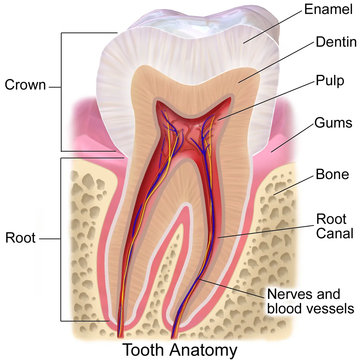
Humans have two dentitions: 20 primary teeth gradually replaced by 32 permanent teeth, arranged in two dental arches on the maxillae and the mandible. Each tooth has an enamel-covered crown and a cementum-covered root held in its socket by the periodontal ligament; inside, dentine surrounds the pulp. The mandible moves at the temporomandibular joints under four muscles of mastication supplied by the mandibular nerve (V3); the teeth receive sensation from the superior and inferior alveolar nerves and blood from the maxillary artery.
Parts
Each part has its own card; press “Show on the model” to fly to it and select it in the 3D model above.
- 01
Incisors
Dentes incisivi
Four per jaw (two central, two lateral), with shovel-shaped crowns and a sharp incisal edge for cutting, and a single root. The upper central incisor is the largest; the lower incisors are the smallest and narrowest teeth of the dentition.
- 02
Canines
Dentes canini
One per quadrant, with a single pointed cusp for tearing and the longest root of the dentition (the upper canine can measure about 27 mm overall). Placed at the “corner” of the arch, it guides the jaw in lateral excursions.
- 03
Premolars
Dentes premolares
Two per quadrant (first and second), usually with two cusps (buccal and lingual), for crushing and tearing. The upper first premolar usually has two roots, the others one. They are absent from the primary dentition.
- 04
Molars
Dentes molares
Three per quadrant with broad occlusal surfaces of four or five cusps for grinding. Upper molars have three roots (two buccal, one palatal), lower molars two (mesial and distal). The first molar erupts at about 6 years; the third (wisdom tooth) at 17–25 and is often impacted.
- 05
Enamel
Enamelum
The hardest tissue in the body, about 96% hydroxyapatite arranged in enamel rods, covering the crown and thickest over the cusps (about 2.5 mm). It is formed by ameloblasts before eruption and cannot regenerate once decayed.
- 06
Dentine
Dentinum
Forms the bulk of the tooth, about 70% mineral, traversed by dentinal tubules holding odontoblast processes and fluid, which is why exposed dentine is sensitive. Odontoblasts keep laying down secondary dentine throughout life and reparative dentine when irritated.
- 07
Dental pulp
Pulpa dentis
Loose connective tissue in the pulp chamber and root canals, with odontoblasts at its periphery and vessels and nerves entering through the apical foramen. Pulpitis within this rigid chamber is very painful and can lead to necrosis requiring root canal treatment.
- 08
Periodontium
Periodontium; Cementum; Gingiva
The supporting apparatus: cementum covering the root, the periodontal ligament (Sharpey fibres about 0.2 mm wide, cushioning and sensing pressure), the alveolar bone and the gingiva. Periodontitis destroying these tissues is the leading cause of tooth loss in adults.
- 09
Primary dentition
Dentes decidui
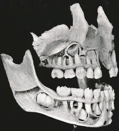
Twenty teeth (per quadrant: 2 incisors, 1 canine, 2 primary molars) erupting from about 6 months to 2.5 years and shed between 6 and 12 years. They have thin enamel, large pulps and widely flared molar roots that cradle the permanent tooth germs.
Dental chart
Select a tooth to see its number in three systems, its size, roots, root canals and age of eruption. The chart faces the patient: the patient's right is on the left of the picture.
- Incisors
- Canines
- Premolars
- Molars
Upper right first molar (16)
- FDI 16 · Universal 3 · Palmer 6┘
- Mean dimensions: crown 7.5 mm, root 13 mm, mesiodistal crown width 10 mm.
- Roots: 3. Root canals: usually 3, fairly often 4.
- Eruption at 6–7 years; root apex complete at about 9–10 years.
The largest crown in the upper arch, with four main cusps (the mesiopalatal is the largest) joined by an oblique ridge. The palatal surface often has an extra cusp of Carabelli. It has three roots: mesiobuccal, distobuccal and palatal. The mesiobuccal root often has a second canal (MB2), so four canals are common. The root tips lie close to the floor of the maxillary sinus.
Sources: PMID 32491475, PMID 30860762, PMID 27578418
Two dentitions, tooth classes and numbering
Human teeth are heterodont: there are four classes, the incisors, canines, premolars and molars. Humans are also diphyodont, with two successive dentitions. The primary dentition has 20 teeth: each quadrant has two incisors, one canine and two primary molars. The primary incisors, canines and molars are replaced in turn by the permanent incisors, canines and premolars. Three permanent molars are added behind them without replacing any tooth, which brings the permanent dentition to 32 teeth.[1]
Each tooth has a crown, the part seen in the mouth, and a root set in a socket of the alveolar process. The boundary between them is the cementoenamel junction. The crown has five surfaces. The outer surface is called labial on anterior teeth and buccal on posterior teeth. The inner surface is called palatal in the upper jaw and lingual in the lower jaw. The mesial surface faces the midline and the distal surface faces away from it. The occlusal surface of posterior teeth corresponds to the incisal edge of anterior teeth.[1]
Three numbering systems are in common use. The FDI system (ISO 3950) uses two digits. The first gives the quadrant: 1 upper right, 2 upper left, 3 lower left, 4 lower right, and 5–8 for primary teeth. The second gives the position from the midline, from 1 for the central incisor to 8 for the wisdom tooth. For example, 36 is the lower left first molar. The Universal system, used in the United States, numbers the teeth 1–32. It starts at the upper right wisdom tooth, runs along the upper arch and comes back along the lower arch; primary teeth are lettered A–T. The Palmer system writes 1–8 (A–E for primary teeth) with an angle mark showing the quadrant.[1]
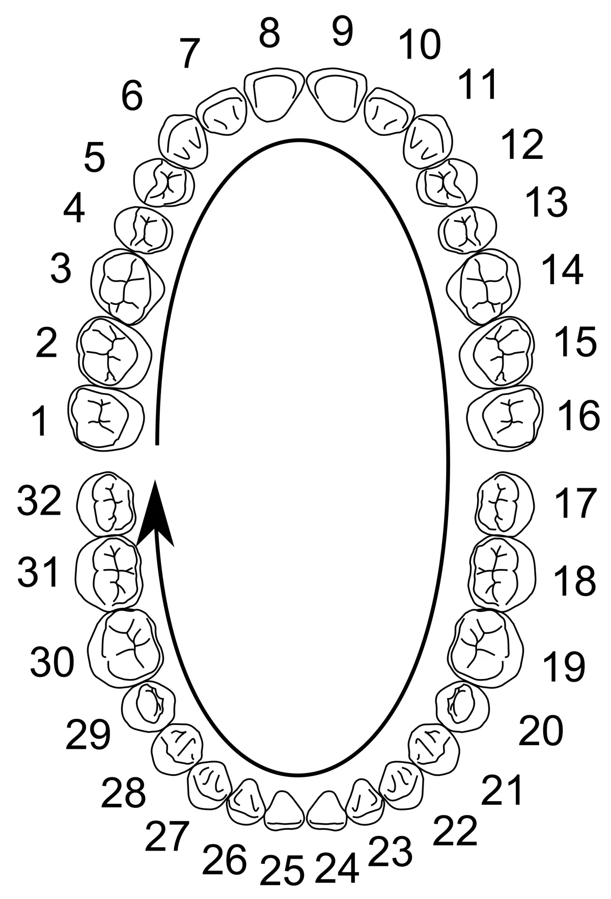
Central incisor
- FDI (q1 · q2 · q3 · q4)
- 11 · 21 · 31 · 41
- Universal
- 8 · 9 · 24 · 25
- Palmer
- 1┘ · └1 · ┌1 · 1┐
- Eruption, years (upper / lower)
- 7–8 / 6–7
Lateral incisor
- FDI (q1 · q2 · q3 · q4)
- 12 · 22 · 32 · 42
- Universal
- 7 · 10 · 23 · 26
- Palmer
- 2┘ · └2 · ┌2 · 2┐
- Eruption, years (upper / lower)
- 8–9 / 7–8
Canine
- FDI (q1 · q2 · q3 · q4)
- 13 · 23 · 33 · 43
- Universal
- 6 · 11 · 22 · 27
- Palmer
- 3┘ · └3 · ┌3 · 3┐
- Eruption, years (upper / lower)
- 11–12 / 9–10
First premolar
- FDI (q1 · q2 · q3 · q4)
- 14 · 24 · 34 · 44
- Universal
- 5 · 12 · 21 · 28
- Palmer
- 4┘ · └4 · ┌4 · 4┐
- Eruption, years (upper / lower)
- 10–11 / 10–12
Second premolar
- FDI (q1 · q2 · q3 · q4)
- 15 · 25 · 35 · 45
- Universal
- 4 · 13 · 20 · 29
- Palmer
- 5┘ · └5 · ┌5 · 5┐
- Eruption, years (upper / lower)
- 10–12 / 11–12
First molar
- FDI (q1 · q2 · q3 · q4)
- 16 · 26 · 36 · 46
- Universal
- 3 · 14 · 19 · 30
- Palmer
- 6┘ · └6 · ┌6 · 6┐
- Eruption, years (upper / lower)
- 6–7 / 6–7
Second molar
- FDI (q1 · q2 · q3 · q4)
- 17 · 27 · 37 · 47
- Universal
- 2 · 15 · 18 · 31
- Palmer
- 7┘ · └7 · ┌7 · 7┐
- Eruption, years (upper / lower)
- 12–13 / 11–13
Third molar
- FDI (q1 · q2 · q3 · q4)
- 18 · 28 · 38 · 48
- Universal
- 1 · 16 · 17 · 32
- Palmer
- 8┘ · └8 · ┌8 · 8┐
- Eruption, years (upper / lower)
- 17–21 / 17–21
| Permanent tooth | FDI (q1 · q2 · q3 · q4) | Universal | Palmer | Eruption, years (upper / lower) |
|---|---|---|---|---|
| Central incisor | 11 · 21 · 31 · 41 | 8 · 9 · 24 · 25 | 1┘ · └1 · ┌1 · 1┐ | 7–8 / 6–7 |
| Lateral incisor | 12 · 22 · 32 · 42 | 7 · 10 · 23 · 26 | 2┘ · └2 · ┌2 · 2┐ | 8–9 / 7–8 |
| Canine | 13 · 23 · 33 · 43 | 6 · 11 · 22 · 27 | 3┘ · └3 · ┌3 · 3┐ | 11–12 / 9–10 |
| First premolar | 14 · 24 · 34 · 44 | 5 · 12 · 21 · 28 | 4┘ · └4 · ┌4 · 4┐ | 10–11 / 10–12 |
| Second premolar | 15 · 25 · 35 · 45 | 4 · 13 · 20 · 29 | 5┘ · └5 · ┌5 · 5┐ | 10–12 / 11–12 |
| First molar | 16 · 26 · 36 · 46 | 3 · 14 · 19 · 30 | 6┘ · └6 · ┌6 · 6┐ | 6–7 / 6–7 |
| Second molar | 17 · 27 · 37 · 47 | 2 · 15 · 18 · 31 | 7┘ · └7 · ┌7 · 7┐ | 12–13 / 11–13 |
| Third molar | 18 · 28 · 38 · 48 | 1 · 16 · 17 · 32 | 8┘ · └8 · ┌8 · 8┐ | 17–21 / 17–21 |
Dental tissues: enamel, dentine, pulp and cementum
A tooth is made of four tissues of different hardness. Enamel covers the crown and is the hardest tissue in the body, about 96% mineral by weight. It consists mainly of carbonated hydroxyapatite arranged in interlocking rods. Enamel is made by ameloblasts, which are epithelial cells, in two phases. In the secretory phase they lay down a protein matrix (amelogenin, enamelin). In the maturation phase they remove almost all the protein and water so that the crystals can grow. The ameloblasts are lost when the tooth erupts, so mature enamel has no cells and cannot regenerate.[1][2][3]
Dentine forms the main mass of the tooth. It is about 70% mineral, 20% organic matter and 10% water. It is softer and more elastic than enamel, so it supports the brittle enamel and absorbs chewing forces. Odontoblasts at the edge of the pulp secrete predentine, a collagen matrix that then mineralises into dentine. Each cell leaves a cytoplasmic process inside a dentinal tubule. Fluid movement in these tubules is the accepted explanation for the sharp pain of exposed dentine. Primary dentine forms until the root is complete. After that, secondary dentine is added slowly throughout life, and tertiary (reactionary or reparative) dentine forms in response to stimuli such as caries.[3][4][5]
Cementum covers the root dentine. Its structure and composition are close to those of bone, but it has no blood vessels. Acellular cementum covers the cervical part of the root and holds the Sharpey's fibres of the periodontal ligament. Cellular cementum is thicker at the apex and in the furcation area. With age many cementocytes die, and small calcified bodies (cementicles) are a common sign of ageing. Enamel comes from ectoderm. Dentine, pulp, cementum and periodontal ligament come from neural crest ectomesenchyme.[3][6][1]
Enamel
- Origin
- Ectoderm (oral epithelium)
- Mineral
- ~96%
- Forming cells
- Ameloblasts, lost at eruption
- Repair
- None
Dentine
- Origin
- Ectomesenchyme (dental papilla)
- Mineral
- ~70%
- Forming cells
- Odontoblasts, kept for life
- Repair
- Secondary and tertiary dentine
Pulp
- Origin
- Ectomesenchyme (dental papilla)
- Mineral
- Not mineralised
- Forming cells
- Fibroblasts, pulp stem cells
- Repair
- Yes, while vital
Cementum
- Origin
- Ectomesenchyme (dental follicle)
- Mineral
- ~45–50%
- Forming cells
- Cementoblasts
- Repair
- Added throughout life
| Tissue | Origin | Mineral | Forming cells | Repair |
|---|---|---|---|---|
| Enamel | Ectoderm (oral epithelium) | ~96% | Ameloblasts, lost at eruption | None |
| Dentine | Ectomesenchyme (dental papilla) | ~70% | Odontoblasts, kept for life | Secondary and tertiary dentine |
| Pulp | Ectomesenchyme (dental papilla) | Not mineralised | Fibroblasts, pulp stem cells | Yes, while vital |
| Cementum | Ectomesenchyme (dental follicle) | ~45–50% | Cementoblasts | Added throughout life |
Pulp space and root canal system
The pulp space is a cavity surrounded by dentine and open only at the apex. It consists of the pulp chamber in the crown and the root canals in the roots. The chamber has a dentine roof, pulp horns reaching under each cusp or incisal edge, and a floor that carries the canal openings at the level of the cementoenamel junction. The canals narrow and often curve, ending at the apical foramen, where vessels and nerves enter. Each chamber usually receives one or two arterioles and one large venule, which form a dense capillary network beneath the odontoblast layer.[7][8]
The pulp is a connective tissue rich in vessels and nerves, organised in layers. Outermost is the odontoblast layer. Next comes the cell-free zone of Weil, rich in capillaries and a nerve plexus, then the cell-rich zone with fibroblasts and undifferentiated mesenchymal cells. Innermost is the pulp core with large vessels and nerve trunks. Sensory fibres from the maxillary and mandibular nerves branch beneath the cell-rich zone to form the plexus of Raschkow. Their unmyelinated endings reach the odontoblasts, and some enter the dentinal tubules. Sympathetic fibres from the superior cervical ganglion control arteriolar tone.[8]
Canal anatomy varies more than the outer shape of the tooth. Besides the main canal there are lateral canals, accessory canals and extra apical foramina. Patterns such as two canals merging into one, one canal splitting into two, or a C-shaped canal are hard to see on two-dimensional radiographs. Cone-beam CT and micro-CT show many patterns that do not fit the older classifications. This is why a new coding system was proposed in 2017, recording the number of roots and the course of the canals tooth by tooth; in 2018 it was extended to anomalies of teeth, roots and canals. With age, secondary dentine narrows the apical foramen, pulp cell density falls by about half between 20 and 70 years, and the chamber shrinks.[9][10][8][11]
Central incisor
- Upper: crown / root (mm)
- 10.5 / 13
- Upper: roots · canals
- 1 · 1
- Lower: crown / root (mm)
- 9 / 12.5
- Lower: roots · canals
- 1 · 1 (2)
Lateral incisor
- Upper: crown / root (mm)
- 9 / 13
- Upper: roots · canals
- 1 · 1
- Lower: crown / root (mm)
- 9.5 / 14
- Lower: roots · canals
- 1 · 1 (2)
Canine
- Upper: crown / root (mm)
- 10 / 17
- Upper: roots · canals
- 1 · 1
- Lower: crown / root (mm)
- 11 / 16
- Lower: roots · canals
- 1 · 1 (2)
First premolar
- Upper: crown / root (mm)
- 8.5 / 14
- Upper: roots · canals
- 2 · 2
- Lower: crown / root (mm)
- 8.5 / 14
- Lower: roots · canals
- 1 · 1 (2)
Second premolar
- Upper: crown / root (mm)
- 8.5 / 14
- Upper: roots · canals
- 1 · 1 (2)
- Lower: crown / root (mm)
- 8 / 14.5
- Lower: roots · canals
- 1 · 1
First molar
- Upper: crown / root (mm)
- 7.5 / 13
- Upper: roots · canals
- 3 · 3 (4)
- Lower: crown / root (mm)
- 7.5 / 14
- Lower: roots · canals
- 2 · 3 (4)
Second molar
- Upper: crown / root (mm)
- 7 / 12
- Upper: roots · canals
- 3 · 3 (4)
- Lower: crown / root (mm)
- 7 / 13
- Lower: roots · canals
- 2 · 3
Third molar
- Upper: crown / root (mm)
- 6.5 / 11
- Upper: roots · canals
- 3 · 3
- Lower: crown / root (mm)
- 7 / 11
- Lower: roots · canals
- 2 · 3
| Tooth | Upper: crown / root (mm) | Upper: roots · canals | Lower: crown / root (mm) | Lower: roots · canals |
|---|---|---|---|---|
| Central incisor | 10.5 / 13 | 1 · 1 | 9 / 12.5 | 1 · 1 (2) |
| Lateral incisor | 9 / 13 | 1 · 1 | 9.5 / 14 | 1 · 1 (2) |
| Canine | 10 / 17 | 1 · 1 | 11 / 16 | 1 · 1 (2) |
| First premolar | 8.5 / 14 | 2 · 2 | 8.5 / 14 | 1 · 1 (2) |
| Second premolar | 8.5 / 14 | 1 · 1 (2) | 8 / 14.5 | 1 · 1 |
| First molar | 7.5 / 13 | 3 · 3 (4) | 7.5 / 14 | 2 · 3 (4) |
| Second molar | 7 / 12 | 3 · 3 (4) | 7 / 13 | 2 · 3 |
| Third molar | 6.5 / 11 | 3 · 3 | 7 / 11 | 2 · 3 |
The periodontium
The periodontium is the attachment apparatus of the tooth: gingiva, periodontal ligament, cementum and alveolar bone. The periodontal ligament is an aligned network of collagen fibres joining the cementum to the alveolar bone. It works as a shock absorber, a barrier against bacteria and a sensor of chewing forces. Its principal fibre groups (alveolar crest, horizontal, oblique, apical and interradicular) are arranged so that biting forces become tension on the socket wall. This architecture organises itself under mechanical loading, which is why it is so hard to rebuild by tissue engineering.[12][13]
Periodontal ligament cells sense mechanical force and turn it into biological signals that drive resorption and formation of the socket bone. This is the biological basis of orthodontic tooth movement. At the neck of the tooth, the junctional epithelium attaches to the enamel and forms the first barrier against plaque bacteria. With inflammation it migrates toward the apex and a periodontal pocket forms. The thickness and width of the keratinised gingiva differ between people (thin or thick periodontal biotype) and affect the risk of gum recession.[14][15][16][3]
Tooth development and eruption
Teeth begin to form in the sixth week of embryonic life, when the oral epithelium thickens into the primary epithelial band. This band splits into a vestibular lamina and a horseshoe-shaped dental lamina. On each arch the dental lamina forms ten thickenings, which become the ten primary tooth buds. Each tooth germ passes through bud, cap and bell stages. The enamel organ gives rise to enamel. The dental papilla gives rise to dentine and pulp. The dental follicle gives rise to cementum, periodontal ligament and alveolar bone. The enamel knot is a signalling centre that sets where the cusps form. Permanent tooth germs start at about week 20, lingual to the primary teeth. The dental lamina near the third molars stays active until about age 15.[1][17][18]
Eruption is the movement of a tooth from where it develops in the jaw to its working position in the mouth. It has pre-eruptive, intraosseous, mucosal, pre-occlusal and post-occlusal phases. The dental follicle coordinates bone resorption above the tooth and bone formation below it, clearing its path. Teeth keep the ability to erupt throughout life, for example when the opposing tooth is lost. A meta-analysis of 42,109 children in 42 studies found clear regional differences in the timing of primary eruption. The lower central incisor erupts first, as early as about 6 months in North America and as late as about 13.5 months in Asia. Sex, jaw and socio-economic status made little difference.[19][20][21]
Primary central incisor
- FDI (q5 · q6 · q7 · q8)
- 51 · 61 · 71 · 81
- Universal
- E · F · O · P
- Eruption, months (upper / lower)
- 8–12 / 6–10
- Shed, years (upper / lower)
- 6–7 / 6–7
Primary lateral incisor
- FDI (q5 · q6 · q7 · q8)
- 52 · 62 · 72 · 82
- Universal
- D · G · N · Q
- Eruption, months (upper / lower)
- 9–13 / 10–16
- Shed, years (upper / lower)
- 7–8 / 7–8
Primary canine
- FDI (q5 · q6 · q7 · q8)
- 53 · 63 · 73 · 83
- Universal
- C · H · M · R
- Eruption, months (upper / lower)
- 16–22 / 17–23
- Shed, years (upper / lower)
- 10–12 / 9–12
Primary first molar
- FDI (q5 · q6 · q7 · q8)
- 54 · 64 · 74 · 84
- Universal
- B · I · L · S
- Eruption, months (upper / lower)
- 13–19 / 14–18
- Shed, years (upper / lower)
- 9–11 / 9–11
Primary second molar
- FDI (q5 · q6 · q7 · q8)
- 55 · 65 · 75 · 85
- Universal
- A · J · K · T
- Eruption, months (upper / lower)
- 25–33 / 23–31
- Shed, years (upper / lower)
- 10–12 / 10–12
| Primary tooth | FDI (q5 · q6 · q7 · q8) | Universal | Eruption, months (upper / lower) | Shed, years (upper / lower) |
|---|---|---|---|---|
| Primary central incisor | 51 · 61 · 71 · 81 | E · F · O · P | 8–12 / 6–10 | 6–7 / 6–7 |
| Primary lateral incisor | 52 · 62 · 72 · 82 | D · G · N · Q | 9–13 / 10–16 | 7–8 / 7–8 |
| Primary canine | 53 · 63 · 73 · 83 | C · H · M · R | 16–22 / 17–23 | 10–12 / 9–12 |
| Primary first molar | 54 · 64 · 74 · 84 | B · I · L · S | 13–19 / 14–18 | 9–11 / 9–11 |
| Primary second molar | 55 · 65 · 75 · 85 | A · J · K · T | 25–33 / 23–31 | 10–12 / 10–12 |
Maxilla and mandible
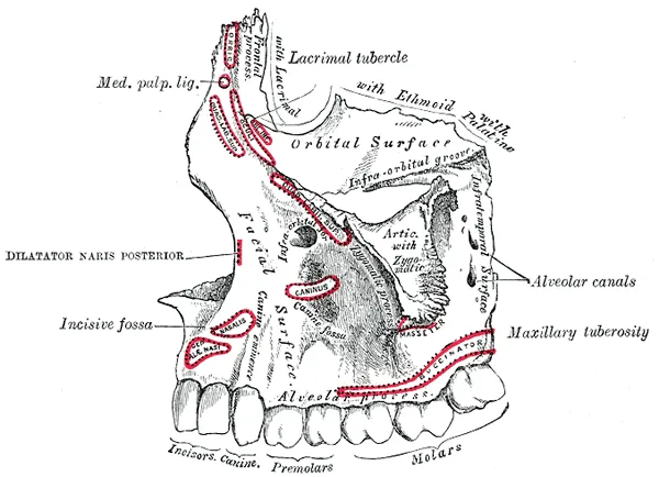
The two maxillae form the middle of the face. Each has a body containing the maxillary sinus and four processes: zygomatic, frontal, palatine and alveolar. The alveolar process carries the upper tooth sockets. Behind the last molar is the maxillary tuberosity, with foramina where the posterior superior alveolar branches enter. The root tips of the upper premolars and molars lie close to the floor of the sinus. This is why periapical infection can spread into the sinus, and why an extraction can open into it.[22]
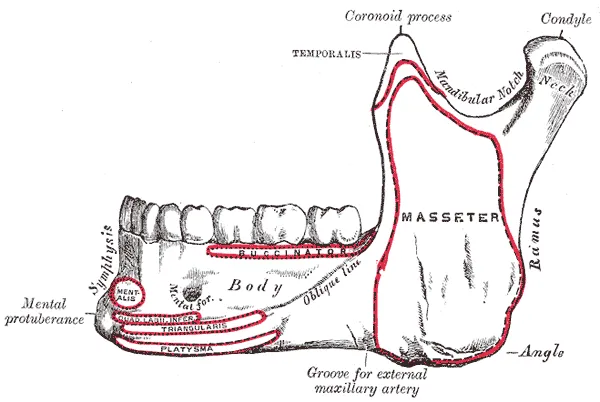
The mandible is the only movable bone of the face. It has a horseshoe-shaped body carrying the alveolar process, and two rami that meet the body at the angle. Each ramus has a coronoid process for the temporalis and a condylar process that articulates with the temporal bone. The mandibular foramen on the inner surface of the ramus, guarded by the lingula, leads into the mandibular canal. The canal runs below the molar roots and opens at the mental foramen, usually below the second premolar. On the inner surface, the mylohyoid line separates the sublingual fossa above from the submandibular fossa below.[23][24]
The mandibular canal has many variants that matter for implants, wisdom tooth removal and orthognathic surgery. A meta-analysis of 8,862 canals on cone-beam CT found a bifid canal in about 38%, a retromolar foramen in 23.6% and an accessory mental foramen in 9.5%. Lingual foramina were present in almost all adults. Before leaving the mental foramen, the inferior alveolar nerve may loop forward and back (the anterior loop) in about 60.8% of cases, with a mean length of 2.09 mm. A safety margin of about 4.75 mm in front of the foramen is therefore advised. In front of the mental foramen, an incisive canal is present in 87–100% of cases, with a mean length of almost 10 mm. It can be damaged by implants longer than 12 mm.[25][26][27][28]
Bifid mandibular canal
- Frequency
- ~38%
- Clinical relevance
- Failed inferior alveolar block, bleeding at third molar removal
Retromolar foramen
- Frequency
- 23.6%
- Clinical relevance
- Incomplete anaesthesia of molars, injury in retromolar incisions
Accessory mental foramen
- Frequency
- 9.5%
- Clinical relevance
- Lip and chin numbness after surgery
Anterior loop of the inferior alveolar nerve
- Frequency
- 60.8%
- Clinical relevance
- Keep a safety margin of about 4.75 mm in front of the foramen
Mandibular incisive canal
- Frequency
- 87–100%
- Clinical relevance
- Injury during implant placement between the mental foramina
| Variant | Frequency | Clinical relevance |
|---|---|---|
| Bifid mandibular canal | ~38% | Failed inferior alveolar block, bleeding at third molar removal |
| Retromolar foramen | 23.6% | Incomplete anaesthesia of molars, injury in retromolar incisions |
| Accessory mental foramen | 9.5% | Lip and chin numbness after surgery |
| Anterior loop of the inferior alveolar nerve | 60.8% | Keep a safety margin of about 4.75 mm in front of the foramen |
| Mandibular incisive canal | 87–100% | Injury during implant placement between the mental foramina |
Temporomandibular joint
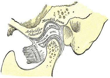
The temporomandibular joint is a hinge-and-glide (ginglymoarthrodial) synovial joint between the mandibular condyle and the mandibular fossa and articular eminence of the temporal bone. The condyle measures about 15–20 mm across and 8–10 mm front to back. The fibrocartilaginous disc is biconcave: about 2 mm thick in front, 3 mm behind, and thinnest in the middle. It divides the joint into two compartments. The lower (disc–condyle) compartment mainly allows rotation. The upper (disc–temporal) compartment allows the forward glide of wide opening. The front of the disc is related to the upper head of the lateral pterygoid; behind it lies the retrodiscal tissue, rich in vessels and nerves.[29][24]
The capsule is reinforced on its outer side by the temporomandibular (lateral) ligament. The sphenomandibular and stylomandibular ligaments lie away from the joint and limit how far the mandible can move. The joint develops from the first pharyngeal arch in three stages: blastemic (weeks 7–8), cavitation (weeks 9–10) and maturation (from week 11). At birth it is still immature, and the condyle keeps growing into adolescence. It is supplied mainly by the superficial temporal, maxillary and masseteric arteries.[29]
Temporomandibular disorders cause jaw and face pain, joint noises and limited opening. Most are muscular in origin, the rest come from the joint. In disc displacement with reduction, the disc sits in front of the condyle when the mouth is closed and snaps back into place on opening, which gives a click on opening and on closing. If the disc does not go back, opening is restricted. Most cases improve with conservative care: patient education, occlusal splints, physiotherapy and painkillers.[30][31]
Muscles of mastication
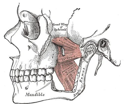
The four muscles of mastication are the masseter, temporalis, medial pterygoid and lateral pterygoid. All four develop from the first pharyngeal arch and receive their motor supply from branches of the mandibular nerve (V3). The masseter, temporalis and medial pterygoid together raise the mandible to bite and grind. The lateral pterygoid is the main muscle that pulls the condyle forward in opening and in moving the jaw to the opposite side. Based on how the nerve branches, the muscles can be grouped into an inner group (the lateral pterygoid) and an outer group (the other three, with their transitional bundles).[32][33][1]
The masseter has superficial and deep parts running from the zygomatic arch to the outer surface of the ramus and angle. An enlarged masseter can make the face look square; ultrasound-guided botulinum toxin injection can then reduce it. The lateral pterygoid has two heads. The lower head works in opening, protrusion and movement to the opposite side. The upper head works in closing and keeps the disc stable on the condyle. The medial pterygoid forms a sling around the angle of the mandible together with the masseter. Chewing is also coordinated by a rhythm generator in the brainstem, guided by feedback from the periodontal ligament and muscle spindles.[34][35][36][37][38]
Masseter
- Origin
- Lower border and inner surface of the zygomatic arch
- Insertion
- Outer surface of the ramus and angle of the mandible
- Action
- Elevates, slightly protrudes
- Nerve
- Masseteric nerve (V3)
Temporalis
- Origin
- Temporal fossa and temporal fascia
- Insertion
- Coronoid process and front of the ramus
- Action
- Elevates; posterior fibres retract
- Nerve
- Deep temporal nerves (V3)
Medial pterygoid
- Origin
- Medial surface of the lateral pterygoid plate, maxillary tuberosity
- Insertion
- Inner surface of the angle of the mandible
- Action
- Elevates, protrudes, moves to the side
- Nerve
- Nerve to medial pterygoid (V3)
Lateral pterygoid
- Origin
- Upper head: infratemporal surface of the greater wing of the sphenoid; lower head: lateral surface of the lateral pterygoid plate
- Insertion
- Pterygoid fovea of the condylar neck, capsule and disc
- Action
- Opens, protrudes, moves to the opposite side
- Nerve
- Nerve to lateral pterygoid (V3)
| Muscle | Origin | Insertion | Action | Nerve |
|---|---|---|---|---|
| Masseter | Lower border and inner surface of the zygomatic arch | Outer surface of the ramus and angle of the mandible | Elevates, slightly protrudes | Masseteric nerve (V3) |
| Temporalis | Temporal fossa and temporal fascia | Coronoid process and front of the ramus | Elevates; posterior fibres retract | Deep temporal nerves (V3) |
| Medial pterygoid | Medial surface of the lateral pterygoid plate, maxillary tuberosity | Inner surface of the angle of the mandible | Elevates, protrudes, moves to the side | Nerve to medial pterygoid (V3) |
| Lateral pterygoid | Upper head: infratemporal surface of the greater wing of the sphenoid; lower head: lateral surface of the lateral pterygoid plate | Pterygoid fovea of the condylar neck, capsule and disc | Opens, protrudes, moves to the opposite side | Nerve to lateral pterygoid (V3) |
Nerve supply of the teeth
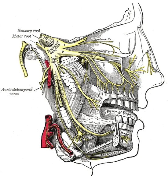
The upper teeth receive sensation from the maxillary nerve (V2). The posterior superior alveolar nerve leaves it in the pterygopalatine fossa and enters the maxillary tuberosity to reach the molars. The middle and anterior superior alveolar nerves come from the infraorbital nerve and supply the premolars and anterior teeth. Not everyone has a middle superior alveolar nerve. The three branches join to form the superior dental plexus. On the palatal side, the greater palatine nerve supplies the gingiva of the posterior teeth, and the nasopalatine nerve supplies the gingiva behind the incisors.[39][1]
A 2026 review brought together imaging and microanatomical studies and proposed a dual innervation model for the upper teeth. Besides the classical buccal pathways, a significant nerve and vessel supply comes from the palatal side. This model explains why some anaesthetic techniques work inconsistently. On this view, the AMSA and P-ASA injections are local palatal infiltrations rather than true nerve blocks. From the model, the authors propose a palatal alveolar foramen injection (PAFI).[40]
The lower teeth receive sensation from the mandibular nerve (V3). The inferior alveolar nerve is the largest branch of its posterior trunk. Before entering the mandibular foramen it gives off the nerve to mylohyoid, which supplies the mylohyoid and the anterior belly of the digastric. Inside the mandibular canal it sends branches to the molars and premolars. Near the second premolar it divides into two terminal branches. The mental nerve leaves through the mental foramen to supply the skin of the chin and lower lip. The incisive nerve continues inside the bone to the canine and incisors. The buccal gingiva of the molars is supplied by the buccal nerve, and the lingual gingiva by the lingual nerve.[41][1]
The lingual nerve carries sensation from the lingual gingiva, the floor of the mouth and the front two-thirds of the tongue. In the wisdom tooth region it runs very close to the lingual cortical plate, so it is easily injured when a lower wisdom tooth is removed. Branches of V3 can be trapped in the infratemporal fossa by an ossified pterygospinous or pterygoalar ligament or by fibres of the pterygoid muscles. This can cause numbness, neuralgia or loss of taste. Injury to the inferior alveolar or lingual nerve is a recognised complication of wisdom tooth removal, implant placement, local anaesthesia and orthognathic surgery.[42][43][44]
Upper incisors and canine
- Pulp
- Anterior superior alveolar
- Buccal / labial gingiva
- Anterior superior alveolar, superior labial
- Palatal / lingual gingiva
- Nasopalatine
Upper premolars
- Pulp
- Middle (or anterior) superior alveolar
- Buccal / labial gingiva
- Middle superior alveolar
- Palatal / lingual gingiva
- Greater palatine
Upper molars
- Pulp
- Posterior superior alveolar
- Buccal / labial gingiva
- Posterior superior alveolar
- Palatal / lingual gingiva
- Greater palatine
Lower incisors and canine
- Pulp
- Incisive (from inferior alveolar)
- Buccal / labial gingiva
- Mental
- Palatal / lingual gingiva
- Lingual
Lower premolars
- Pulp
- Inferior alveolar
- Buccal / labial gingiva
- Mental
- Palatal / lingual gingiva
- Lingual
Lower molars
- Pulp
- Inferior alveolar
- Buccal / labial gingiva
- Buccal
- Palatal / lingual gingiva
- Lingual
| Teeth | Pulp | Buccal / labial gingiva | Palatal / lingual gingiva |
|---|---|---|---|
| Upper incisors and canine | Anterior superior alveolar | Anterior superior alveolar, superior labial | Nasopalatine |
| Upper premolars | Middle (or anterior) superior alveolar | Middle superior alveolar | Greater palatine |
| Upper molars | Posterior superior alveolar | Posterior superior alveolar | Greater palatine |
| Lower incisors and canine | Incisive (from inferior alveolar) | Mental | Lingual |
| Lower premolars | Inferior alveolar | Mental | Lingual |
| Lower molars | Inferior alveolar | Buccal | Lingual |
Blood supply of the teeth and jaws
The maxillary artery is a large terminal branch of the external carotid artery. It arises behind the neck of the condyle and runs through the infratemporal fossa to the pterygopalatine fossa. It has three parts: mandibular, pterygoid and pterygopalatine. The mandibular part gives the inferior alveolar artery. This artery runs in the mandibular canal, supplies the molars and premolars, and divides at the mental foramen into mental and incisive branches. The pterygoid part supplies the four muscles of mastication. The pterygopalatine part gives the posterior superior alveolar artery and the infraorbital artery, which in turn gives the middle and anterior superior alveolar branches. The greater palatine artery supplies the palatal mucosa and gingiva.[45][46][1]
The course of the maxillary artery relative to the lateral pterygoid varies, which matters for surgery and anaesthesia in the infratemporal fossa. In 500 sides studied by CT angiography, the artery ran lateral to the muscle in 64.2%, medial in 29.6% and through its fibres in 6.2%. The course was the same on both sides in 77.6% of people. Pooling 32 studies with 5,938 arteries, the lateral course was found in 79.6%. The alveolar veins drain into the pterygoid venous plexus. This plexus communicates with the cavernous sinus, so dental infection can, rarely, spread inside the skull. Normal dental pulp seems to have no lymphatic vessels, but they may appear when the pulp is inflamed.[47][48][1]
Full-text reading (StatPearls)
The StatPearls chapters on this topic, reproduced verbatim. Their CC BY-NC-ND 4.0 licence allows unmodified copies only, so they are not translated or shortened.
Endodontics, Pulp Space Anatomy and Access Cavity of Anterior TeethChauhan PR, Daley JO, Chauhan G · StatPearls 2025 · NBK618377 · 32k characters
Loading…
Chauhan PR, Daley JO, Chauhan G. Endodontics, Pulp Space Anatomy and Access Cavity of Anterior Teeth. StatPearls [Internet]. Treasure Island (FL): StatPearls Publishing; 2025. NBK618377 · PMID 40991764 · CC BY-NC-ND 4.0. Reproduced verbatim, unmodified.
Anatomy, Head and Neck, TeethMorris AL, Tadi P · StatPearls 2023 · NBK557543 · 24k characters
Loading…
Morris AL, Tadi P. Anatomy, Head and Neck, Teeth. StatPearls [Internet]. Treasure Island (FL): StatPearls Publishing; 2023. NBK557543 · PMID 32491475 · CC BY-NC-ND 4.0. Reproduced verbatim, unmodified.
Anatomy, Head and Neck, Tooth EruptionJain P, Rathee M · StatPearls 2023 · NBK549878 · 22k characters
Loading…
Jain P, Rathee M. Anatomy, Head and Neck, Tooth Eruption. StatPearls [Internet]. Treasure Island (FL): StatPearls Publishing; 2023. NBK549878 · PMID 31751068 · CC BY-NC-ND 4.0. Reproduced verbatim, unmodified.
Anatomy, Head and Neck, Temporomandibular JointBordoni B, Brizuela M · StatPearls 2025 · NBK538486 · 21k characters
Loading…
Bordoni B, Brizuela M. Anatomy, Head and Neck, Temporomandibular Joint. StatPearls [Internet]. Treasure Island (FL): StatPearls Publishing; 2025. NBK538486 · PMID 30860721 · CC BY-NC-ND 4.0. Reproduced verbatim, unmodified.
Anatomy, Head and Neck, MaxillaSoriano RM, Brizuela M · StatPearls 2025 · NBK538527 · 17k characters
Loading…
Soriano RM, Brizuela M. Anatomy, Head and Neck, Maxilla. StatPearls [Internet]. Treasure Island (FL): StatPearls Publishing; 2025. NBK538527 · PMID 30860762 · CC BY-NC-ND 4.0. Reproduced verbatim, unmodified.
Anatomy, Head and Neck, Maxillary ArteryBrizuela M, Al Khalili Y · StatPearls 2025 · NBK542301 · 15k characters
Loading…
Brizuela M, Al Khalili Y. Anatomy, Head and Neck, Maxillary Artery. StatPearls [Internet]. Treasure Island (FL): StatPearls Publishing; 2025. NBK542301 · PMID 31194441 · CC BY-NC-ND 4.0. Reproduced verbatim, unmodified.
Anatomy, Head and Neck, Maxillary NerveShafique S, Das JM · StatPearls 2023 · NBK542277 · 15k characters
Loading…
Shafique S, Das JM. Anatomy, Head and Neck, Maxillary Nerve. StatPearls [Internet]. Treasure Island (FL): StatPearls Publishing; 2023. NBK542277 · PMID 31194417 · CC BY-NC-ND 4.0. Reproduced verbatim, unmodified.
Anatomy, Head and Neck, Dental PulpBrizuela M, Bordoni B · StatPearls 2025 · NBK537112 · 12k characters
Loading…
Brizuela M, Bordoni B. Anatomy, Head and Neck, Dental Pulp. StatPearls [Internet]. Treasure Island (FL): StatPearls Publishing; 2025. NBK537112 · PMID 30725797 · CC BY-NC-ND 4.0. Reproduced verbatim, unmodified.
Anatomy, Head and Neck, MandibleBreeland G, Aktar A, Patel BC · StatPearls 2023 · NBK532292 · 12k characters
Loading…
Breeland G, Aktar A, Patel BC. Anatomy, Head and Neck, Mandible. StatPearls [Internet]. Treasure Island (FL): StatPearls Publishing; 2023. NBK532292 · PMID 30335325 · CC BY-NC-ND 4.0. Reproduced verbatim, unmodified.
Anatomy, Head and Neck, Mandibular NerveGhatak RN, Helwany M, Ginglen JG · StatPearls 2023 · NBK507820 · 11k characters
Loading…
Ghatak RN, Helwany M, Ginglen JG. Anatomy, Head and Neck, Mandibular Nerve. StatPearls [Internet]. Treasure Island (FL): StatPearls Publishing; 2023. NBK507820 · PMID 29939597 · CC BY-NC-ND 4.0. Reproduced verbatim, unmodified.
Anatomy, Head and Neck, Lateral Pterygoid MuscleRathee M, Jain P · StatPearls 2024 · NBK549799 · 11k characters
Loading…
Rathee M, Jain P. Anatomy, Head and Neck, Lateral Pterygoid Muscle. StatPearls [Internet]. Treasure Island (FL): StatPearls Publishing; 2024. NBK549799 · PMID 31747206 · CC BY-NC-ND 4.0. Reproduced verbatim, unmodified.
Histology, ToothFarci F, Soni A · StatPearls 2023 · NBK572055 · 11k characters
Loading…
Farci F, Soni A. Histology, Tooth. StatPearls [Internet]. Treasure Island (FL): StatPearls Publishing; 2023. NBK572055 · PMID 34283421 · CC BY-NC-ND 4.0. Reproduced verbatim, unmodified.
Anatomy, Head and Neck: Inferior Alveolar ArteriesNguyen JD, Duong H · StatPearls 2023 · NBK547736 · 9k characters
Loading…
Nguyen JD, Duong H. Anatomy, Head and Neck: Inferior Alveolar Arteries. StatPearls [Internet]. Treasure Island (FL): StatPearls Publishing; 2023. NBK547736 · PMID 31613516 · CC BY-NC-ND 4.0. Reproduced verbatim, unmodified.
Anatomy, Head and Neck, Masseter MuscleCorcoran NM, Goldman EM · StatPearls 2023 · NBK539869 · 9k characters
Loading…
Corcoran NM, Goldman EM. Anatomy, Head and Neck, Masseter Muscle. StatPearls [Internet]. Treasure Island (FL): StatPearls Publishing; 2023. NBK539869 · PMID 30969691 · CC BY-NC-ND 4.0. Reproduced verbatim, unmodified.
Anatomy, Head and Neck, Medial (Internal) Pterygoid NerveJain P, Rathee M · StatPearls 2023 · NBK547712 · 8k characters
Loading…
Jain P, Rathee M. Anatomy, Head and Neck, Medial (Internal) Pterygoid Nerve. StatPearls [Internet]. Treasure Island (FL): StatPearls Publishing; 2023. NBK547712 · PMID 31613492 · CC BY-NC-ND 4.0. Reproduced verbatim, unmodified.
Anatomy, Head and Neck, Mastication MusclesBasit H, Tariq MA, Siccardi MA · StatPearls 2023 · NBK541027 · 8k characters
Loading…
Basit H, Tariq MA, Siccardi MA. Anatomy, Head and Neck, Mastication Muscles. StatPearls [Internet]. Treasure Island (FL): StatPearls Publishing; 2023. NBK541027 · PMID 31082071 · CC BY-NC-ND 4.0. Reproduced verbatim, unmodified.
Anatomy, Head and Neck, Medial Pterygoid MuscleJain P, Rathee M · StatPearls 2023 · NBK546588 · 7k characters
Loading…
Jain P, Rathee M. Anatomy, Head and Neck, Medial Pterygoid Muscle. StatPearls [Internet]. Treasure Island (FL): StatPearls Publishing; 2023. NBK546588 · PMID 31536196 · CC BY-NC-ND 4.0. Reproduced verbatim, unmodified.
Neuroanatomy, Pterygoid PlexusGolub B, Bordoni B · StatPearls 2023 · NBK555896 · 7k characters
Loading…
Golub B, Bordoni B. Neuroanatomy, Pterygoid Plexus. StatPearls [Internet]. Treasure Island (FL): StatPearls Publishing; 2023. NBK555896 · PMID 32310356 · CC BY-NC-ND 4.0. Reproduced verbatim, unmodified.
Organs in this system
References
Sources for the values and descriptions on this page, indexed in PubMed (US National Library of Medicine).
- [1]Anatomy, Head and Neck, TeethMorris AL et al., StatPearls 2023PMID 32491475
- [2]DENTAL ENAMEL FORMATION AND IMPLICATIONS FOR ORAL HEALTH AND DISEASELacruz RS et al., Physiological reviews 2017PMID 28468833
- [3]Histology, ToothFarci F et al., StatPearls 2023PMID 34283421
- [4]Dentin: structure, composition and mineralizationGoldberg M et al., Frontiers in bioscience 2011PMID 21196346
- [5]Odontoblast physiologyBleicher F, Experimental cell research 2014PMID 24361392
- [6]On the discovery of cementumFoster BL, Journal of periodontal research 2017PMID 28261800
- [7]Endodontics, Pulp Space Anatomy and Access Cavity of Anterior TeethChauhan PR et al., StatPearls 2025PMID 40991764
- [8]Anatomy, Head and Neck, Dental PulpBrizuela M et al., StatPearls 2025PMID 30725797
- [9]A new system for classifying root and root canal morphologyAhmed HMA et al., International endodontic journal 2017PMID 27578418
- [10]A new system for classifying tooth, root and canal anomaliesAhmed HMA et al., International endodontic journal 2018PMID 29023779
- [11]Age-related morphological, histological and functional changes in teethCarvalho TS et al., Journal of oral rehabilitation 2017PMID 28032898
- [12]The intricate anatomy of the periodontal ligament and its development: Lessons for periodontal regenerationde Jong T et al., Journal of periodontal research 2017PMID 28635007
- [13]Exploring the mechanical and biological interplay in the periodontal ligamentWen X et al., International journal of oral science 2025PMID 40169537
- [14]Periodontal cell mechanotransductionChukkapalli SS et al., Open biology 2018PMID 30209038
- [15]The junctional epithelium: from health to diseaseBosshardt DD et al., Journal of dental research 2005PMID 15615869
- [16]Characteristics of periodontal biotype, its dimensions, associations and prevalence: a systematic reviewZweers J et al., Journal of clinical periodontology 2014PMID 24836578
- [17]Molecular and cellular mechanisms of tooth development, homeostasis and repairYu T et al., Development 2020PMID 31980484
- [18]The Morphogenesis, Pathogenesis, and Molecular Regulation of Human Tooth Development-A Histological ReviewNovacescu D et al., International journal of molecular sciences 2025PMID 40649989
- [19]How do teeth erupt?Stonehouse-Smith D et al., British dental journal 2024PMID 39123030
- [20]Anatomy, Head and Neck, Tooth EruptionJain P et al., StatPearls 2023PMID 31751068
- [21]Global variations in eruption chronology of primary teeth: A systematic review and meta-analysisMuthu MS et al., Archives of oral biology 2024PMID 38128337
- [22]Anatomy, Head and Neck, MaxillaSoriano RM et al., StatPearls 2025PMID 30860762
- [23]Anatomy, Head and Neck, MandibleBreeland G et al., StatPearls 2023PMID 30335325
- [24]Anatomy of the Mandible, Temporomandibular Joint, and DentitionHatcher DC, Neuroimaging clinics of North America 2022PMID 36244721
- [25]Systematic review and meta-analysis of mandibular canal variations on cone beam computed tomographyShan S et al., Oral radiology 2022PMID 35503592
- [26]The anterior loop of the inferior alveolar nerve within the mentonian foramen: a scoping reviewBezerra AA et al., Surgical and radiologic anatomy 2024PMID 38888832
- [27]The Anatomy of the Mandibular Incisive Canal and Its Influence in Implant Therapy: A Scoping ReviewPeña-Cardelles JF et al., The International journal of periodontics & restorative dentistry 2024PMID 38198437
- [28]The clinical anatomy of accessory mental nerves and foraminaIwanaga J et al., Clinical anatomy 2015PMID 26201838
- [29]Anatomy, Head and Neck, Temporomandibular JointBordoni B et al., StatPearls 2025PMID 30860721
- [30]Temporomandibular joint disc displacement with reduction: a review of mechanisms and clinical presentationPoluha RL et al., Journal of applied oral science 2019PMID 30810641
- [31]Diagnosis and treatment of temporomandibular disordersGauer RL et al., American family physician 2015PMID 25822556
- [32]Anatomy, Head and Neck, Mastication MusclesBasit H et al., StatPearls 2023PMID 31082071
- [33]Masticatory Muscles and Branches of Mandibular Nerve: Positional Relationships Between Various Muscle Bundles and Their Innervating BranchesAkita K et al., Anatomical record 2019PMID 30312011
- [34]Anatomy, Head and Neck, Masseter MuscleCorcoran NM et al., StatPearls 2023PMID 30969691
- [35]Anatomy, Head and Neck, Lateral Pterygoid MuscleRathee M et al., StatPearls 2024PMID 31747206
- [36]Anatomy, Head and Neck, Medial Pterygoid MuscleJain P et al., StatPearls 2023PMID 31536196
- [37]Ultrasound-Guided Botulinum Toxin-A Injections into the Masseter Muscle for Both Medical and Aesthetic PurposesPopescu MN et al., Toxins 2024PMID 39453189
- [38]The coordination of chewingFalardeau D et al., Current opinion in neurobiology 2023PMID 37913688
- [39]Anatomy, Head and Neck, Maxillary NerveShafique S et al., StatPearls 2023PMID 31194417
- [40]Innervation of the Maxillary Teeth: Integrating Classical Anatomy With the Dual Innervation ModelHino K et al., Clinical anatomy 2026PMID 41797295
- [41]Anatomy, Head and Neck, Mandibular NerveGhatak RN et al., StatPearls 2023PMID 29939597
- [42]Lingual nerve revisited-A comprehensive review Part I: Anatomy and variationsKitagawa N et al., Clinical anatomy 2025PMID 39118432
- [43]Functional anatomy of the mandibular nerve: consequences of nerve injury and entrapmentPiagkou M et al., Clinical anatomy 2011PMID 21322036
- [44]Dentoalveolar nerve injuryAuyong TG et al., Oral and maxillofacial surgery clinics of North America 2011PMID 21798439
- [45]Anatomy, Head and Neck, Maxillary ArteryBrizuela M et al., StatPearls 2025PMID 31194441
- [46]Anatomy, Head and Neck: Inferior Alveolar ArteriesNguyen JD et al., StatPearls 2023PMID 31613516
- [47]Mapping the Maxillary Artery and Lateral Pterygoid Muscle Relationship: Insights from Radiological and Meta-Analytic EvidencePiagkou M et al., Medicina 2025PMID 40731831
- [48]Neuroanatomy, Pterygoid PlexusGolub B et al., StatPearls 2023PMID 32310356
- [49]Gastrointestinal mean transit times in young and middle-aged healthy subjectsPMID 11318834Supports: Gastrointestinal transit time
- [50]Revised Estimates for the Number of Human and Bacteria Cells in the BodySender R, Fuchs S, Milo R, 2016PMID 27541692Supports: Colonic bacterial count