Lower limb
Model
Layers
Z-Anatomy anatomical model based on BodyParts3D, licensed CC BY-SA 4.0.
Overview
A regional model of the (right) lower limb: gluteal region, thigh, leg and foot; muscle compartments, the hip, knee, ankle and foot joints with their ligaments, the femoral, popliteal and tibial arteries, great and small saphenous veins, and the femoral, sciatic, tibial and fibular nerves.
Boundaries and subdivisions
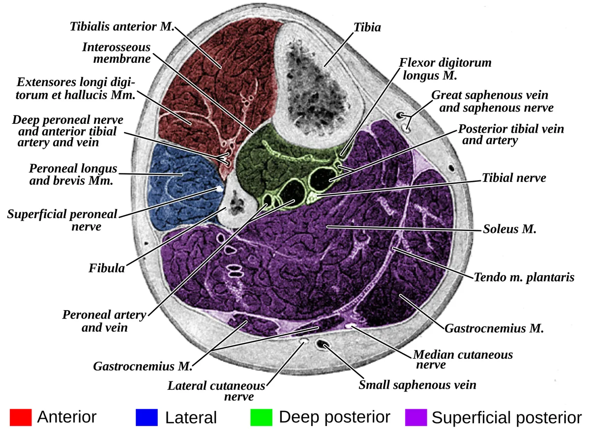
The lower limb comprises the gluteal region, thigh, knee, leg and foot, attached to the trunk through the hip joint. The femur is the longest and strongest bone in the body. The thigh has three compartments: anterior (quadriceps, sartorius; femoral nerve), medial (adductors; obturator nerve) and posterior (hamstrings; sciatic nerve). The leg has four: anterior (tibialis anterior, extensor hallucis longus, extensor digitorum longus, fibularis tertius; deep fibular nerve, anterior tibial artery), lateral (the two fibularis muscles; superficial fibular nerve), superficial posterior (gastrocnemius, soleus, plantaris) and deep posterior (tibialis posterior, flexor digitorum longus, flexor hallucis longus; tibial nerve).[1][2][3][4]
Layers from superficial to deep
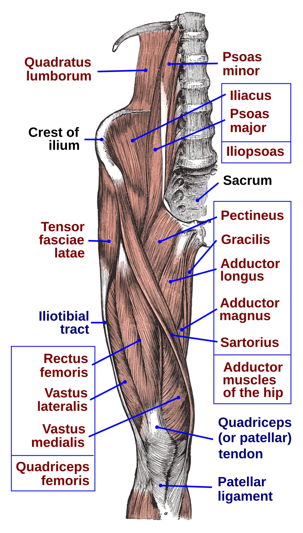
The superficial fascia of the lower limb carries two large superficial veins: the great saphenous vein passes in front of the medial malleolus, up the medial leg and thigh, and joins the femoral vein at the saphenous opening below the inguinal ligament; the small saphenous vein passes behind the lateral malleolus and drains into the popliteal vein. The deep fascia of the thigh is the fascia lata, thickened laterally as the iliotibial tract; in the leg the deep fascia, intermuscular septa and interosseous membrane form four closed, poorly distensible compartments. Quadriceps femoris comprises rectus femoris and the three vasti, converging on the quadriceps tendon, which encloses the patella and continues as the patellar ligament to the tibial tuberosity.[2][5][3]
Vessels, nerves and lymphatics
The femoral artery continues the external iliac below the inguinal ligament, gives the deep femoral artery (which supplies most of the thigh through perforating branches and the two circumflex femoral arteries), then passes through the adductor canal and adductor hiatus to become the popliteal artery. The popliteal artery divides into anterior and posterior tibial arteries; the posterior tibial gives the fibular artery and is palpable behind the medial malleolus at the ankle. The femoral nerve (L2–L4) supplies the anterior thigh; the sciatic nerve (L4–S3), the largest nerve in the body, descends in the back of the thigh and usually divides into the tibial and common fibular nerves at the apex of the popliteal fossa.[1][2][6][4]
Embryology
The lower limb bud appears on the ventrolateral body wall at about week 4, a few days after the upper limb, and the limbs are well differentiated by weeks 8 to 10. The bud is somatopleuric mesenchyme capped by an apical ectodermal ridge; cells from the hypaxial myotome migrate into it to form muscle, while connective tissue comes from somatopleuric mesoderm. The mesenchyme of the proximal thigh separates into distinct masses: the adductor group is visible in an embryo of about 11 mm, individual muscles at 14 mm though their tendons are not yet formed, and gracilis is complete by about week 28. The posterior thigh muscles derive from paraxial mesoderm. The sciatic nerve, like all peripheral nerves, develops from neural crest after neurulation begins in week 3, and its myelinating Schwann cells also come from the neural crest.[7][2][8][9][10][11]
Anatomical variants
The sciatic nerve usually leaves the greater sciatic foramen below piriformis as a single trunk; six patterns are described. The commonest variant is a nerve divided above piriformis, the common fibular part passing through the muscle and the tibial part below it; overall, about 10% of people have a sciatic nerve passing through or above piriformis. An adductor minimus separate from adductor magnus was found in more than half of 20 adult cadavers and in all 5 dissected fetuses. The pes anserinus shows several tendon arrangements (sartorius not covering gracilis, accessory gracilis or semitendinosus). A tensor of vastus intermedius may be present, with four morphological types. The femoral nerve occasionally splits in the abdomen and rejoins just above the inguinal canal. Women have a larger vastus medialis and smaller gracilis and sartorius than men, one factor in their higher rate of anterior cruciate ligament injury.[11][8][12][2][13][9]
Clinical correlations
The anterior compartment of the leg is the commonest site of acute compartment syndrome, an emergency requiring fasciotomy. An intracapsular femoral neck fracture tends to disrupt the deep branch of the medial circumflex femoral artery, causing avascular necrosis of the head. Injury to the common fibular nerve where it winds around the fibular neck causes foot drop. Saphenous varicose veins and deep vein thrombosis are common vascular disorders of the lower limb.[3][1]
Surgical and procedural considerations
Many thigh approaches run through intermuscular planes, so sound anatomy minimises neurovascular injury and helps locate complications quickly. The femoral artery is the access route for many procedures, including aortic aneurysm repair; the femoral nerve must be identified first, since its injury paralyses the anterior compartment (the branches to rectus femoris number one to four). Gracilis and semitendinosus tendons are common autografts for anterior cruciate ligament reconstruction, and gracilis is also transferred as a functioning muscle flap for facial palsy, brachial plexus injury or loss of finger movement. Distal quadriceps tendon rupture usually occurs after 40 years of age at the superior pole of the patella and should be repaired early. After total knee arthroplasty, adductor canal block is preferred to femoral nerve block because it gives similar analgesia with less quadriceps weakness. Most sciatica has a spinal cause (disc herniation compressing the L5 or S1 root); non-spinal causes such as piriformis syndrome are less common.[7][13][9][2][12][11]
Key structures
Compartimentum femoris anterius
- Name
- Anterior compartment of thigh
- Position, role
- Quadriceps, sartorius, iliopsoas; femoral nerve
Compartimentum femoris mediale
- Name
- Medial compartment of thigh
- Position, role
- Adductors, gracilis, pectineus; obturator nerve
Compartimentum femoris posterius
- Name
- Posterior compartment of thigh
- Position, role
- Hamstrings; sciatic nerve
Canalis adductorius
- Name
- Adductor canal (Hunter)
- Position, role
- Femoral artery and vein and saphenous nerve
Tractus iliotibialis
- Name
- Iliotibial tract
- Position, role
- Thickened lateral part of the fascia lata
V. saphena magna
- Name
- Great saphenous vein
- Position, role
- In front of the medial malleolus; joins the femoral vein
Compartimentum cruris anterius
- Name
- Anterior compartment of leg
- Position, role
- Extensors; deep fibular nerve; prone to compartment syndrome
| Terminologia Anatomica | Name | Position, role |
|---|---|---|
| Compartimentum femoris anterius | Anterior compartment of thigh | Quadriceps, sartorius, iliopsoas; femoral nerve |
| Compartimentum femoris mediale | Medial compartment of thigh | Adductors, gracilis, pectineus; obturator nerve |
| Compartimentum femoris posterius | Posterior compartment of thigh | Hamstrings; sciatic nerve |
| Canalis adductorius | Adductor canal (Hunter) | Femoral artery and vein and saphenous nerve |
| Tractus iliotibialis | Iliotibial tract | Thickened lateral part of the fascia lata |
| V. saphena magna | Great saphenous vein | In front of the medial malleolus; joins the femoral vein |
| Compartimentum cruris anterius | Anterior compartment of leg | Extensors; deep fibular nerve; prone to compartment syndrome |
What each layer contains
SkinCutis33
- Distal transverse arch of foot
- Proximal transverse arch of foot
- Dorsum of foot
- Hallucial eminence
- Dorsal surfaces of digits of foot
- Plantar surfaces of digits of foot
- Popliteal fossa
- Nail plate (foot)
- Lateral malleolus
- Medial malleolus
- Lateral border of foot
- Medial border of foot
- Lateral part of longitudinal arch of foot
- Medial part of longitudinal arch of foot
- Perionyx (foot)
- Sole
- Anterior region of leg
- Anterior region of thigh
- Anterior region of knee
- Anterior region of ankle
- Heel region
- Hip region
- Gluteal region
- Inguinal region
- Metatarsal region
- Posterior region of leg
- Posterior region of thigh
- Posterior region of knee
- Lateral retromalleolar region
- Medial retromalleolar region
- Sacral region
- Gluteal fold
- Femoral triangle
MusclesMusculi62
- (Adductor minimus)
- (Opponens digiti minimi muscle of foot)
- Abductor digiti minimi of foot
- Abductor hallucis
- Adductor brevis
- Adductor longus
- Adductor magnus
- Short head of biceps femoris
- Lateral head of flexor hallucis brevis
- Lateral head of gastrocnemius
- Long head of biceps femoris
- Medial head of flexor hallucis brevis
- Medial head of gastrocnemius
- Oblique head of adductor hallucis
- Transverse head of adductor hallucis
- Extensor digitorum brevis
- Extensor hallucis brevis
- Extensor digitorum longus
- Extensor hallucis longus
- Flexor digitorum brevis
- Flexor digiti minimi of foot
- Flexor hallucis longus
- Inguinal ligament
- Dorsal interossei muscles of foot
- Plantar interossei muscles
- Lumbrical muscles of foot
- Fibularis brevis muscle
- Fibularis longus muscle
- Fibularis tertius muscle
- Flexor digitorum longus
- Inferior gemellus muscle
- Superior gemellus muscle
- Gluteus maximus muscle
- Gluteus medius muscle
- Gluteus minimus muscle
- Gracilis muscle
- Iliacus muscle
- Pectineus muscle
- Piriformis muscle
- Plantaris muscle
- Popliteus muscle
- Quadratus femoris muscle
- Quadratus plantae muscle
- Rectus femoris muscle
- Sartorius muscle
- Semimembranosus muscle
- Semitendinosus muscle
- Soleus muscle
- Tibialis anterior muscle
- Tibialis posterior muscle
- Vastus intermedius muscle
- Vastus lateralis muscle
- Vastus medialis muscle
- Obturator externus
- Obturator internus
- Psoas major
- Lateral patellar retinaculum
- Medial patellar retinaculum
- Calcaneal tendon
- Tendon of extensor digitorum longus
- Tensor fasciae latae
- Iliotibial tract
Bones and cartilageOssa et cartilagines32
- Calcaneus
- Fibula
- Hip bone
- Cuboid bone
- Intermediate cuneiform bone
- Lateral cuneiform bone
- Medial cuneiform bone
- Femur
- Navicular bone
- First metatarsal bone
- Fourth metatarsal bone
- Fifth metatarsal bone
- Second metatarsal bone
- Talus
- Third metatarsal bone
- Sesamoid bones of foot
- Patella
- Distal phalanx of first finger of foot
- Distal phalanx of fourth finger of foot
- Distal phalanx of fifth finger of foot
- Distal phalanx of second finger of foot
- Distal phalanx of third finger of foot
- Middle phalanx of fourth finger of foot
- Middle phalanx of fifth finger of foot
- Middle phalanx of second finger of foot
- Middle phalanx of third finger of foot
- Proximal phalanx of first finger of foot
- Proximal phalanx of fourth finger of foot
- Proximal phalanx of fifth finger of foot
- Proximal phalanx of second finger of foot
- Proximal phalanx of third finger of foot
- Tibia
Joints and ligamentsJuncturae et ligamenta84
- Articular capsule of superior tibiofibular joint
- Articular capsule of interphalangeal joint of great toe
- Articular capsule of hip joint
- Articular capsule of knee joint
- Articular capsules of metatarsophalangeal joints
- Articular capsules of proximal interphalangeal joints of foot
- Articular capsules of distal interphalangeal joints of foot
- Triradiate cartilage
- Infrapatellar fat pad
- Frenula capsulae
- Acetabular labrum
- Cuneometatarsal interosseous ligaments
- Dorsal cuneonavicular ligaments
- Plantar cuneonavicular ligaments
- Dorsal intercuneiform ligaments
- Intercuneiform interosseous ligaments
- Plantar intercuneiform ligaments
- Collateral interphalangeal ligaments of foot
- Plantar interphalangeal ligaments
- Dorsal metatarsal ligaments
- Metatarsal interosseous ligaments
- Plantar metatarsal ligaments
- Collateral metatarsophalangeal ligaments
- Plantar metatarsophalangeal ligaments
- Dorsal tarsometatarsal ligaments
- Plantar tarsometatarsal ligaments
- Anterior ligament of fibular head
- Calcaneocuboid ligament
- Dorsal calcaneocuboid ligament
- Plantar calcaneocuboid ligament
- Calcaneofibular ligament
- Calcaneonavicular ligament
- Plantar calcaneonavicular ligament
- Tibiocalcaneal ligament
- Ligament of head of femur
- Fibular collateral ligament
- Anterior cruciate ligament
- Dorsal cuboideonavicular ligament
- Plantar cuboideonavicular ligament
- Dorsal cuneocuboid ligament
- Cuneocuboid interosseous ligament
- Plantar cuneocuboid ligament
- Posterior cruciate ligament
- Intersesamoid ligament
- Ischiofemoral ligament
- Meniscopatellar ligament
- Anterior meniscotibial ligament (Lateral meniscus)
- Anterior meniscotibial ligament (Medial meniscus)
- Posterior meniscotibial ligament (Lateral meniscus)
- Posterior meniscotibial ligament (Medial meniscus)
- Deep transverse metatarsal ligament
- Long plantar ligament
- Popliteofibular ligament
- Arcuate popliteal ligament
- Oblique popliteal ligament
- Posterior ligament of fibular head
- Pubofemoral ligament
- Anterior sacro-iliac ligament
- Interosseous sacro-iliac ligament
- Posterior sacro-iliac ligament
- Anterior talocalcaneal ligament
- Talocalcaneal interosseous ligament
- Lateral talocalcaneal ligament
- Medial talocalcaneal ligament
- Posterior talocalcaneal ligament
- Anterior talofibular ligament
- Posterior talofibular ligament
- Talonavicular ligament
- Posterior tibiotalar ligament
- Anterior tibiofibular ligament
- Posterior tibiofibular ligament
- Transverse tibiofibular ligament
- Tibionavicular ligament
- Transverse acetabular ligament
- Transverse ligament of knee
- Interosseous membrane of leg
- Obturator membrane
- Lateral meniscus
- Medial meniscus
- Descending part of iliofemoral ligament
- Deep part of tibial collateral ligament
- Superficial part of tibial collateral ligament
- Transverse part of iliofemoral ligament
- Zona orbicularis
ArteriesArteriae43
- Plantar arch
- Appendicular artery
- Arcuate artery
- Lateral circumflex femoral artery
- Medial circumflex femoral artery
- Dorsalis pedis artery
- Inferior epigastric artery
- Superficial epigastric artery
- Femoral artery
- Fibular artery
- Inferior gluteal artery
- Superior gluteal artery
- External iliac artery
- Inferior lateral genicular artery
- Inferior medial genicular artery
- Obturator artery
- Lateral plantar artery
- Medial plantar artery
- Deep plantar artery
- Popliteal artery
- Deep femoral artery
- Deep external pudendal artery
- Internal pudendal artery
- Superior lateral genicular artery
- Superior medial genicular artery
- Lateral tarsal artery
- Anterior tibial artery
- Posterior tibial artery
- Dorsal digital arteries of foot
- Common plantar digital arteries
- Proper plantar digital arteries
- Dorsal metatarsal arteries
- Plantar metatarsal arteries
- Perforating femoral arteries
- Middle genicular artery
- Calcaneal branches of fibular artery
- Calcaneal branches of posterior tibial artery
- Perforating branches of plantar metatarsal arteries
- Descending branch of lateral circumflex femoral artery
- Iliacus branch of iliolumbar artery
- Lumbar branch of iliolumbar artery
- Superficial branch of medial plantar artery
- Patellar anastomosis
VeinsVenae25
- Dorsal venous arch of foot
- Plantar venous arch
- Superficial epigastric vein
- Femoral vein
- Iliolumbar vein
- Popliteal vein
- Deep femoral vein
- Great saphenous vein
- Small saphenous vein
- Lateral circumflex femoral veins
- Medial circumflex femoral veins
- Dorsal digital veins of foot
- Plantar digital veins
- Fibular veins
- Genicular veins
- Inferior gluteal veins
- Superior gluteal veins
- Intercapitular veins of foot
- Dorsal metatarsal veins
- Plantar metatarsal veins
- Perforating veins
- Lateral plantar veins
- Medial plantar veins
- Anterior tibial veins
- Posterior tibial veins
NervesNervi33
- Intermediate dorsal cutaneous nerve of foot
- Lateral femoral cutaneous nerve
- Medial dorsal cutaneous nerve of foot
- Medial sural cutaneous nerve
- Posterior femoral cutaneous nerve
- Femoral nerve
- Common fibular nerve
- Deep fibular nerve
- Superficial fibular nerve
- Superior gluteal nerve
- Ilio-inguinal nerve
- Sciatic nerve
- Nerve to piriformis muscle
- Nerve to quadratus femoris muscle
- Obturator nerve
- Lateral plantar nerve
- Medial plantar nerve
- Saphenous nerve
- Sural nerve
- Tibial nerve
- Anterior cutaneous branches of femoral nerve
- Medial crural cutaneous branches of saphenous nerve
- Dorsal digital branches of deep fibular nerve
- Dorsal digital branches of superficial fibular nerve
- Common plantar digital branches of lateral plantar nerve
- Common plantar digital branches of medial plantar nerve
- Proper plantar digital branches of lateral plantar nerve
- Proper plantar digital branches of medial plantar nerve
- Muscular branches of deep fibular nerve
- Sural communicating branch of common fibular nerve
- Femoral branch of genitofemoral nerve
- Infrapatellar branch of saphenous nerve
- Posterior branch of obturator nerve
Full-text reading (StatPearls)
The StatPearls chapters on this topic, reproduced verbatim. Their CC BY-NC-ND 4.0 licence allows unmodified copies only, so they are not translated or shortened.
Anatomy, Bony Pelvis and Lower Limb: Thigh Quadriceps MuscleBordoni B, Varacallo MA · StatPearls 2023 · NBK513334 · 35k characters
Loading…
Bordoni B, Varacallo MA. Anatomy, Bony Pelvis and Lower Limb: Thigh Quadriceps Muscle. StatPearls [Internet]. Treasure Island (FL): StatPearls Publishing; 2023. NBK513334 · PMID 30020706 · CC BY-NC-ND 4.0. Reproduced verbatim, unmodified.
Anatomy, Bony Pelvis and Lower Limb: Thigh Femoral NerveRefai NA, Black AC, Tadi P · StatPearls 2023 · NBK556065 · 15k characters
Loading…
Refai NA, Black AC, Tadi P. Anatomy, Bony Pelvis and Lower Limb: Thigh Femoral Nerve. StatPearls [Internet]. Treasure Island (FL): StatPearls Publishing; 2023. NBK556065 · PMID 32310525 · CC BY-NC-ND 4.0. Reproduced verbatim, unmodified.
Anatomy, Bony Pelvis and Lower Limb: Thigh Gracilis MuscleKhan IA, Bordoni B, Varacallo MA · StatPearls 2023 · NBK538229 · 13k characters
Loading…
Khan IA, Bordoni B, Varacallo MA. Anatomy, Bony Pelvis and Lower Limb: Thigh Gracilis Muscle. StatPearls [Internet]. Treasure Island (FL): StatPearls Publishing; 2023. NBK538229 · PMID 30855817 · CC BY-NC-ND 4.0. Reproduced verbatim, unmodified.
Anatomy, Sciatic NerveGiuffre BA, Black AC, Jeanmonod R · StatPearls 2023 · NBK482431 · 13k characters
Loading…
Giuffre BA, Black AC, Jeanmonod R. Anatomy, Sciatic Nerve. StatPearls [Internet]. Treasure Island (FL): StatPearls Publishing; 2023. NBK482431 · PMID 29494038 · CC BY-NC-ND 4.0. Reproduced verbatim, unmodified.
Anatomy, Bony Pelvis and Lower Limb: Thigh MusclesAttum B, Varacallo MA · StatPearls 2022 · NBK482445 · 12k characters
Loading…
Attum B, Varacallo MA. Anatomy, Bony Pelvis and Lower Limb: Thigh Muscles. StatPearls [Internet]. Treasure Island (FL): StatPearls Publishing; 2022. NBK482445 · PMID 29494052 · CC BY-NC-ND 4.0. Reproduced verbatim, unmodified.
Anatomy, Bony Pelvis and Lower Limb: Medial Thigh MusclesRamage JL, Varacallo MA · StatPearls 2023 · NBK534775 · 11k characters
Loading…
Ramage JL, Varacallo MA. Anatomy, Bony Pelvis and Lower Limb: Medial Thigh Muscles. StatPearls [Internet]. Treasure Island (FL): StatPearls Publishing; 2023. NBK534775 · PMID 30521196 · CC BY-NC-ND 4.0. Reproduced verbatim, unmodified.
Anatomy, Bony Pelvis and Lower Limb: Thigh Adductor Magnus MuscleJeno SH, Launico MV, Schindler GS · StatPearls 2023 · NBK534842 · 11k characters
Loading…
Jeno SH, Launico MV, Schindler GS. Anatomy, Bony Pelvis and Lower Limb: Thigh Adductor Magnus Muscle. StatPearls [Internet]. Treasure Island (FL): StatPearls Publishing; 2023. NBK534842 · PMID 30521263 · CC BY-NC-ND 4.0. Reproduced verbatim, unmodified.
Anatomy, Bony Pelvis and Lower Limb: Posterior Thigh MusclesVaughn JE, Cohen-Levy WB · StatPearls 2023 · NBK542215 · 10k characters
Loading…
Vaughn JE, Cohen-Levy WB. Anatomy, Bony Pelvis and Lower Limb: Posterior Thigh Muscles. StatPearls [Internet]. Treasure Island (FL): StatPearls Publishing; 2023. NBK542215 · PMID 31194372 · CC BY-NC-ND 4.0. Reproduced verbatim, unmodified.
Anatomy, Bony Pelvis and Lower Limb: Thigh NervesGlenesk NL, Lopez PP · StatPearls 2023 · NBK482225 · 10k characters
Loading…
Glenesk NL, Lopez PP. Anatomy, Bony Pelvis and Lower Limb: Thigh Nerves. StatPearls [Internet]. Treasure Island (FL): StatPearls Publishing; 2023. NBK482225 · PMID 29489256 · CC BY-NC-ND 4.0. Reproduced verbatim, unmodified.
Anatomy, Bony Pelvis and Lower Limb: Posterior ThighAnderson TB, Vilella RC · StatPearls 2023 · NBK554598 · 9k characters
Loading…
Anderson TB, Vilella RC. Anatomy, Bony Pelvis and Lower Limb: Posterior Thigh. StatPearls [Internet]. Treasure Island (FL): StatPearls Publishing; 2023. NBK554598 · PMID 32119485 · CC BY-NC-ND 4.0. Reproduced verbatim, unmodified.
Anatomy, Bony Pelvis and Lower Limb: Anterior Thigh MusclesKhan A, Arain A · StatPearls 2023 · NBK538425 · 9k characters
Loading…
Khan A, Arain A. Anatomy, Bony Pelvis and Lower Limb: Anterior Thigh Muscles. StatPearls [Internet]. Treasure Island (FL): StatPearls Publishing; 2023. NBK538425 · PMID 30860696 · CC BY-NC-ND 4.0. Reproduced verbatim, unmodified.
Anatomy, Bony Pelvis and Lower Limb: Thigh Semitendinosus MuscleMathew K, Pillarisetty LS · StatPearls 2023 · NBK539862 · 7k characters
Loading…
Mathew K, Pillarisetty LS. Anatomy, Bony Pelvis and Lower Limb: Thigh Semitendinosus Muscle. StatPearls [Internet]. Treasure Island (FL): StatPearls Publishing; 2023. NBK539862 · PMID 30969684 · CC BY-NC-ND 4.0. Reproduced verbatim, unmodified.
Anatomy, Bony Pelvis and Lower Limb: Thigh Sartorius MuscleWalters BB, Varacallo MA · StatPearls 2023 · NBK532889 · 7k characters
Loading…
Walters BB, Varacallo MA. Anatomy, Bony Pelvis and Lower Limb: Thigh Sartorius Muscle. StatPearls [Internet]. Treasure Island (FL): StatPearls Publishing; 2023. NBK532889 · PMID 30422484 · CC BY-NC-ND 4.0. Reproduced verbatim, unmodified.
Regional anatomy
References
Sources for the values and descriptions on this page, indexed in PubMed (US National Library of Medicine).
- [1]Anatomy, Bony Pelvis and Lower Limb: FemurChang A et al., StatPearls 2023PMID 30422577
- [2]Anatomy, Bony Pelvis and Lower Limb: Thigh Quadriceps MuscleBordoni B et al., StatPearls 2023PMID 30020706
- [3]Anatomy, Bony Pelvis and Lower Limb: Leg Anterior CompartmentLezak B et al., StatPearls 2026PMID 30969547
- [4]Anatomy, Bony Pelvis and Lower Limb: Tibial NerveDesai SS et al., StatPearls 2026PMID 30725713
- [5]Anatomy, Bony Pelvis and Lower Limb, Knee PatellaCox CF et al., StatPearls 2023PMID 30137819
- [6]Anatomy, Bony Pelvis and Lower Limb: Posterior Tibial ArteryLezak B et al., StatPearls 2026PMID 30725666
- [7]Anatomy, Bony Pelvis and Lower Limb: Thigh MusclesAttum B et al., StatPearls 2022PMID 29494052
- [8]Anatomy, Bony Pelvis and Lower Limb: Thigh Adductor Magnus MuscleJeno SH et al., StatPearls 2023PMID 30521263
- [9]Anatomy, Bony Pelvis and Lower Limb: Thigh Gracilis MuscleKhan IA et al., StatPearls 2023PMID 30855817
- [10]Anatomy, Bony Pelvis and Lower Limb: Posterior Thigh MusclesVaughn JE et al., StatPearls 2023PMID 31194372
- [11]Anatomy, Sciatic NerveGiuffre BA et al., StatPearls 2023PMID 29494038
- [12]Anatomy, Bony Pelvis and Lower Limb: Medial Thigh MusclesRamage JL et al., StatPearls 2023PMID 30521196
- [13]Anatomy, Bony Pelvis and Lower Limb: Thigh Femoral NerveRefai NA et al., StatPearls 2023PMID 32310525