Pancreas
Model
Hover or tap to see names, click to read a description; click a name in the legend to fly to it. Procedurally built to histological proportions; colours follow illustration conventions.
Description
A mixed retroperitoneal gland. The exocrine part secretes enzyme-rich pancreatic juice; the islets of Langerhans (about 1–2% of pancreatic mass) secrete insulin, glucagon and somatostatin.
Parts
Each part has its own card; press “Show on the model” to fly to it and select it in the 3D model above.
- 01
Head and uncinate process
Caput pancreatis; Processus uncinatus
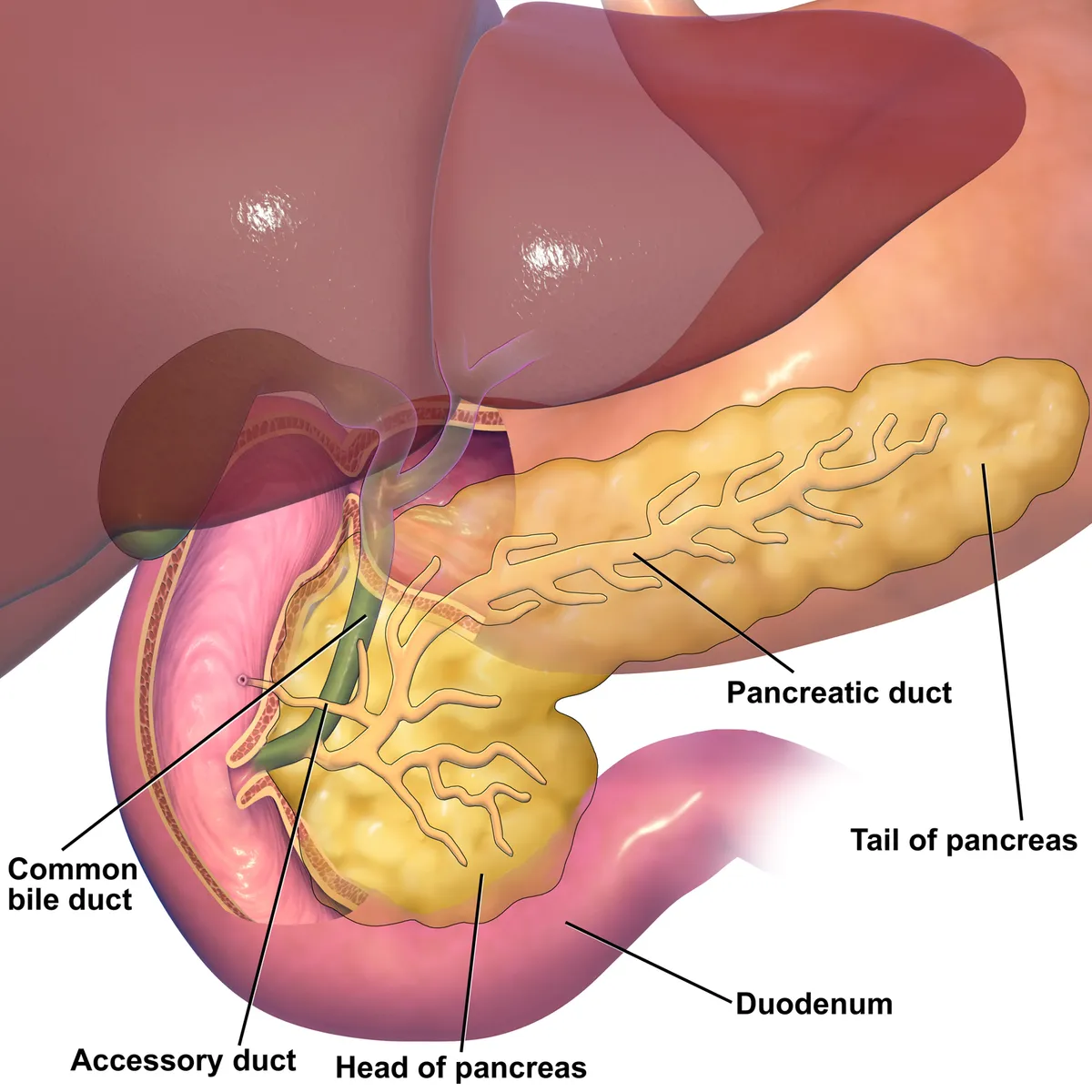
Lies within the C-loop of the duodenum to the right of the superior mesenteric vessels; the uncinate process hooks behind the superior mesenteric vein. The bile duct runs in or behind the head, so a head tumour causes painless obstructive jaundice with a palpable gallbladder (Courvoisier sign).
- 02
Neck
Collum pancreatis
A narrow segment about 2 cm long in front of the confluence where the superior mesenteric and splenic veins form the portal vein. It is the line of transection in the Whipple operation, and tumour invasion of the veins here affects resectability.
- 03
Body
Corpus pancreatis
Crosses in front of the aorta and the L1–L2 vertebrae, behind the lesser sac and stomach, with the splenic vein on its posterior surface. Draped over the spine, it is easily crushed in blunt abdominal trauma (seat belt, handlebar).
- 04
Tail
Cauda pancreatis
The mobile part lying in the splenorenal ligament and reaching the splenic hilum; islet density is highest here. It is at risk during splenectomy, which can cause a pancreatic fistula.
- 05
Main and accessory pancreatic ducts
Ductus pancreaticus; Ductus pancreaticus accessorius
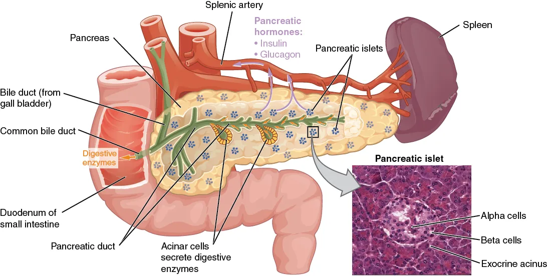
The main duct (of Wirsung) runs from tail to head and joins the bile duct at the major papilla; the accessory duct (of Santorini) opens at the minor papilla. When the embryonic ducts fail to fuse (pancreas divisum, about 10% of people), most pancreatic juice drains through the minor papilla.
- 06
Exocrine acini
Acini pancreatici
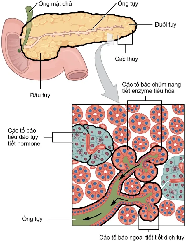
About 98% of the gland; acinar cells secrete enzymes (trypsinogen, lipase, amylase) as zymogen granules and duct cells secrete bicarbonate under secretin, some 1.5 L of juice a day. Premature activation of enzymes within the gland causes acute pancreatitis.
- 07
Pancreatic islets (of Langerhans)
Insulae pancreaticae
About a million islets, only 1–2% of pancreatic mass but receiving about 10% of its blood. β cells (about 60–70%) secrete insulin, α cells glucagon, δ cells somatostatin and PP cells pancreatic polypeptide. Autoimmune destruction of β cells causes type 1 diabetes.
Position and relations
The pancreas lies retroperitoneally across the posterior abdominal wall at L1–L2. Its head sits within the duodenal curve, its body crosses in front of the aorta and inferior vena cava, and its tail reaches the splenic hilum. The superior mesenteric and splenic veins join behind the neck of the pancreas to form the portal vein.
Form and structure
About 98% of the pancreas consists of exocrine acini that secrete enzymes into the main pancreatic duct (of Wirsung), which opens with the common bile duct at the major duodenal papilla. Some 1–2% consists of scattered islets of Langerhans, most numerous in the tail, containing α (glucagon), β (insulin), δ (somatostatin) and PP cells.[1]
Physiology
The endocrine and exocrine pancreas both arise from the “tip–trunk” domain of the pancreatic bud and interact throughout life. Under some conditions exocrine cells can transdifferentiate into insulin-secreting endocrine-like cells; diseases such as pancreatitis, pancreatic cancer and diabetes often affect both parts.[1]
Endocrine–exocrine crosstalk
The pancreas has dual functions: its endocrine part (islets of Langerhans) regulates blood glucose through insulin and glucagon, and its exocrine part secretes digestive enzymes. Both arise from the “tip–trunk” domain of the embryonic pancreatic epithelium and interact directly and indirectly; disease of one (chronic pancreatitis) often affects the other (pancreatogenic diabetes).[1][2]
Human β-cell development is directed by a cascade of transcription factors that partly differs from the mouse; understanding it underpins the generation of β-like cells from embryonic or induced pluripotent stem cells for diabetes therapy. In type 2 diabetes, endoplasmic reticulum stress and loss of β-cell identity with dedifferentiation (possibly partly reversible) contribute to failing insulin secretion.[3][4][5]
Type 1 diabetes is autoimmune destruction of β cells; treatment replaces insulin, and new approaches include encapsulated β-cell transplantation to avoid rejection.[6][7]
Full-text reading (StatPearls)
The StatPearls chapters on this topic, reproduced verbatim. Their CC BY-NC-ND 4.0 licence allows unmodified copies only, so they are not translated or shortened.
Anatomy, Abdomen and Pelvis, PancreasTalathi SS, Omole AE, Young M · StatPearls 2025 · NBK532912 · 11k characters
Loading…
Talathi SS, Omole AE, Young M. Anatomy, Abdomen and Pelvis, Pancreas. StatPearls [Internet]. Treasure Island (FL): StatPearls Publishing; 2025. NBK532912 · PMID 30422507 · CC BY-NC-ND 4.0. Reproduced verbatim, unmodified.
Organs in this system
References
Sources for the values and descriptions on this page, indexed in PubMed (US National Library of Medicine).
- [1]Pancreatic endocrine and exocrine signaling and crosstalk in physiological and pathological statusHu C et al., Signal transduction and targeted therapy 2025PMID 39948335
- [2]Chronic PancreatitisStram M et al., Surgical pathology clinics 2016PMID 27926364
- [3]Revealing transcription factors during human pancreatic β cell developmentConrad E et al., Trends in endocrinology and metabolism 2014PMID 24831984
- [4]Diabetes mellitus and the key role of endoplasmic reticulum stress in pancreatic β cellsLytrivi M et al., Nature reviews. Endocrinology 2025PMID 40467970
- [5]Loss of β-cell identity and dedifferentiation, not an irreversible process?Patel S et al., Frontiers in endocrinology 2024PMID 38915897
- [6]Type 1 Diabetes: A ReviewJacobsen LM et al., JAMA 2026PMID 41697686
- [7]Emerging Strategies for Beta Cell Encapsulation for Type 1 Diabetes TherapyToftdal MS et al., Advanced healthcare materials 2024PMID 38452393
- [8]Physiology, Pituitary HormonesStatPearls (NCBI Bookshelf)PMID 32491488Supports: Pituitary and endocrine axes
- [9]Physiology, Anterior PituitaryStatPearls (NCBI Bookshelf)PMID 29763073