Reproductive system
Model
Z-Anatomy anatomical model based on BodyParts3D, licensed CC BY-SA 4.0.
Overview
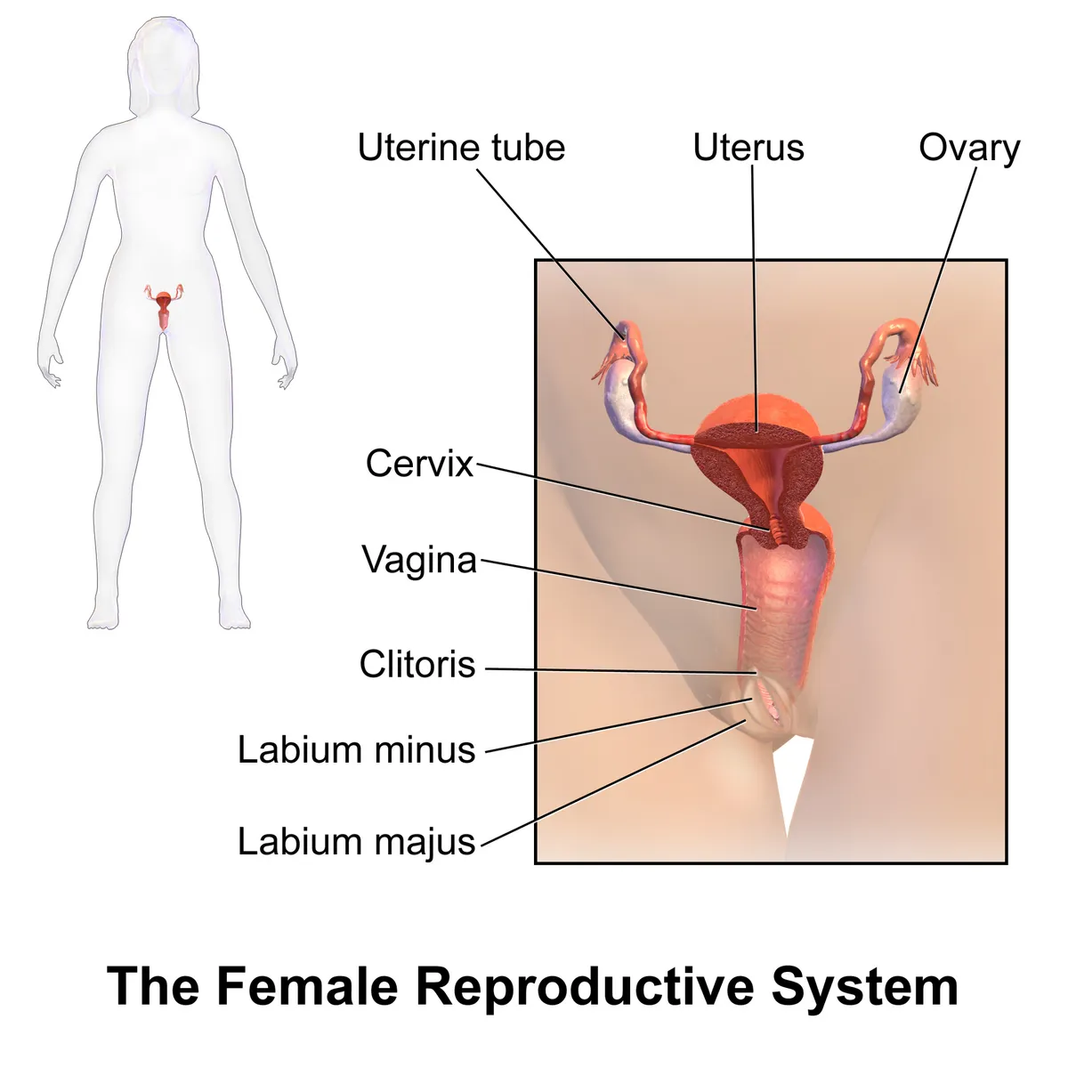
The reproductive system comprises the gonads (testes in the male, ovaries in the female), the genital ducts, the accessory glands and the external genitalia. The gonads produce gametes and secrete sex hormones; in the female, the uterus receives the implanting embryo and houses the developing fetus.
Functions
- 01Gametogenesis: spermatogenesis in the testes, follicle development and ovulation in the ovaries
- 02Secretion of sex hormones: testosterone, estradiol, progesterone
- 03Fertilisation, implantation of the embryo and gestation
Development
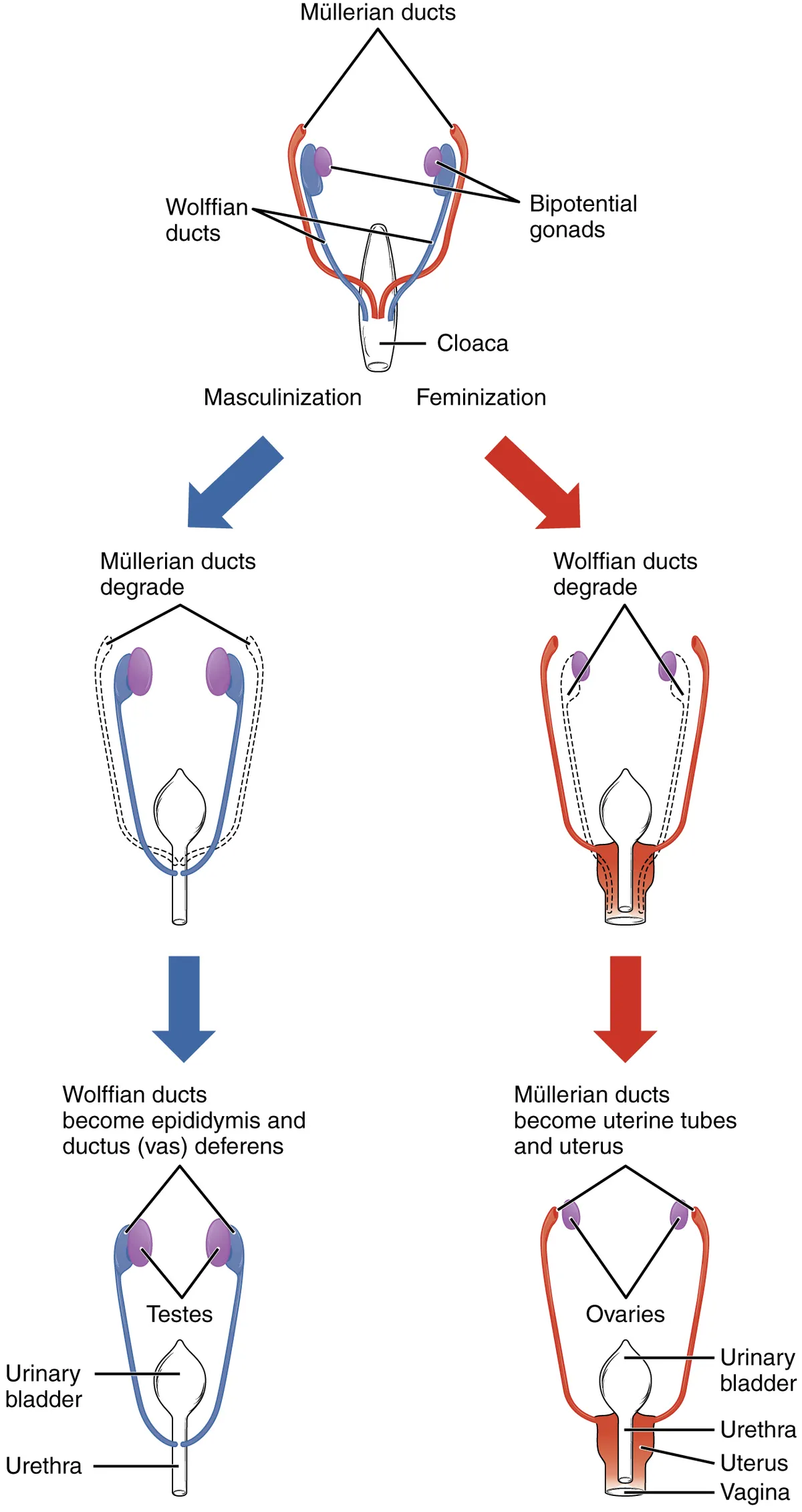
The gonads develop from the genital ridges on the posterior abdominal wall and are initially indifferent. In the male embryo, the SRY gene on the Y chromosome triggers testis differentiation; Sertoli cells secrete anti-Müllerian hormone, which regresses the paramesonephric ducts, while testosterone from Leydig cells maintains the mesonephric ducts as the epididymis, ductus deferens and seminal glands. In the absence of these signals, the paramesonephric ducts form the uterine tubes, uterus and upper vagina.
Physiology
The ovarian cycle is a dynamic endocrine system that includes ovarian events, hypothalamic–pituitary interactions, changes in the endometrium and myometrium during implantation and pregnancy, cervical changes and breast development; ovarian steroids also influence the risk of epithelial cancers of the ovary, uterus, cervix and breast.[1]
Reproductive function is governed by the hypothalamic–pituitary–gonadal axis: pulsatile GnRH from the hypothalamus drives the anterior pituitary to release LH and FSH. In the male, LH stimulates Leydig cells to secrete testosterone, and FSH together with testosterone supports Sertoli cells in spermatogenesis. In the female, FSH drives follicle growth and estradiol secretion; the mid-cycle LH surge triggers ovulation, after which the corpus luteum secretes progesterone to prepare the endometrium for implantation.
Development of the genital ducts and uterus
By about week 8 embryos of both sexes have two pairs of ducts: the paramesonephric (Müllerian) and the mesonephric (Wolffian). Each Müllerian duct has three segments: a vertical part lateral to and parallel with the Wolffian duct, a horizontal part crossing in front of the Wolffian duct of the same side, and a second vertical part that fuses with its partner in the midline and meets the posterior wall of the urogenital sinus (Müllerian tubercle). The first two segments become the uterine tube; the fused segment canalises to form the body and cervix of the uterus and roughly the upper fifth of the vagina. Without a Y chromosome and testicular testosterone, the Müllerian ducts develop and the Wolffian ducts regress; failures of fusion or septal resorption cause Müllerian anomalies (didelphys, bicornuate or septate uterus).[2]
The ovary arises from intermediate mesoderm. Primordial germ cells originate in the dorsal endoderm of the yolk sac, migrate along the hindgut to the gonadal ridge (formed by week 4), dividing as they go, and become oogonia; they are surrounded by cells that will become granulosa cells, forming primordial follicles. The ovary also descends but stops in the pelvis because the gubernaculum is attached to the uterus; its remnants are the proper ovarian ligament and the round ligament of the uterus. In the male embryo, the prostate grows as epithelial buds from the wall of the urogenital sinus, which branch and canalise into ducts and acini under androgen from Leydig cells.[3][4]
Uterus and ovary: position, relations and blood supply
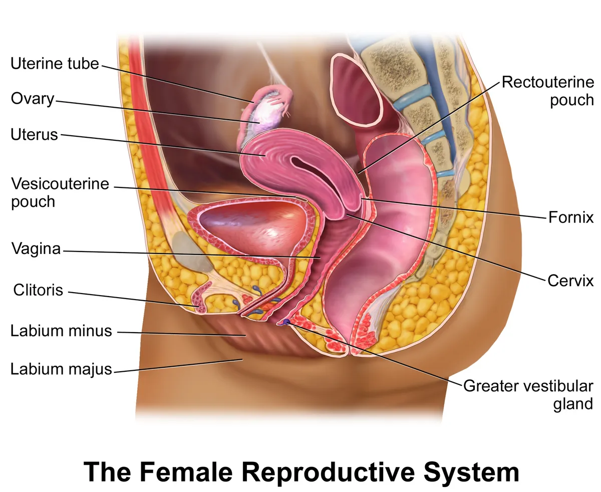
The axis of the cervix usually does not align with that of the body: the body bends forward over the cervix (anteflexion), and in about 80% of women the uterus is anteverted, nearly at right angles to the vagina; in about 20% it is retroverted. The cervix-to-body ratio is 2:1 before puberty, 1:2 in the reproductive years and returns to 2:1 after menopause with atrophy. The ovary (about 3.5 × 2 × 1 cm) lies in the ovarian fossa at the division of the external and internal iliac arteries, with the ureter behind; its volume rises from 0.7 mL at age 2 to a peak of 7.7 mL at 20 and falls to about 2.8 mL at menopause. The suspensory (infundibulopelvic) ligament carries the ovarian artery, vein and nerve plexus; the proper ligament joins the ovary to the uterine angle and contains no vessels.[2][3]
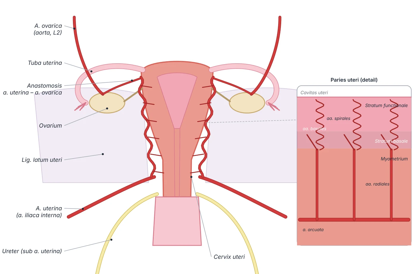
The uterus is supplied mainly by the uterine artery (anterior division of the internal iliac), which anastomoses in the broad ligament with the ovarian artery (from the abdominal aorta near L2, below the renal artery). In the myometrium the uterine artery gives arcuate arteries, then radial arteries, which at the endometrium divide into basal arteries (supplying the permanent basal layer) and spiral arteries (supplying the functional layer, constricting at menstruation and remodelled by trophoblast in pregnancy). The left ovarian vein drains into the left renal vein, the right directly into the inferior vena cava. Lymph drains segmentally: the fundus and ovaries to the para-aortic nodes, part along the round ligament to the superficial inguinal nodes, and the lower uterus to the external and internal iliac nodes, all important routes for gynaecological cancer spread.[2][3]
The corpus luteum: a temporary endocrine gland
After ovulation the follicle collapses into a corpus haemorrhagicum and then the corpus luteum, made of luteinised granulosa and theca cells linked by gap junctions. The corpus luteum has the highest blood flow per unit of tissue of any organ, thanks to vigorous angiogenesis: the follicular basement membrane breaks down and endothelial cells proliferate and migrate into the granulosa layer under angiogenic growth factors. The rate-limiting step of progesterone synthesis is the transfer of cholesterol into mitochondria by the StAR protein.[5]
The corpus luteum has two fates. If fertilisation and implantation occur, from about day 9 the syncytiotrophoblast secretes hCG (the hormone detected by pregnancy tests), which maintains the corpus luteum of pregnancy and its progesterone secretion, keeping the endometrium until about week 12, when the placenta takes over; it also secretes relaxin, which softens the pubic symphysis. Otherwise the corpus luteum degenerates into a corpus albicans, progesterone falls and menstruation follows. Luteal phase dysfunction (from weight loss, hyperprolactinaemia, stress, hyperandrogenism or intense training) is a common cause of infertility and the basis of luteal support with progestins in assisted reproduction; combined oral contraceptives suppress FSH and LH, so there is neither ovulation nor a corpus luteum.[5]
The prostate: relations and zones
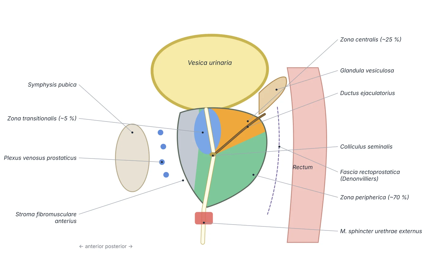
The prostate is a dense fibromuscular gland shaped like an inverted cone: its base above surrounds the bladder neck and its apex below rests on the external urethral sphincter. In front lies the pubic symphysis, separated by retropubic fat and the prostatic venous plexus; behind lies the rectum, separated by Denonvilliers fascia (which is why the gland can be felt on rectal examination); laterally lies levator ani. Textbooks divide the gland into anterior, posterior, two lateral and a median lobe, but the histological zones are more useful clinically: the peripheral zone, the largest (about 70%) and posterior, where most cancers arise; the central zone (about 25%) around the ejaculatory ducts; the small transition zone (about 5%) around the prostatic urethra, where benign hyperplasia arises; and the non-glandular anterior fibromuscular stroma.[4]
The main artery is the inferior vesical artery, with branches from the middle rectal and internal pudendal arteries; the veins form the prostatic plexus draining to the internal iliac veins, and lymph goes to the internal iliac and sacral nodes. Benign prostatic hyperplasia is a nodular (not diffuse) enlargement that compresses the urethra, causing frequency, nocturia and a weak stream; alpha-1 blockers relax smooth muscle of the bladder neck and prostatic urethra, while 5-alpha-reductase inhibitors block conversion of testosterone to dihydrotestosterone and shrink the gland. Prostate cancer is usually an adenocarcinoma of the peripheral zone, associated with family history, high BMI, smoking and radiation.[4]
Peripheral
- Share of volume
- ~70%
- Position
- Posteroinferior, palpable per rectum
- Typical disease
- Adenocarcinoma
Central
- Share of volume
- ~25%
- Position
- Base of the gland, around the ejaculatory ducts
- Typical disease
- Rarely diseased
Transition
- Share of volume
- ~5%
- Position
- Around the proximal prostatic urethra
- Typical disease
- Benign hyperplasia
Anterior fibromuscular stroma
- Share of volume
- Non-glandular
- Position
- Anterior surface
- Typical disease
- —
| Zone | Share of volume | Position | Typical disease |
|---|---|---|---|
| Peripheral | ~70% | Posteroinferior, palpable per rectum | Adenocarcinoma |
| Central | ~25% | Base of the gland, around the ejaculatory ducts | Rarely diseased |
| Transition | ~5% | Around the proximal prostatic urethra | Benign hyperplasia |
| Anterior fibromuscular stroma | Non-glandular | Anterior surface | — |
Principal organs
A cone-shaped gland below the bladder that surrounds the prostatic urethra and weighs about 20 g in young men. It has a peripheral zone (where most carcinomas arise), a central zone and a transition zone (the site of benign hyperplasia); its secretion contains PSA and forms part of the seminal fluid.
TestesTestisPaired ovoid glands in the scrotum, about 4–5 cm long, covered by the tunica albuginea, with the epididymis along the posterior border. The seminiferous tubules (with Sertoli cells) produce spermatozoa; interstitial Leydig cells secrete testosterone. Scrotal temperature, a few degrees below core temperature, is needed for spermatogenesis.
UterusUterusA thick-walled, pear-shaped hollow organ in the lesser pelvis between the bladder and the rectum, usually anteverted and anteflexed; about 7–8 cm long in nulliparous women. It has a fundus, body and cervix, and its wall has three layers: endometrium, myometrium and perimetrium.
Uterine tubesTuba uterinaPaired ducts about 10–12 cm long in the upper border of the broad ligament, linking the uterine cornua to the ovaries. From lateral to medial they have four parts: the fimbriated infundibulum, the ampulla (the usual site of fertilisation), the isthmus and the intramural part within the uterine wall. Cilia and peristalsis carry the oocyte and embryo towards the uterine cavity.
OvariesOvariumPaired almond-shaped glands, about 3 × 2 × 1 cm, lying in the ovarian fossa on the lateral pelvic wall and attached to the broad ligament by the mesovarium. The cortex contains the follicles; the ovary produces oocytes and secretes oestrogens and progesterone.
Reference values
- In humans, spermatogenesis from spermatogonium to spermatozoon takes about 74 days.
- The ovaries hold about 1–2 million primordial follicles at birth, yet only some 400–500 oocytes are ovulated during reproductive life.
References
Sources for the values and descriptions on this page, indexed in PubMed (US National Library of Medicine).
- [1]The Ovarian CycleRichards JS, Vitamins and hormones 2018PMID 29544627
- [2]Anatomy, Abdomen and Pelvis: UterusAmeer MA et al., StatPearls 2025PMID 29262069
- [3]Anatomy, Abdomen and Pelvis, OvaryGibson E et al., StatPearls 2023PMID 31424771
- [4]Anatomy, Abdomen and Pelvis, ProstateSingh O et al., StatPearls 2023PMID 31082031
- [5]Anatomy, Abdomen and Pelvis, Ovary Corpus LuteumOliver R et al., StatPearls 2023PMID 30969526