Thorax
Model
Layers
Z-Anatomy anatomical model based on BodyParts3D, licensed CC BY-SA 4.0.
Overview
A regional model of the thorax: the chest wall (pectoral and intercostal muscles, rib cage, costovertebral joints), the heart and great vessels, the lungs and tracheobronchial tree, the mediastinum (oesophagus, thymus, thoracic duct, lymph nodes), intercostal nerves, sympathetic trunk and thoracic spinal cord. Peel each layer from skin to mediastinum.
Boundaries and subdivisions
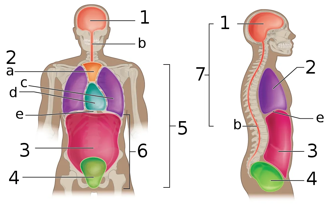
The thoracic cage consists of the 12 thoracic vertebrae, 12 pairs of ribs with their costal cartilages, and the sternum (manubrium, body, xiphoid process). The superior thoracic aperture, bounded by T1, the first ribs and the upper border of the manubrium, is the passage to the neck for the trachea, oesophagus, great vessels and nerves; the inferior aperture is closed by the diaphragm. The thoracic cavity contains two pleural cavities laterally and the mediastinum centrally; a horizontal plane through the sternal angle (manubriosternal joint, at the T4–T5 disc) separates the superior from the inferior mediastinum, which is subdivided into anterior, middle (heart within the pericardium) and posterior parts.[1][2][3]
Layers from superficial to deep
From outside in: skin and the breast (on pectoralis major, from the second to the sixth rib), pectoralis major and minor, and serratus anterior on the lateral wall. Between the ribs lie three layers, the external, internal and innermost intercostal muscles; the intercostal neurovascular bundle runs in the costal groove along the lower border of each rib, from above down vein, artery, nerve, between the internal and innermost layers. Deeper come the endothoracic fascia and parietal pleura, then the lungs covered by visceral pleura. The diaphragm, the main respiratory muscle, is dome-shaped with three large openings for the inferior vena cava (T8), the oesophagus (T10) and the aorta (T12).[4][5][1][6][7]
Vessels, nerves and lymphatics
The superior mediastinum contains the aortic arch and its three branches (brachiocephalic trunk, left common carotid, left subclavian), the superior vena cava formed by the two brachiocephalic veins, the trachea, oesophagus, thoracic duct, and the phrenic and vagus nerves; the left recurrent laryngeal nerve hooks under the arch. The posterior mediastinum holds the descending thoracic aorta, oesophagus, azygos system, thoracic duct and sympathetic trunks. The chest wall is supplied by posterior intercostal arteries (from the aorta) and anterior intercostal arteries (from the internal thoracic artery). Lymph from the breast drains mostly to the axillary nodes and the remainder to the parasternal chain.[8][9][10][4][11]
Embryology
The three-layered chest wall develops from somatopleuric mesoderm by about week 9. The three intercostal muscle layers differentiate from hypaxial myotomes supplied by the ventral rami of T1–T11, the intercostal nerves; twelve ribs grow from segmental sclerotomes and divide the muscular wall into eleven intercostal spaces, each named after the rib above. Cutaneous sensation follows overlapping dermatomes, so anaesthetising one area requires blocking at least three adjacent nerves. The lymphatic system starts in week 6 as buds near the base of the neck and forms six lymph sacs (cisterna chyli, two jugular, two iliac and a retroperitoneal sac); the adult thoracic duct arises from paired embryonic right and left ducts joined by anastomoses, keeping the lower part of the right duct and the upper part of the left.[7][11]
Anatomical variants
The textbook thoracic duct is found in only about half of people. A cisterna chyli is present in roughly 50%; when the embryonic lymphatic trunks converge above T12, it is usually absent and replaced by dilated lumbar trunks. Common variants are a bifid lower duct (failure of the embryonic right and left ducts to fuse) or a lower plexus of lymphatic channels that forms a single duct higher in the mediastinum; rarer are complete bilateral ducts or a duct ending on the right. The superior intercostal artery usually arises from the costocervical trunk but may come from the thyrocervical trunk, aortic arch, axillary or vertebral artery (in which case it passes through the C7 transverse foramen). In the mediastinum the brachiocephalic trunk may be absent, with the right common carotid and subclavian arising directly from the arch, and cardiac dominance depends on which coronary artery gives the posterior interventricular branch.[11][12][3]
Clinical correlations
Pleural aspiration or chest drain insertion is done just above the upper border of the rib below, to avoid the intercostal neurovascular bundle. Rib fractures most often involve the middle ribs; fractures of the lower ribs may accompany liver or spleen injury. Axillary dissection for breast cancer can injure the long thoracic nerve and cause a winged scapula. Hiatal hernia through the oesophageal opening of the diaphragm is a common cause of gastro-oesophageal reflux.[1][4][6]
Surgical and procedural considerations
The site of a thoracotomy is chosen to avoid the underlying viscera, nerves and vessels; the costodiaphragmatic recess, the largest part of the pleural cavity not occupied by lung, is the usual target for aspiration and drains. The intercostal nerves are at constant risk during thoracotomy and chest drain insertion; some authors advocate blunt dissection of the space with identification of the nerve rather than a stab technique. The thoracic duct is long, variable and close to the oesophagus, so it can be injured in oesophageal, cardiac or neck surgery and even during central line placement, producing a chylothorax; first-line treatment is a low-fat diet with medium-chain triglycerides. Superior intercostal artery aneurysms are rare, linked to aortic coarctation and neurofibromatosis type 1, and are best treated by endovascular embolisation, which is tolerated because the anterior intercostal arteries supply collateral flow. Spontaneous pneumothorax typically affects young male smokers through rupture of apical blebs.[7][13][11][12][14]
Key structures
Angulus sterni
- Name
- Sternal angle (of Louis)
- Position, role
- Level of the 2nd costal cartilage and T4–T5 disc; superior/inferior mediastinum boundary
Mediastinum superius
- Name
- Superior mediastinum
- Position, role
- Aortic arch, superior vena cava, trachea, oesophagus, thymus
Mediastinum medium
- Name
- Middle mediastinum
- Position, role
- Heart within the pericardium, roots of the great vessels
Mediastinum posterius
- Name
- Posterior mediastinum
- Position, role
- Descending aorta, oesophagus, azygos vein, thoracic duct
Sulcus costae
- Name
- Costal groove
- Position, role
- Holds the intercostal vein, artery and nerve (from above down)
Diaphragma
- Name
- Diaphragm
- Position, role
- Caval (T8), oesophageal (T10) and aortic (T12) openings
| Terminologia Anatomica | Name | Position, role |
|---|---|---|
| Angulus sterni | Sternal angle (of Louis) | Level of the 2nd costal cartilage and T4–T5 disc; superior/inferior mediastinum boundary |
| Mediastinum superius | Superior mediastinum | Aortic arch, superior vena cava, trachea, oesophagus, thymus |
| Mediastinum medium | Middle mediastinum | Heart within the pericardium, roots of the great vessels |
| Mediastinum posterius | Posterior mediastinum | Descending aorta, oesophagus, azygos vein, thoracic duct |
| Sulcus costae | Costal groove | Holds the intercostal vein, artery and nerve (from above down) |
| Diaphragma | Diaphragm | Caval (T8), oesophageal (T10) and aortic (T12) openings |
What each layer contains
SkinCutis15
- Infraclavicular fossa
- Lesser supraclavicular fossa
- Interscapular region
- Epigastric region
- Hypochondriac region
- Inframammary region
- Infrascapular region
- Lateral region of thorax
- Mammary region
- Pectoral region
- Presternal region
- Scapular region
- Vertebral region
- Triangle of auscultation
- Deltopectoral triangle
MusclesMusculi36
- (Abdominal part of pectoralis major muscle)
- Diaphragm
- Levatores breves costarum
- Levatores longi costarum
- External intercostal muscles
- Internal intercostal muscles
- Innermost intercostal muscles
- Interspinales thoracis muscles
- Iliocostalis colli muscle
- Iliocostalis thoracis muscle
- Infraspinatus muscle
- Latissimus dorsi muscle
- Longissimus colli muscle
- Longissimus thoracis muscle
- Multifidus thoracis muscle
- Pectoralis minor muscle
- Rhomboid major muscle
- Rhomboid minor muscle
- Semispinalis thoracis muscle
- Serratus anterior muscle
- Serratus posterior inferior muscle
- Serratus posterior superior muscle
- Spinalis thoracis muscle
- Splenius colli muscle
- Subclavius muscle
- Subscapularis muscle
- Supraspinatus muscle
- Teres major muscle
- Teres minor muscle
- Transversus thoracis muscle
- Clavicular part of deltoid muscle
- Clavicular head of pectoralis major muscle
- Descending part of trapezius muscle
- Sternocostal head of pectoralis major muscle
- Transverse part of trapezius muscle
- Rotatores
Bones and cartilageOssa et cartilagines39
- Costal cartilage of tenth rib
- Costal cartilage of eighth rib
- Costal cartilage of first rib
- Costal cartilage of fourth rib
- Costal cartilage of fifth rib
- Costal cartilage of second rib
- Costal cartilage of seventh rib
- Costal cartilage of sixth rib
- Costal cartilage of third rib
- Clavicle
- Body of sternum
- Costal cartilage of ninth rib
- First rib
- Second rib
- Tenth rib
- Twelfth rib
- Manubrium of sternum
- Ninth rib
- Eighth rib
- Xiphoid process
- Fourth rib
- Fifth rib
- Scapula
- Seventh rib
- Sixth rib
- Third rib
- Eleventh rib
- Vertebra L1
- Vertebra T2
- Vertebra T3
- Vertebra T4
- Vertebra T9
- Vertebra T5
- Vertebra T6
- Vertebra T7
- Vertebra T8
- Vertebra T10
- Vertebra T11
- Vertebra T12
Joints and ligamentsJuncturae et ligamenta50
- Articular capsule of acromioclavicular joint
- Articular capsule of sternoclavicular joint
- Articular disc of sternoclavicular joint
- Intervertebral disc L1-L2
- Intervertebral disc T1-T2
- Intervertebral disc T10-T11
- Intervertebral disc T11-T12
- Intervertebral disc T12-L1
- Intervertebral disc T2-T3
- Intervertebral disc T3-T4
- Intervertebral disc T4-T5
- Intervertebral disc T5-T6
- Intervertebral disc T6-T7
- Intervertebral disc T7-T8
- Intervertebral disc T8-T9
- Intervertebral disc T9-T10
- Glenoid labrum
- Ligamenta flava
- Interspinous ligaments
- Intertransverse ligaments
- Conoid ligament
- Coraco-acromial ligament
- Costoclavicular ligament
- Costotransverse ligament
- Interclavicular ligament
- Intra-articular ligament of head of rib
- Anterior longitudinal ligament
- Posterior longitudinal ligament
- Radiate ligament of head of rib
- Anterior sternoclavicular ligament
- Posterior sternoclavicular ligament
- Supraspinous ligament
- Inferior transverse scapular ligament
- Superior transverse scapular ligament
- Trapezoid ligament
- External intercostal membrane
- Internal intercostal membrane
- Nucleus pulposus L1-L2
- Nucleus pulposus T1-T2
- Nucleus pulposus T10-T11
- Nucleus pulposus T11-T12
- Nucleus pulposus T12-L1
- Nucleus pulposus T2-T3
- Nucleus pulposus T3-T4
- Nucleus pulposus T4-T5
- Nucleus pulposus T5-T6
- Nucleus pulposus T6-T7
- Nucleus pulposus T7-T8
- Nucleus pulposus T8-T9
- Nucleus pulposus T9-T10
HeartCor29
- ????????
- Right inferolateral branch of right coronary artery
- Circumflex artery of heart
- Right coronary artery
- Left coronary artery
- Anterior interventricular artery
- Right atrium
- Left atrium
- Inferior leaflet of right atrioventricular valve
- Posterior leaflet of left atrioventricular valve
- Septal leaflet of right atrioventricular valve
- Inferior vein of left ventricle (//Posterior '')
- Anterior papillary muscle of right ventricle
- Inferior papillary muscle of right ventricle
- Inferior papillary muscle of left ventricle
- Septal papillary muscle of right ventricle
- Septal branches of anterior interventricular artery
- Coronary sinus
- Right coronary leaflet
- Left coronary leaflet
- Non-coronary leaflet
- Anterior semilunar leaflet of pulmonary valve
- Right semilunar leaflet of pulmonary valve
- Left semilunar leaflet of pulmonary valve
- Inferior vein of left ventricle
- Great cardiac vein
- Middle cardiac vein
- Right ventricle
- Left ventricle
Lungs and airwaysPulmones et arbor bronchialis34
- (Anteromedial basal segmental bronchus of left lung)
- Intermediate bronchus
- Right inferior lobar bronchus
- Left inferior lobar bronchus
- Middle lobar bronchus
- Right superior lobar bronchus
- Left superior lobar bronchus
- Right main bronchus
- Left main bronchus
- Superior lingular segmental bronchus of left lung (BIV)
- Anterior segmental bronchus of right lung (BIII)
- Anterior segmental bronchus of left lung (BIII)
- Apical segmental bronchus of right lung (BI)
- Apicoposterior segmental bronchus of left lung (BI+BII)
- Anterior basal segmental bronchus of right lung (BVIII)
- Anterior basal segmental bronchus of left lung (BVIII)
- Lateral basal segmental bronchus of right lung (BIX)
- Lateral basal segmental bronchus of left lung (BIX)
- Medial basal segmental bronchus of right lung (BVII)
- Medial basal segmental bronchus of left lung (BVII)
- Posterior basal segmental bronchus of right lung (BX)
- Posterior basal segmental bronchus of left lung (BX)
- Lateral segmental bronchus of right lung (BIV)
- Inferior lingular segmental bronchus of left lung (BV)
- Medial segmental bronchus of right lung (BV)
- Posterior segmental bronchus of right lung (BII)
- Superior segmental bronchus of right lung (BVI)
- Superior segmental bronchus of left lung (BVI)
- Inferior lobe of right lung
- Inferior lobe of left lung
- Middle lobe of right lung
- Superior lobe of right lung
- Superior lobe of left lung
- Trachea
Oesophagus and glandsOesophagus et glandulae2
- Suprarenal gland
- Oesophagus
ArteriesArteriae78
- Right inferolateral branch of right coronary artery
- (Deep branch of transverse cervical artery)
- (Superficial branch of transverse cervical artery)
- Abdominal aorta
- Ascending aorta
- Thoracic aorta
- Aortic arch
- Axillary artery
- Circumflex artery of heart
- Circumflex scapular artery
- Middle colic artery
- Right coronary artery
- Left coronary artery
- Superior epigastric artery
- Left gastric artery
- Gastroduodenal artery
- Common hepatic artery
- Proper hepatic artery
- First posterior intercostal artery
- Second posterior intercostal artery
- Supreme intercostal artery
- Anterior interventricular artery
- Inferior lingular artery of left lung
- Superior lingular artery of left lung
- Inferior lobar artery of right lung
- Middle lobar artery of right lung
- Superior lobar artery of right lung
- Musculophrenic artery
- Inferior pancreaticoduodenal artery
- Anterior inferior pancreaticoduodenal artery
- Inferior phrenic artery
- Right pulmonary artery
- Left pulmonary artery
- Right renal artery
- Left renal artery
- Anterior segmental artery of right lung
- Anterior segmental artery of left lung
- Apical segmental artery of right lung
- Apical segmental artery of left lung
- Anterior basal segmental artery of right lung
- Anterior basal segmental artery of left lung
- Lateral basal segmental artery of right lung
- Lateral basal segmental artery of left lung
- Medial basal segmental artery of right lung
- Medial basal segmental artery of left lung
- Posterior basal segmental artery of right lung
- Posterior basal segmental artery of left lung
- Lateral segmental artery of right lung
- Medial segmental artery of right lung
- Posterior segmental artery of right lung
- Posterior segmental artery of left lung
- Superior segmental artery of right lung
- Superior segmental artery of left lung
- Splenic artery
- Right subclavian artery
- Left subclavian artery
- Subcostal artery
- Subscapular artery
- Inferior suprarenal artery
- Suprascapular artery
- Internal thoracic artery
- Lateral thoracic artery
- Thoraco-acromial artery
- Thoracodorsal artery
- Posterior intercostal arteries
- Intrarenal arteries of right kidney
- Intrarenal arteries of left kidney
- Superior phrenic arteries
- Bifurcation of pulmonary trunk
- Pectoral branches of thoraco-acromial artery
- Septal branches of anterior interventricular artery
- Anterior branch of renal artery
- Posterior branch of renal artery
- Brachiocephalic trunk
- Coeliac trunk
- Costocervical trunk
- Pulmonary trunk
- Thyrocervical trunk
VeinsVenae60
- ????????
- Inferior vein of left ventricle (//Posterior '')
- Coronary sinus
- Anterior vein of right lung
- Anterior vein of left lung
- Apical vein of right lung
- Apicoposterior vein of left lung
- Axillary vein
- Azygos vein
- Inferior basal vein of right lung
- Inferior basal vein of left lung
- Superior basal vein of right lung
- Superior basal vein of left lung
- Right brachiocephalic vein
- Left brachiocephalic vein
- Inferior vena cava (abdominal part)
- Inferior vena cava (thoracic part)
- Superior vena cava
- Circumflex scapular vein
- Middle colic vein
- Right gastro-omental vein
- Left gastro-omental vein
- Hemi-azygos vein
- Accessory hemi-azygos vein
- Inferior vein of left ventricle
- Right superior intercostal vein
- Left superior intercostal vein
- Lateral vein of right lung
- Inferior lingular vein of left lung
- Lingular vein of left lung
- Superior lingular vein of left lung
- Great cardiac vein
- Middle cardiac vein
- Medial vein of right lung
- Right superior phrenic vein
- Hepatic portal vein
- Posterior vein of right lung
- Right inferior pulmonary vein
- Right superior pulmonary vein
- Left inferior pulmonary vein
- Left superior pulmonary vein
- Right renal vein
- Left renal vein
- Splenic vein
- Right subclavian vein
- Left subclavian vein
- Right subcostal vein
- Left subcostal vein
- Subscapular vein
- Superior vein of right lung
- Superior vein of left lung
- Suprascapular vein
- Lateral thoracic vein
- Thoracodorsal vein
- Superior epigastric veins
- Hepatic veins
- Intrarenal veins of right kidney
- Intrarenal veins of left kidney
- Musculophrenic veins
- Internal thoracic veins
NervesNervi55
- Central canal
- Anterior horn of spinal cord
- Posterior horn of spinal cord
- Anterior division of inferior trunk of brachial plexus
- Anterior division of middle trunk of brachial plexus
- Anterior division of superior trunk of brachial plexus
- Posterior division of inferior trunk of brachial plexus
- Posterior division of middle trunk of brachial plexus
- Posterior division of superior trunk of brachial plexus
- Gracile fasciculus
- Posterior cord of brachial plexus
- Posterolateral tract
- Anterior fasciculus proprius
- Lateral fasciculus proprius
- Posterior fasciculus proprius
- Ganglia of sympathetic trunk
- Spinal ganglion
- Intercostal nerves
- Sympathetic nerves
- Accessory nerve (XI)
- Axillary nerve
- Medial brachial cutaneous nerve
- Dorsal scapular nerve
- Lateral pectoral nerve
- Medial pectoral nerve
- Subclavian nerve
- Inferior subscapular nerve
- Superior subscapular nerve
- Suprascapular nerve
- Long thoracic nerve
- Thoracodorsal nerve
- Vagus nerve (X)
- Intermediolateral nucleus
- Intermediomedial nucleus
- Nucleus proprius
- Spinal reticular process
- Anterior root of spinal nerve
- Posterior root of spinal nerve
- White matter of spinal cord
- Lateral intermediate substance
- Anterior corticospinal tract
- Lateral corticospinal tract
- Lateral reticulospinal tract
- Medial reticulospinal tract
- Rubrospinal tract
- Anterior spinocerebellar tract
- Posterior spinocerebellar tract
- Spinotectal tract
- Anterior spinothalamic tract
- Lateral spinothalamic tract
- Tectospinal tract
- Lateral vestibulospinal tract
- Medial vestibulospinal tract
- Inferior trunk of brachial plexus
- Sympathetic trunk
LymphaticsSystema lymphoideum46
- (Retropyloric nodes)
- (Subpyloric nodes)
- (Suprapyloric node)
- Spleen
- Right lobe of thymus
- Left lobe of thymus
- Node of arch of azygos vein
- Node of ligamentum arteriosum
- Lateral aortic nodes
- Anterior axillary nodes
- Apical axillary nodes
- Central axillary nodes
- Brachiocephalic nodes
- Lateral caval nodes
- Inferior deep lateral cervical nodes
- Coeliac nodes
- Right colic nodes
- Middle colic nodes
- Right gastric nodes
- Right gastro-omental nodes
- Ileocolic nodes
- Infraclavicular nodes
- Intercostal nodes
- Interpectoral nodes
- Intrapulmonary nodes
- Juxta-oesophageal nodes
- Intermediate lumbar nodes
- Inferior pancreatic nodes
- Superior pancreatic nodes
- Superior pancreaticoduodenal nodes
- Parasternal nodes
- Paratracheal thoracic nodes
- Lateral pericardial nodes
- Inferior diaphragmatic nodes
- Superior diaphragmatic nodes
- Pre-aortic nodes
- Precaval nodes
- Prepericardial nodes
- Prevertebral nodes
- Retro-aortic nodes
- Retrocaval nodes
- Splenic nodes
- Supraclavicular nodes
- Inferior tracheobronchial nodes
- Superior tracheobronchial nodes
- Cystic node
Full-text reading (StatPearls)
The StatPearls chapters on this topic, reproduced verbatim. Their CC BY-NC-ND 4.0 licence allows unmodified copies only, so they are not translated or shortened.
Anatomy, Thorax, MediastinumStoddard N, Heil JR, Lowery DR · StatPearls 2023 · NBK539819 · 12k characters
Loading…
Stoddard N, Heil JR, Lowery DR. Anatomy, Thorax, Mediastinum. StatPearls [Internet]. Treasure Island (FL): StatPearls Publishing; 2023. NBK539819 · PMID 30969641 · CC BY-NC-ND 4.0. Reproduced verbatim, unmodified.
Anatomy, Thoracotomy and the Collateral Intercostal Neurovascular BundleBoaz NT, Bernor RL, Meshida K, Lui F · StatPearls 2022 · NBK544368 · 12k characters
Loading…
Boaz NT, Bernor RL, Meshida K, Lui F. Anatomy, Thoracotomy and the Collateral Intercostal Neurovascular Bundle. StatPearls [Internet]. Treasure Island (FL): StatPearls Publishing; 2022. NBK544368 · PMID 31335088 · CC BY-NC-ND 4.0. Reproduced verbatim, unmodified.
Anatomy, Thorax, Mediastinum Superior and Great VesselsRizvi S, Wehrle CJ, Law MA · StatPearls 2023 · NBK519576 · 10k characters
Loading…
Rizvi S, Wehrle CJ, Law MA. Anatomy, Thorax, Mediastinum Superior and Great Vessels. StatPearls [Internet]. Treasure Island (FL): StatPearls Publishing; 2023. NBK519576 · PMID 30137860 · CC BY-NC-ND 4.0. Reproduced verbatim, unmodified.
Anatomy, Thorax, Superior Intercostal ArteriesGranger CJ, Martin AR · StatPearls 2023 · NBK549847 · 8k characters
Loading…
Granger CJ, Martin AR. Anatomy, Thorax, Superior Intercostal Arteries. StatPearls [Internet]. Treasure Island (FL): StatPearls Publishing; 2023. NBK549847 · PMID 31751037 · CC BY-NC-ND 4.0. Reproduced verbatim, unmodified.
Anatomy, Thorax, Intercostal NervesGlenesk NL, Rahman S, Lopez PP · StatPearls 2023 · NBK538238 · 7k characters
Loading…
Glenesk NL, Rahman S, Lopez PP. Anatomy, Thorax, Intercostal Nerves. StatPearls [Internet]. Treasure Island (FL): StatPearls Publishing; 2023. NBK538238 · PMID 30855826 · CC BY-NC-ND 4.0. Reproduced verbatim, unmodified.
Anatomy, Thorax, Thoracic DuctIlahi M, St Lucia K, Ilahi TB · StatPearls 2023 · NBK513227 · 7k characters
Loading…
Ilahi M, St Lucia K, Ilahi TB. Anatomy, Thorax, Thoracic Duct. StatPearls [Internet]. Treasure Island (FL): StatPearls Publishing; 2023. NBK513227 · PMID 30020599 · CC BY-NC-ND 4.0. Reproduced verbatim, unmodified.
Anatomy, Thorax, Lung Pleura And MediastinumMahabadi N, Goizueta AA, Bordoni B · StatPearls 2024 · NBK519048 · 7k characters
Loading…
Mahabadi N, Goizueta AA, Bordoni B. Anatomy, Thorax, Lung Pleura And Mediastinum. StatPearls [Internet]. Treasure Island (FL): StatPearls Publishing; 2024. NBK519048 · PMID 30085590 · CC BY-NC-ND 4.0. Reproduced verbatim, unmodified.
Regional anatomy
References
Sources for the values and descriptions on this page, indexed in PubMed (US National Library of Medicine).
- [1]Anatomy, Thorax, RibsSafarini OA et al., StatPearls 2026PMID 30855912
- [2]Anatomy, Thorax, SternumAltalib AA et al., StatPearls 2023PMID 31082185
- [3]Anatomy, Thorax, MediastinumStoddard N et al., StatPearls 2023PMID 30969641
- [4]Anatomy, Thorax, BreastRivard AB et al., StatPearls 2023PMID 30137859
- [5]Anatomy, Thorax, Pectoralis Major MajorSolari F et al., StatPearls 2023PMID 30252247
- [6]Anatomy, Thorax: DiaphragmBains KNS et al., StatPearls 2023PMID 30137842
- [7]Anatomy, Thoracotomy and the Collateral Intercostal Neurovascular BundleBoaz NT et al., StatPearls 2022PMID 31335088
- [8]Anatomy, Thorax, Heart Great VesselsBamalan OA et al., StatPearls 2023PMID 31613460
- [9]Anatomy, Thorax, Superior Vena CavaWhite HJ et al., StatPearls 2023PMID 31424839
- [10]Anatomy, Thorax, EsophagusOmole AE et al., StatPearls 2026PMID 29494119
- [11]Anatomy, Thorax, Thoracic DuctIlahi M et al., StatPearls 2023PMID 30020599
- [12]Anatomy, Thorax, Superior Intercostal ArteriesGranger CJ et al., StatPearls 2023PMID 31751037
- [13]Anatomy, Thorax, Intercostal NervesGlenesk NL et al., StatPearls 2023PMID 30855826
- [14]Anatomy, Thorax, Lung Pleura And MediastinumMahabadi N et al., StatPearls 2024PMID 30085590