Cerebral arteries
Model
Layers
Z-Anatomy anatomical model based on BodyParts3D, licensed CC BY-SA 4.0.
Description
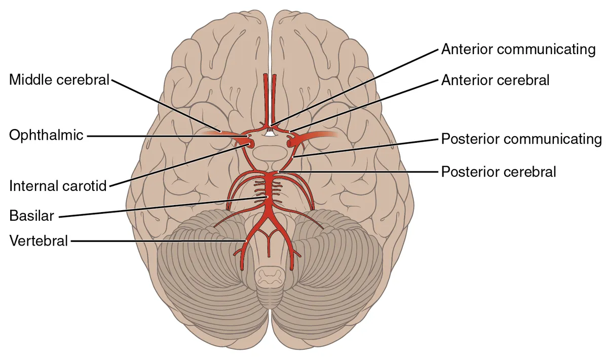
The brain is supplied by two systems: the internal carotid arteries (anterior circulation) and the vertebrobasilar system (posterior circulation). They anastomose in the cerebral arterial circle (of Willis) at the base of the brain, which gives off the anterior, middle and posterior cerebral arteries.
Parts
Each part has its own card; press “Show on the model” to fly to it and select it in the 3D model above.
- 01
Internal carotid artery
Arteria carotis interna
Supplies about 80% of the brain. It passes through the carotid canal and cavernous sinus (the siphon), pierces the dura, gives off the ophthalmic, posterior communicating and anterior choroidal arteries, and divides into the anterior and middle cerebral arteries.
- 02
Ophthalmic artery
Arteria ophthalmica
The first intradural branch of the internal carotid, entering the orbit through the optic canal. It gives the central retinal, ciliary, lacrimal, anterior and posterior ethmoidal, supra-orbital and supratrochlear arteries; occlusion of the central retinal artery causes sudden monocular blindness.
- 03
Anterior cerebral artery
Arteria cerebri anterior
Runs into the interhemispheric fissure and curves round the corpus callosum as the pericallosal and callosomarginal arteries. It supplies the medial frontal and parietal cortex, including the leg area, so occlusion weakens and numbs the opposite leg.
- 04
Middle cerebral artery
Arteria cerebri media
The largest terminal branch of the internal carotid: M1 (giving the lenticulostriate arteries to the internal capsule and basal nuclei), M2 over the insula, M3 in the opercula and M4 over the lateral cortex. Occlusion causes face- and arm-predominant hemiplegia and hemianaesthesia, with aphasia in the dominant hemisphere.
- 05
Vertebral artery
Arteria vertebralis
Arises from the subclavian artery, ascends through the transverse foramina of C6–C1, loops over the posterior arch of the atlas and enters the skull through the foramen magnum. It gives the anterior spinal and posterior inferior cerebellar (PICA) arteries before joining its fellow at the lower border of the pons.
- 06
Basilar artery
Arteria basilaris
Runs in the basilar groove on the front of the pons, giving the anterior inferior cerebellar (AICA), pontine and superior cerebellar arteries, and ends by dividing into the two posterior cerebral arteries. Basilar occlusion can cause locked-in syndrome or coma.
- 07
Posterior cerebral artery
Arteria cerebri posterior
Winds round the cerebral peduncle to supply the occipital lobe (visual cortex), the inferior temporal lobe, thalamus and midbrain. Occlusion causes contralateral homonymous hemianopia, often with macular sparing.
- 08
Cerebral arterial circle (of Willis)
Circulus arteriosus cerebri
An anastomotic ring at the base of the brain around the optic chiasm: the anterior cerebral arteries are joined by the anterior communicating artery, and each internal carotid joins the posterior cerebral by a posterior communicating artery. A complete circle is present in only about half of people, and its branch points are the commonest sites of saccular aneurysms.
Anterior circulation: internal carotid
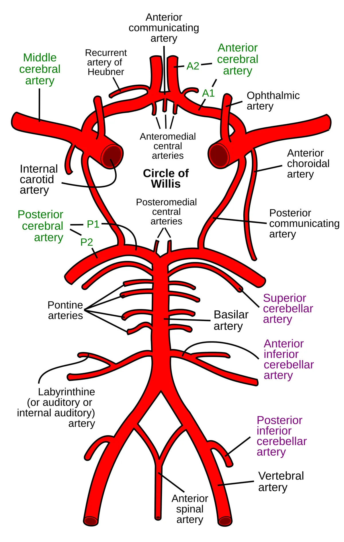
The internal carotid artery arises from the common carotid at the upper border of the thyroid cartilage, gives no branches in the neck, passes through the carotid canal of the temporal bone and the cavernous sinus, and enters the subarachnoid space. Its first intracranial branch is the ophthalmic artery, followed by the posterior communicating and anterior choroidal arteries; it then divides into the anterior and middle cerebral arteries.
The anterior cerebral artery runs in the longitudinal fissure around the corpus callosum and supplies the medial surface of the frontal and parietal lobes, including the motor and sensory areas of the lower limb. The middle cerebral artery, the largest terminal branch, runs in the lateral sulcus and supplies most of the lateral surface of the hemisphere (face and upper-limb motor and sensory areas, language areas of the dominant hemisphere) and, through its lenticulostriate branches, the basal nuclei and internal capsule.
Posterior circulation: vertebrobasilar system
The two vertebral arteries arise from the subclavian arteries, ascend through the transverse foramina of C6 to C1, enter the skull through the foramen magnum and give off the posterior inferior cerebellar and spinal arteries. At the lower border of the pons they unite to form the basilar artery, which runs in the basilar groove, gives the anterior inferior cerebellar arteries, pontine branches and superior cerebellar arteries, and divides into the two posterior cerebral arteries.
The posterior cerebral artery winds around the cerebral peduncle and supplies the occipital lobe (visual cortex), the inferior surface of the temporal lobe and, through perforating branches, the thalamus and midbrain. The vertebrobasilar system therefore supplies the brainstem, cerebellum, occipital lobe and posterior thalamus.
Cerebral arterial circle (of Willis)
At the base of the brain, around the optic chiasm and pituitary stalk, the two systems join in a ring: the anterior communicating artery links the two anterior cerebral arteries, and the two posterior communicating arteries link each internal carotid to the posterior cerebral artery. The ring can compensate when one source narrows, but the complete textbook form is found only in part of the population; hypoplastic or absent segments are common variants.
Veins and flow regulation
Venous blood drains through the superficial and deep cerebral veins (the great cerebral vein of Galen) into the dural sinuses (superior sagittal, straight, transverse and sigmoid) and out through the internal jugular vein.
Cerebral blood flow is adjusted locally to neuronal activity. This neurovascular coupling is carried out by the neurovascular unit, made up of neurons, astrocytes, endothelial cells of the blood–brain barrier, smooth muscle cells, pericytes and extracellular matrix, which detect demand and trigger vasodilation or vasoconstriction accordingly.[1]
Clinical correlations
The signs of ischaemic stroke reflect the territory that is occluded: middle cerebral artery occlusion causes contralateral face- and arm-predominant weakness and sensory loss, with aphasia if the dominant hemisphere is affected; anterior cerebral artery occlusion causes contralateral leg weakness; posterior cerebral artery occlusion causes contralateral homonymous hemianopia; occlusion of a lenticulostriate branch causes a lacunar infarct of the internal capsule.
The neurovascular unit
Central nervous system vessels have two features absent in peripheral organs: the blood–brain barrier, which selectively controls traffic into the parenchyma, and neurovascular coupling, which raises regional blood flow as soon as neurons become active, to supply nutrients and remove waste.[2]
The neurovascular unit was formalised in 2001. Neurovascular coupling is now seen as multidimensional: mediators from several cell types (neurons, astrocytes, pericytes, endothelium) engage distinct pathways across the whole cerebrovascular network, from capillaries back up to pial arteries, rather than a single neuron–astrocyte signal to a local vessel. Its dysfunction appears early in Alzheimer’s disease.[3][4]
Clinical correlations
Cerebral small vessel disease is a disorder of brain microvessels seen on imaging as white matter hyperintensities, recent small subcortical infarcts and lacunes; it is a common cause of stroke and vascular dementia. Mechanisms centre on microvascular endothelial dysfunction: blood–brain barrier leakage, impaired vasodilation, stiffening, disturbed flow and interstitial drainage, followed by ischaemia, inflammation and myelin damage.[5]
Migraine involves the trigeminovascular pathway: sensory fibres of the trigeminal nerve innervate meningeal and cerebral vessels and convey pain to the trigeminal nuclei in the brainstem, then to the thalamus and cortex. Activation of this pathway and related cortical and subcortical regions explains the headache and associated neurological symptoms.[6]
Full-text reading (StatPearls)
The StatPearls chapters on this topic, reproduced verbatim. Their CC BY-NC-ND 4.0 licence allows unmodified copies only, so they are not translated or shortened.
Neuroanatomy, Circle of WillisRosner J, Reddy V, Lui F · StatPearls 2023 · NBK534861 · 9k characters
Loading…
Rosner J, Reddy V, Lui F. Neuroanatomy, Circle of Willis. StatPearls [Internet]. Treasure Island (FL): StatPearls Publishing; 2023. NBK534861 · PMID 30521282 · CC BY-NC-ND 4.0. Reproduced verbatim, unmodified.
Organs in this system
References
Sources for the values and descriptions on this page, indexed in PubMed (US National Library of Medicine).
- [1]The neurovascular unit - concept reviewMuoio V et al., Acta physiologica 2014PMID 24629161
- [2]Neuronal regulation of the blood-brain barrier and neurovascular couplingKaplan L et al., Nature reviews. Neuroscience 2020PMID 32636528
- [3]The Neurovascular Unit Coming of Age: A Journey through Neurovascular Coupling in Health and DiseaseIadecola C, Neuron 2017PMID 28957666
- [4]Cerebral blood flow regulation and neurovascular dysfunction in Alzheimer diseaseKisler K et al., Nature reviews. Neuroscience 2017PMID 28515434
- [5]Small vessel disease: mechanisms and clinical implicationsWardlaw JM et al., The Lancet. Neurology 2019PMID 31097385
- [6]Migraine pathophysiology: anatomy of the trigeminovascular pathway and associated neurological symptoms, cortical spreading depression, sensitization, and modulation of painNoseda R et al., Pain 2013PMID 23891892
- [7]The human brain in numbers: a linearly scaled-up primate brainHerculano-Houzel S, 2009PMID 19915731Supports: Neuron count
- [8]Determinants of conduction velocity in myelinated nerve fibersWaxman SG, 1980PMID 6245357Supports: Conduction velocity
- [9]Quantitative imaging of energy expenditure in human brainPMID 22487547Supports: Cerebral energy expenditure