Nervous system
Model
Z-Anatomy anatomical model based on BodyParts3D, licensed CC BY-SA 4.0.
Overview
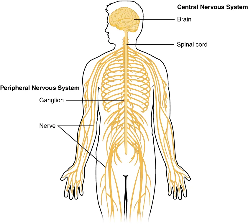
The nervous system detects, integrates and responds to stimuli arising inside and outside the body. Anatomically it comprises the central nervous system (brain and spinal cord) and the peripheral nervous system (cranial and spinal nerves and their ganglia).
Functions
- 01Sensory function: detection of stimuli by sensory receptors
- 02Integrative function: processing and storage of information and selection of responses
- 03Motor function: control of skeletal and smooth muscle contraction and of glandular secretion
- 04Homeostatic regulation through the autonomic (sympathetic and parasympathetic) divisions
Microscopic organisation
Nervous tissue consists of neurons and supporting glial cells (neuroglia). Astrocytes are the most abundant glial cells of the central nervous system: they maintain ionic and neurotransmitter balance around synapses, contribute to the blood–brain barrier and support neurons during development, normal function and ageing.[1]
Neurons, astrocytes, the endothelial cells of the blood–brain barrier, smooth muscle cells, pericytes and the extracellular matrix together form the neurovascular unit. This unit senses neuronal metabolic demand and adjusts local arteriolar calibre (vasodilation or vasoconstriction), so that cerebral blood flow rises precisely where activity occurs; this coupling is the physiological basis of functional MRI.[2]
Pathways and cranial nerves
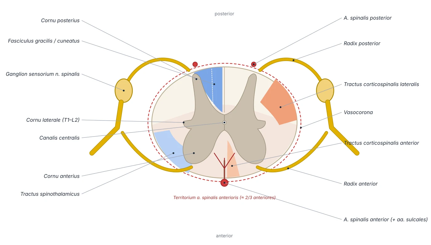
Pain is detected by peripheral nociceptors, carried by Aδ and C fibres to the dorsal horn of the spinal cord, then crosses and ascends in the spinothalamic tract to the thalamus and somatosensory cortex. Along the way, inhibitory and excitatory systems (from the periaqueductal grey and the brainstem raphe nuclei) modulate the signal; their dysfunction contributes to chronic pain.[3]
All twelve pairs of cranial nerves (nervi craniales) arise within the cranial cavity, except the accessory nerve, part of whose roots come from the cervical cord. Each passes through its own foramen or canal in the skull base; clinically, MRI shows the nerve itself while CT assesses the bony foramina it traverses.[4]
Neural tube, neural crest and the plan of the cord
At the end of week 3 the notochord induces the overlying ectoderm to thicken into the neural plate; the plate folds into a groove, the neural folds close into the neural tube, and cells at its edges separate as the neural crest. The cranial end swells into three primary brain vesicles, while the caudal end stays tubular and becomes the spinal cord; maternal folic acid is important for tube closure. The neuroepithelium forms central neurons, ependymal cells, oligodendrocytes and astrocytes; microglia have a different origin. The neural crest gives peripheral neurons and glia: dorsal root ganglia, sympathetic ganglia (and the adrenal medulla), Schwann and satellite cells, cranial parasympathetic ganglia and the enteric nervous system (from vagal crest, migrating from rostral to caudal and reaching the whole colon by weeks 12–13). The brain reaches 90% of adult volume by about age 6.[5][6][7]
The cord tube differentiates from inside out into ependymal, mantle and marginal layers. The mantle layer proliferates into grey matter, divided by the sulcus limitans into a ventral basal plate (motor: anterior and lateral horns) and a dorsal alar plate (sensory: posterior horn); the lumen narrows into the central canal. Axons occupy the marginal layer from week 4 and bundle into the anterior, lateral and posterior white columns; the cervical and lumbar enlargements appear with the limb buds in weeks 5–6, tracts form from the third month and the corticospinal tract appears around the fifth. In adults the cord is about 42 cm long in men and 39 cm in women, with a cervical enlargement (C4–T2, widest at C5) for the brachial plexus and a lumbosacral enlargement (L2–S3) for the lumbosacral plexus, tapering into the conus medullaris, whose filum terminale (pia) attaches to the coccyx.[6]
Blood supply of the spinal cord and brain
The spinal cord is supplied by three longitudinal channels: one anterior spinal artery in the anterior median fissure and two posterior spinal arteries in the posterolateral sulci, all arising from the vertebral arteries. Vertebral blood suffices only for the cervical segments; further down, these channels are reinforced by radicular arteries from the vertebral, cervical, intercostal, lumbar and sacral arteries. The largest is the artery of Adamkiewicz, usually from a left posterior intercostal artery between T9 and T12, which joins the anterior spinal artery. The branches anastomose into a pial vasocorona, but most of the cord (roughly the anterior two-thirds) depends on the anterior spinal artery, so its occlusion causes paralysis and loss of pain and temperature with preserved deep sensation. The veins form six longitudinal channels (anteromedian, posteromedian, two anterolateral and two posterolateral).[6]
The brain is only about 2% of body weight but receives about 15% of cardiac output. The two internal carotid arteries, entering the skull through the carotid canal in the petrous temporal bone, provide about 80% of its blood and divide into the anterior cerebral artery (medial frontal and parietal lobes: contralateral leg motor and sensory areas) and the middle cerebral artery (most of the lateral hemisphere, site of up to 80% of ischaemic strokes); the vertebrobasilar system supplies the rest. The circle of Willis links the two systems. In brain capillaries the blood–brain barrier comprises non-fenestrated endothelium with extensive tight junctions and very few pinocytic vesicles, a basement membrane, pericytes and astrocyte end-feet. It blocks paracellular diffusion of hydrophilic substances, actively transports nutrients in, pumps hydrophobic molecules and drugs out and controls the passage of leucocytes and pathogens; it forms through angiogenesis (driven by VEGF), differentiation and maturation and is fully functional by the third trimester.[5][8]
Sympathetic and parasympathetic outflow
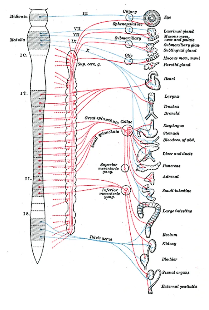
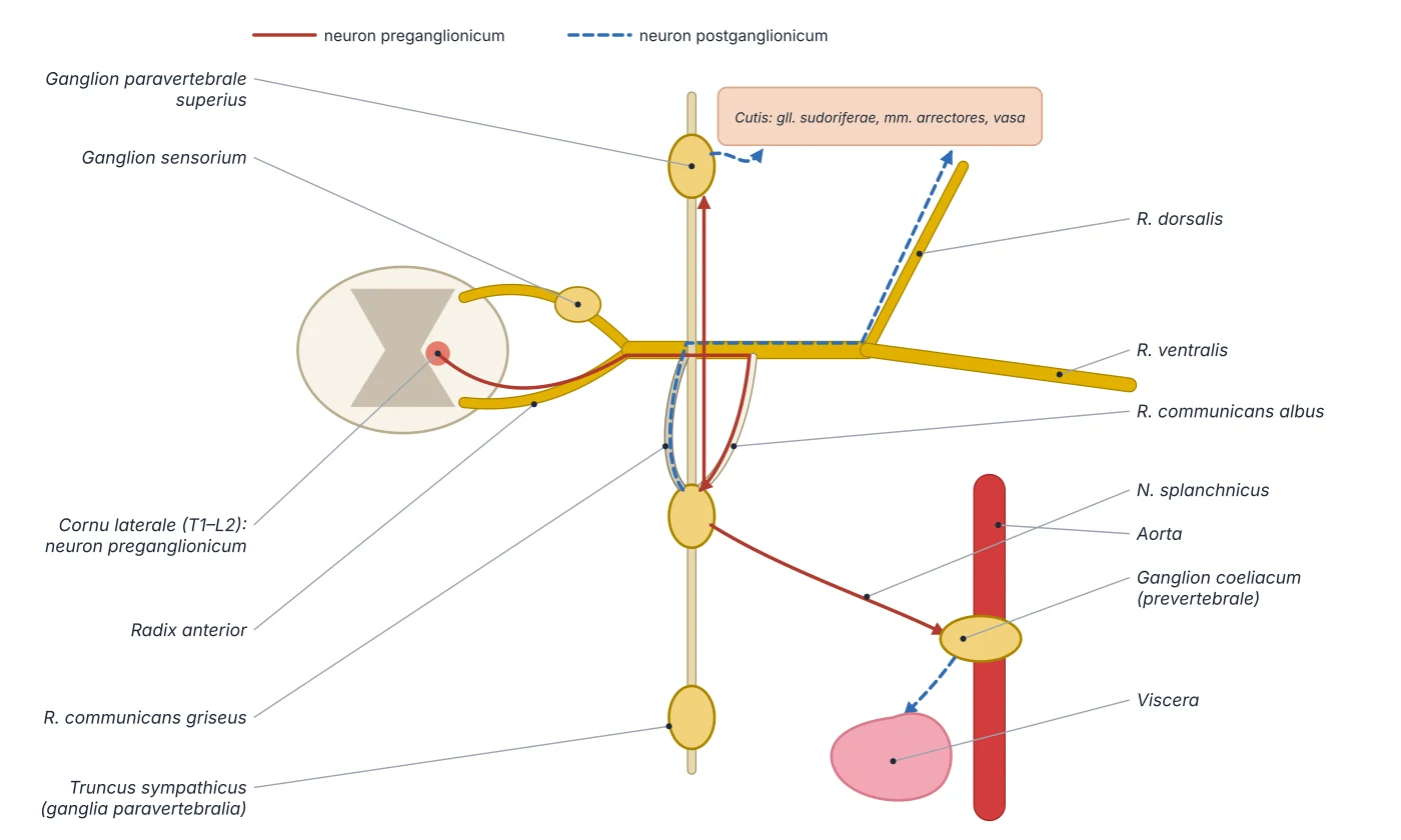
Preganglionic sympathetic cell bodies lie in the intermediolateral column (lateral horn) of cord segments T1–L2. Their fibres leave through the anterior roots, enter the anterior rami of the spinal nerves and reach the sympathetic chain through white rami communicantes. There a fibre may ascend or descend the chain to synapse in a higher or lower paravertebral ganglion, synapse at its own level and send postganglionic fibres back to the spinal nerve through a grey ramus communicans (to supply sweat glands, arrector pili muscles and cutaneous vessels of the body wall and limbs), or pass through the chain without synapsing in the abdominopelvic splanchnic nerves to the prevertebral ganglia (coeliac, superior and inferior mesenteric). Because the ganglia lie close to the midline, sympathetic preganglionic fibres are generally short and postganglionic fibres long. The chain usually has 3 cervical (superior, middle, inferior), 12 thoracic, 4 lumbar and 5 sacral ganglia; the inferior cervical ganglion often fuses with the first thoracic as the stellate ganglion.[7]
The parasympathetic outflow, by contrast, is craniosacral: brainstem nuclei through cranial nerves III, VII, IX and X, and sacral segments S2–S4 through the pelvic splanchnic nerves. Parasympathetic ganglia lie next to or within the wall of the target organ, so preganglionic fibres are long and postganglionic fibres short. The vagus carries most of the parasympathetic outflow to the heart, lungs and gut as far as about the splenic flexure; the pelvic splanchnic nerves supply the rest of the colon, the bladder and the genital organs. Visceral sensation travels back with both systems: visceral pain follows sympathetic fibres to the corresponding cord segments, so it is vague and referred to skin of the same segments (cardiac pain to the left arm, early appendicitis pain around the umbilicus).[7]
Central origin
- Sympathetic
- Lateral horn of T1–L2
- Parasympathetic
- Brainstem (III, VII, IX, X) and S2–S4
Ganglia
- Sympathetic
- Paravertebral (chain) and prevertebral
- Parasympathetic
- Next to or within the organ wall
Pre-/postganglionic fibres
- Sympathetic
- Short / long
- Parasympathetic
- Long / short
Skin of trunk and limbs
- Sympathetic
- Yes (sweat glands, arrectores pilorum, vessels)
- Parasympathetic
- No
Typical effect
- Sympathetic
- Faster heart, bronchodilation, reduced motility
- Parasympathetic
- Slower heart, bronchoconstriction, increased motility and secretion
| Feature | Sympathetic | Parasympathetic |
|---|---|---|
| Central origin | Lateral horn of T1–L2 | Brainstem (III, VII, IX, X) and S2–S4 |
| Ganglia | Paravertebral (chain) and prevertebral | Next to or within the organ wall |
| Pre-/postganglionic fibres | Short / long | Long / short |
| Skin of trunk and limbs | Yes (sweat glands, arrectores pilorum, vessels) | No |
| Typical effect | Faster heart, bronchodilation, reduced motility | Slower heart, bronchoconstriction, increased motility and secretion |
Cerebral cortex: lobes and functional areas
The frontal lobe is the largest, lying in front of the central sulcus; its lateral surface is divided into prefrontal, premotor and primary motor cortex, and damage causes contralateral weakness and impaired execution of movement. In the dominant hemisphere (usually the left), Broca area (Brodmann 44 and 45) in the inferior frontal gyrus governs expressive language; damage causes non-fluent aphasia. The orbitofrontal and medial frontal cortex regulate emotion, social behaviour and personality and contain the cortical micturition centre; damage causes disinhibition, poor concentration and judgement and re-emergence of primitive reflexes. The frontal eye field controls saccades: a lesion deviates the eyes toward its side, whereas a seizure there drives them away.[5]
The temporal lobe turns sensory input into meaning for the retention of emotion, visual memory and language comprehension (Wernicke area in the dominant hemisphere); the parietal lobe holds the somatosensory cortex and integrates space; the occipital lobe holds the visual cortex. The main motor pathway, the corticospinal (pyramidal) tract, arises mainly from area 4 with contributions from areas 6, 3-1-2 and 5, passes through the posterior limb of the internal capsule, the cerebral peduncle and pons, mostly crosses in the medulla and descends in the lateral column of the cord. A lesion of the motor cortex or internal capsule therefore causes contralateral hemiplegia, and the territories of the anterior and middle cerebral arteries explain why a middle cerebral stroke usually weakens the face and arm more than the leg.[5][6]
Principal organs
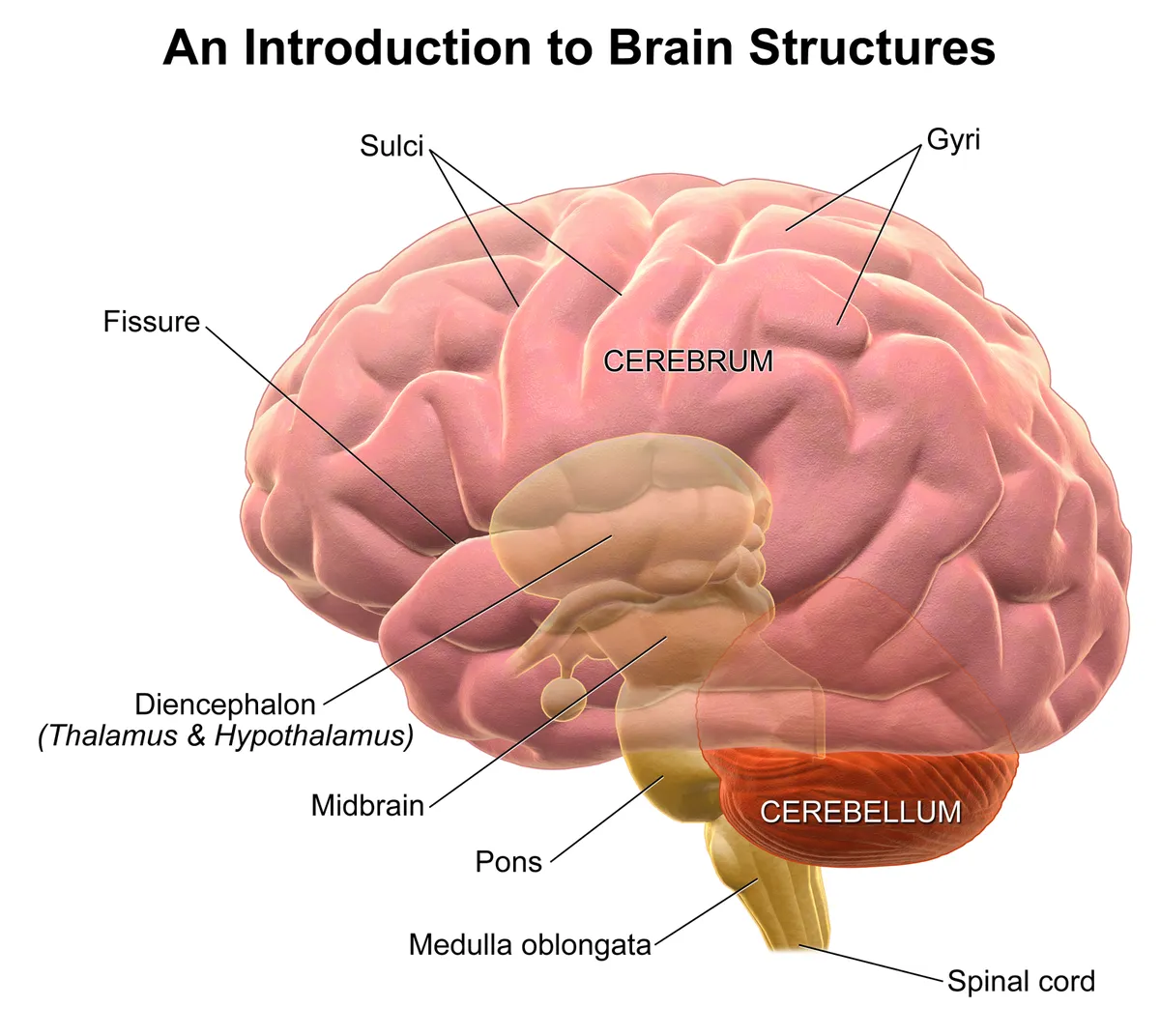
The part of the central nervous system enclosed in the cranium, comprising the cerebrum, diencephalon, cerebellum and brainstem. Mean adult mass is about 1.3–1.4 kg; the brain accounts for roughly 20% of resting energy expenditure.
Spinal cordMedulla spinalisLies within the vertebral canal, about 42–45 cm long, extending from the foramen magnum to the level of the L1–L2 vertebrae. It conducts signals between the brain and the periphery and integrates spinal reflexes.
Meninges and ventriclesMeninges et ventriculi encephaliThree membranes (dura, arachnoid and pia mater) wrap the brain and spinal cord; dural folds form the falx cerebri and tentorium cerebelli. The ventricular system (two lateral ventricles, the third ventricle, the cerebral aqueduct and the fourth ventricle) holds about 150 ml of cerebrospinal fluid secreted by the choroid plexus.
Cerebral arteriesArteriae encephaliThe brain is supplied by two systems: the internal carotid arteries (anterior circulation) and the vertebrobasilar system (posterior circulation). They anastomose in the cerebral arterial circle (of Willis) at the base of the brain, which gives off the anterior, middle and posterior cerebral arteries.
Cranial nervesNervi cranialesTwelve pairs of nerves that arise directly from the brain, numbered I–XII from front to back. They carry the special senses (smell, vision, hearing, taste), supply the muscles of the eye, face, tongue, pharynx and larynx, and convey parasympathetic fibres (III, VII, IX, X).
Spinal nerves and plexusesNervi spinales et plexusThirty-one pairs of spinal nerves (8 cervical, 12 thoracic, 5 lumbar, 5 sacral, 1 coccygeal), each formed by an anterior (motor) and a posterior (sensory, with the spinal ganglion) root. The anterior rami of the cervical, lumbar and sacral nerves interweave into the cervical, brachial, lumbar and sacral plexuses that supply the neck and limbs.
Autonomic nervous systemPars autonomica systematis nervosi periphericiThe division of the nervous system that controls smooth muscle, cardiac muscle and glands involuntarily. The sympathetic part arises from the thoracolumbar cord (T1–L2) through the paravertebral sympathetic trunk; the parasympathetic part arises from the brainstem (nerves III, VII, IX, X) and the sacral cord (S2–S4).
EyeOculus et structurae pertinentesThe organ of vision: the eyeball within the orbit and its accessory structures, namely six extra-ocular muscles, the eyelids, conjunctiva and lacrimal apparatus. The eyeball, about 24 mm across, has three coats (fibrous, vascular and neural) enclosing the transparent media: aqueous humour, lens and vitreous body.
EarAurisThe organ of hearing and balance: the external ear (auricle, external acoustic meatus), the middle ear (tympanic cavity with the tympanic membrane and three ossicles) and the internal ear in the petrous temporal bone (the cochlea for hearing; the vestibule and three semicircular canals for balance).
Reference values
- The human brain contains approximately 86 billion neurons.
- Large myelinated axons conduct impulses at velocities of up to about 120 m/s.
References
Sources for the values and descriptions on this page, indexed in PubMed (US National Library of Medicine).
- [1]AstrocytesHasel P et al., Current biology 2021PMID 33848482
- [2]The neurovascular unit - concept reviewMuoio V et al., Acta physiologica 2014PMID 24629161
- [3]Basic anatomy and physiology of pain pathwaysBourne S et al., Neurosurgery clinics of North America 2014PMID 25240653
- [4]Cranial Nerve AnatomyTraylor KS et al., Neuroimaging clinics of North America 2022PMID 35843663
- [5]Neuroanatomy, Cerebral CortexJaved K et al., StatPearls 2023PMID 30725932
- [6]Neuroanatomy, Spinal CordKhan YS et al., StatPearls 2023PMID 32644482
- [7]Anatomy, Autonomic Nervous SystemWaxenbaum JA et al., StatPearls 2025PMID 30969667
- [8]Anatomy, Head and Neck: Blood Brain BarrierDotiwala AK et al., StatPearls 2023PMID 30137840
- [9]The human brain in numbers: a linearly scaled-up primate brainHerculano-Houzel S, 2009PMID 19915731Supports: Neuron count
- [10]Determinants of conduction velocity in myelinated nerve fibersWaxman SG, 1980PMID 6245357Supports: Conduction velocity
- [11]Quantitative imaging of energy expenditure in human brainPMID 22487547Supports: Cerebral energy expenditure