Vertebral column
Model
Hover or tap to see names, click to read a description; click a name in the legend to fly to it. Procedurally built to histological proportions; colours follow illustration conventions.
Description
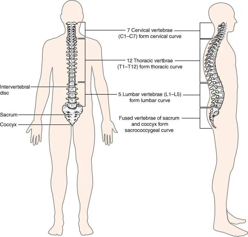
The axial skeleton of the trunk, made of 33–34 vertebrae (7 cervical, 12 thoracic, 5 lumbar, the sacrum and coccyx) joined by intervertebral discs and ligaments. It supports the head and trunk, allows movement and protects the spinal cord.
Parts
Each part has its own card; press “Show on the model” to fly to it and select it in the 3D model above.
- 01
Cervical vertebrae (C1–C7)
Vertebrae cervicales
The seven smallest vertebrae, with foramina in the transverse processes for the vertebral artery (C1–C6) and bifid spinous processes. C7 has the long, easily felt spinous process (vertebra prominens). The cervical segment is lordotic and the most mobile, hence vulnerable to whiplash.
- 02
Atlas and axis
Atlas et axis
The atlas (C1) is a ring with no body that carries the skull at the atlanto-occipital joints (nodding). The axis (C2) has the dens, projecting into the ring of C1 and held by the transverse ligament, forming the atlanto-axial joint that provides about half of head rotation. A fractured dens or torn transverse ligament can compress the medulla.
- 03
Thoracic vertebrae (T1–T12)
Vertebrae thoracicae
Twelve vertebrae with costal facets on the bodies and transverse processes and long, downward-sloping overlapping spinous processes. The thoracic segment is kyphotic and stiffened by the rib cage. The canal is narrowest here, so injuries more often cause complete cord lesions.
- 04
Lumbar vertebrae (L1–L5)
Vertebrae lumbales
The five largest vertebrae, with kidney-shaped weight-bearing bodies and square horizontal spinous processes. The lumbar segment is lordotic and flexes and extends freely. The adult cord ends at L1–L2, so lumbar puncture is done at the L3–L4 or L4–L5 interspace. The L4–L5 and L5–S1 discs are the most common sites of herniation.
- 05
Sacrum
Os sacrum
Five fused sacral vertebrae forming a triangular block that articulates on each side with the hip bone at the sacro-iliac joint, transferring the weight of the trunk to the lower limbs. Its pelvic surface has four pairs of anterior sacral foramina for the ventral rami; the sacral canal opens below at the sacral hiatus, the route for caudal epidural anaesthesia.
- 06
Coccyx
Os coccygis
Three to five small fused coccygeal vertebrae, the remnant of a tail. It anchors levator ani, gluteus maximus and the anococcygeal ligament; a fall on the buttocks can cause persistent coccygeal pain.
- 07
Intervertebral discs
Disci intervertebrales
Twenty-three fibrocartilaginous discs join the vertebral bodies from C2 to S1 and make up about a quarter of the column’s length. Each has a tough outer annulus fibrosus and a water-rich nucleus pulposus acting as a hydraulic cushion. The annulus is thinnest posterolaterally, where the nucleus usually herniates and compresses a nerve root.
- 08
Curvatures
Curvaturae columnae vertebralis
Seen from the side, the adult column has four curves: cervical lordosis, thoracic kyphosis, lumbar lordosis and sacrococcygeal kyphosis. The thoracic and sacral curves are primary (present in the fetus); the cervical curve appears when the infant holds up the head and the lumbar curve with walking. The curves add resistance to axial load and absorb shocks. A lateral curvature is scoliosis.
Position and relations
In profile the adult spine has four curves: cervical and lumbar lordoses (secondary, appearing as the infant holds up its head and stands) and thoracic and sacral kyphoses (primary). The atlas (C1) and axis (C2) form joints for nodding and rotation of the head; the vertebral canal contains the spinal cord down to about L1–L2, below which lies the cauda equina.
Form and structure
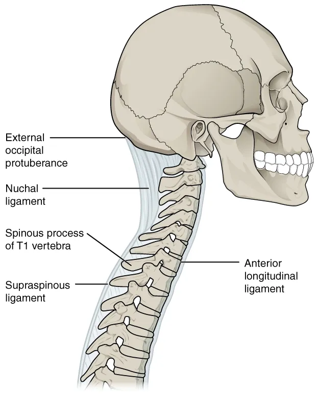
A typical vertebra has a body in front and an arch behind (pedicles, laminae, spinous process, two transverse processes and four articular processes) around the vertebral foramen. Each disc has a central nucleus pulposus and a surrounding annulus fibrosus; herniation usually occurs posterolaterally and compresses a nerve root. The anterior and posterior longitudinal ligaments, ligamenta flava, interspinous and supraspinous ligaments stabilise the column.
Vessels and nerves
Vertebrae are supplied by posterior intercostal, lumbar, vertebral and ascending cervical branches; the valveless vertebral venous plexus (Batson) communicates with pelvic veins, explaining spinal metastasis from prostate and breast cancer. Spinal nerves leave through the intervertebral foramina between adjacent pedicles.
Vertebrae and intervertebral discs
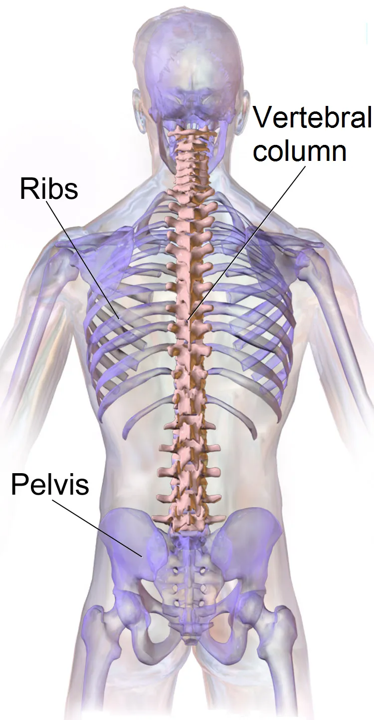
The vertebral column supports the body and the nervous system, enabling movement and sensation. In humans it has 33 vertebrae: 7 cervical, 12 thoracic, 5 lumbar, 5 sacral and 4 coccygeal, which together with the skull, ribs and sternum form the axial skeleton. It is the defining feature of the subphylum Vertebrata.[1]
Intervertebral discs link the vertebral bodies. Each has a proteoglycan-rich nucleus pulposus, a collagen-rich lamellar annulus fibrosus and cartilaginous endplates, providing support and shock absorption. The disc is nearly avascular: blood reaches only the endplates, and the nerve supply comes mainly from the sinuvertebral nerve. Its key constituents are collagen, elastin and aggrecan.[2][3]
With ageing the nucleus loses osmotic pressure, dehydrates and the disc loses height; nociceptive nuclear material leaks through the outer annulus, the main source of discogenic pain. The lowered disc alters the ligamentum flavum, facet joints and foraminal shape, causing spinal stenosis and radicular pain in older people. Disc degeneration has been defined as an aberrant cell-mediated response to progressive structural failure; defects such as endplate fracture, radial fissures and herniation are not inevitable with age, relate to pain more closely than any other ageing feature and are irreversible because adult discs heal poorly.[2][4]
Full-text reading (StatPearls)
The StatPearls chapters on this topic, reproduced verbatim. Their CC BY-NC-ND 4.0 licence allows unmodified copies only, so they are not translated or shortened.
Anatomy, Back, Vertebral ColumnDeSai C, Reddy V, Agarwal A · StatPearls 2023 · NBK525969 · 12k characters
Loading…
DeSai C, Reddy V, Agarwal A. Anatomy, Back, Vertebral Column. StatPearls [Internet]. Treasure Island (FL): StatPearls Publishing; 2023. NBK525969 · PMID 30247844 · CC BY-NC-ND 4.0. Reproduced verbatim, unmodified.
Organs in this system
References
Sources for the values and descriptions on this page, indexed in PubMed (US National Library of Medicine).
- [1]Anatomy, Back, Vertebral ColumnDeSai C et al., StatPearls 2023PMID 30247844
- [2]Intervertebral disc: anatomy-physiology-pathophysiology-treatmentRaj PP, Pain practice 2008PMID 18211591
- [3]Discogenic Low Back Pain: Anatomy, Pathophysiology and Treatments of Intervertebral Disc DegenerationMohd Isa IL et al., International journal of molecular sciences 2022PMID 36613651
- [4]What is intervertebral disc degeneration, and what causes it?Adams MA et al., Spine 2006PMID 16915105
- [5]Anatomy, BonesStatPearls (NCBI Bookshelf)PMID 30725884Supports: Number of bones
- [6]Calcium metabolism in health and diseasePeacock M, 2010PMID 20089499Supports: Skeletal calcium store