Back
Model
Layers
Z-Anatomy anatomical model based on BodyParts3D, licensed CC BY-SA 4.0.
Overview
A regional model of the back from the nape to the sacrum: the superficial muscles linking the upper limb to the spine (trapezius, latissimus dorsi, rhomboids, levator scapulae), the intermediate serratus posterior muscles, the deep intrinsic back muscles (erector spinae, transversospinales, suboccipital muscles), the three-layered thoracolumbar fascia, the vertebral column with its discs and ligaments, the spinal cord, the posterior rami of spinal nerves and the posterior intercostal and lumbar arteries.
Boundaries and subdivisions
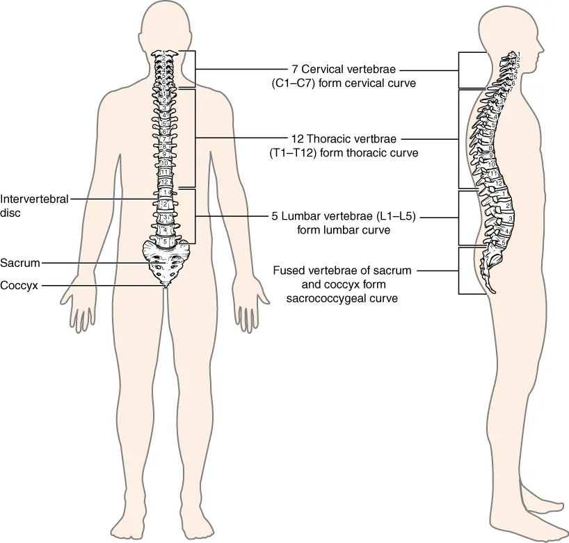
The back extends from the superior nuchal line down to the iliac crests and the sacrum, bounded laterally by the medial borders of the scapulae and the posterior axillary lines. Its axis is the vertebral column of 33 vertebrae: 7 cervical, 12 thoracic, 5 lumbar, 5 sacral (fused into the sacrum) and 4 coccygeal. In the adult it shows four curves: cervical lordosis, thoracic kyphosis, lumbar lordosis and the sacral curve. On the surface the spinous processes serve as levels: the C7 spine is the most prominent at the nape, the line joining the iliac crests crosses about L4, and the inferior angle of the scapula lies at about T7.[1]
Layers from superficial to deep
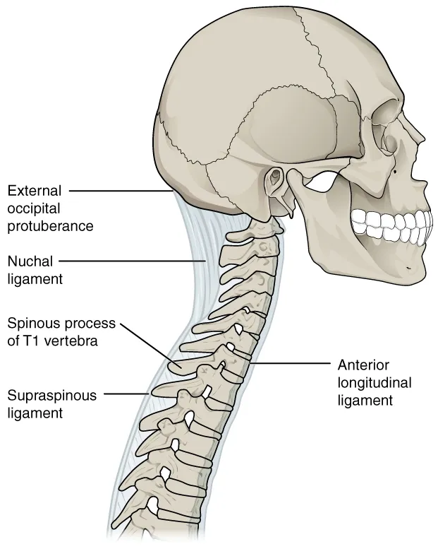
The back muscles lie in three layers. The superficial layer links the upper limb to the spine: trapezius, latissimus dorsi, levator scapulae and the two rhomboids, supplied by anterior rami of spinal nerves (and nerve XI for trapezius). The intermediate layer is serratus posterior superior and inferior, attaching to the ribs. The deep layer consists of the intrinsic back muscles supplied by posterior rami: the splenius muscles, erector spinae with its three columns iliocostalis, longissimus and spinalis, the transversospinales (semispinalis, multifidus, rotatores) and the small interspinales and intertransversarii. Erector spinae extends the spine and maintains its curves. The thoracolumbar fascia wraps the deep muscles in three layers (anterior, middle, posterior) and gives attachment to latissimus dorsi and transversus abdominis. Deepest lies the vertebral column with its intervertebral discs (annulus fibrosus around the nucleus pulposus), the anterior and posterior longitudinal ligaments and ligamenta flava, and the spinal cord in the vertebral canal.[1][2][3][4]
Vessels, nerves and lymphatics
Each spinal nerve divides into anterior and posterior rami; the posterior rami run backwards to supply the deep muscles and the skin of the back segment by segment. The back muscles and skin are supplied by the posterior branches of the posterior intercostal and lumbar arteries arising from the aorta. The spinal cord receives one anterior and two posterior spinal arteries, reinforced by radiculomedullary arteries; the largest is the artery of Adamkiewicz, which supplies the lumbar cord. The cord ends at the conus medullaris, usually at L1–L2; below this level the subarachnoid space holds only the cauda equina in the lumbar cistern. The vertebral veins form valveless internal and external vertebral venous plexuses.[5][6][7]
Embryology
Paraxial mesoderm segments into somites under a “segmentation clock” of oscillating Notch, Wnt and FGF signalling. Each somite splits into a sclerotome, which forms vertebrae and ribs, and a dermomyotome, which forms skeletal muscle and dermis. Sclerotome cells migrate around the notochord and neural tube and resegment: the caudal half of one sclerotome joins the cranial half of the next to form a vertebral body, so spinal nerves exit between vertebrae and the back muscles bridge the joints. The intervertebral disc has two origins: the nucleus pulposus from the notochord, the annulus fibrosus and cartilaginous endplates from sclerotome. Vertebrae begin to ossify at about 8 weeks from three primary centres (the centrum and two neural-arch halves), which fuse between 3 and 6 years; five secondary centres (spinous tip, both transverse tips, both vertebral body surfaces) appear at puberty. Errors in neural tube closure produce defects ranging from spina bifida to anencephaly.[8][9][10][11]
Anatomical variants
Vertebral and rib counts are not fixed: about 3.4% of people have only 11 rib pairs, and thoracolumbar transitional vertebrae occur in about 11%, so counting ribs is not a reliable way to number levels. Lumbosacral transitional vertebrae (sacralisation of L5 or lumbarisation of S1) are common and graded by the Castellvi classification; when they cause low back pain the picture is called Bertolotti syndrome. Bifid spinous processes are consistent at C2–C4 but almost never at C7 (about 0.3%), so spinous process shape alone cannot identify the level during surgery. The artery of Adamkiewicz enters the dura at T9–T12 in 75% of cases, T5–T8 in 15% and L1–L2 in 10%; in an “Adamkiewicz-dominant” circulation, its injury causes severe paraplegia. The left vertebral artery arises directly from the aortic arch in 2–5% of people.[8][9][12][10][13]
Clinical correlations
Disc herniation occurs when the nucleus pulposus protrudes beyond the annulus fibrosus, most often in the lumbar and cervical spine, compressing nerve roots and causing pain, numbness and weakness in the corresponding limb. Lumbar puncture is performed in the lumbar cistern below the conus medullaris (usually at the L3–L4 or L4–L5 interspace) to sample cerebrospinal fluid for meningitis or subarachnoid haemorrhage, or to measure opening pressure. In thoracoabdominal aortic surgery, damage to the artery of Adamkiewicz can cause spinal cord ischaemia and paraplegia. Spinal tuberculosis may present with back pain, kyphosis and leg weakness.[2][6][5][1]
Surgical and procedural considerations
The transverse foramina of the cervical vertebrae carry the vertebral artery, so a misdirected lateral mass or C1–C2 transarticular screw can cause catastrophic bleeding and posterior-circulation stroke; because the artery is variable in origin, dominance and course, preoperative 3D CT angiography is mandatory when instrumentation approaches the foramen. In the thoracic spine the canal is narrow, the pedicles slim, the aorta lies against the left side from T3 to T7, the sympathetic chain runs from T1 to T12 and the azygos vein crosses at T4–T5, so the cord tolerates retraction and rotation poorly. Surgery for disc herniation requires clinicoradiological concordance, because degenerative discs are common in asymptomatic adults; cauda equina syndrome and progressive motor deficit are urgent indications. Lumbar operations include laminectomy, microdiscectomy and interbody fusion by posterior, transforaminal, lateral, anterior or oblique approaches.[10][13][8][9][12]
Key structures
M. trapezius
- Name
- Trapezius
- Position, role
- Superficial layer; elevates, retracts and rotates the scapula; nerve XI
M. latissimus dorsi
- Name
- Latissimus dorsi
- Position, role
- From the thoracolumbar fascia to the intertubercular groove; thoracodorsal nerve
M. erector spinae
- Name
- Erector spinae
- Position, role
- Iliocostalis, longissimus and spinalis columns; extends the spine
Mm. transversospinales
- Name
- Transversospinales
- Position, role
- Semispinalis, multifidus, rotatores; segmental stabilisers
Mm. suboccipitales
- Name
- Suboccipital muscles
- Position, role
- Bound the suboccipital triangle with the vertebral artery
Fascia thoracolumbalis
- Name
- Thoracolumbar fascia
- Position, role
- Three layers enclosing the deep muscles; attachment for latissimus and transversus
Discus intervertebralis
- Name
- Intervertebral disc
- Position, role
- Annulus fibrosus around the nucleus pulposus; shock absorber between bodies
Conus medullaris
- Name
- Conus medullaris
- Position, role
- Lower end of the spinal cord, usually at L1–L2
| Terminologia Anatomica | Name | Position, role |
|---|---|---|
| M. trapezius | Trapezius | Superficial layer; elevates, retracts and rotates the scapula; nerve XI |
| M. latissimus dorsi | Latissimus dorsi | From the thoracolumbar fascia to the intertubercular groove; thoracodorsal nerve |
| M. erector spinae | Erector spinae | Iliocostalis, longissimus and spinalis columns; extends the spine |
| Mm. transversospinales | Transversospinales | Semispinalis, multifidus, rotatores; segmental stabilisers |
| Mm. suboccipitales | Suboccipital muscles | Bound the suboccipital triangle with the vertebral artery |
| Fascia thoracolumbalis | Thoracolumbar fascia | Three layers enclosing the deep muscles; attachment for latissimus and transversus |
| Discus intervertebralis | Intervertebral disc | Annulus fibrosus around the nucleus pulposus; shock absorber between bodies |
| Conus medullaris | Conus medullaris | Lower end of the spinal cord, usually at L1–L2 |
What each layer contains
SkinCutis7
- Interscapular region
- Infrascapular region
- Lumbar region
- Sacral region
- Scapular region
- Vertebral region
- Triangle of auscultation
Muscles, fasciae and tendon sheathsMusculi, fasciae et vaginae tendinum69
- Sciatic bursa of obturator internus
- Subacromial bursa
- Subdeltoid bursa
- Subtendinous bursa of infraspinatus muscle
- Subtendinous bursa of teres major muscle
- Subtendinous bursa of trapezius muscle
- Subtendinous bursa of obturator internus
- Long head of triceps brachii
- Deltoid fascia
- Superficial investing cervical fascia
- Piriformis fascia
- Anterior layer of thoracolumbar fascia
- Middle layer of thoracolumbar fascia
- Posterior layer of thoracolumbar fascia
- Levator scapulae
- Levatores breves costarum
- Levatores longi costarum
- Interspinales colli muscles
- Interspinales lumborum muscles
- Interspinales thoracis muscles
- Coccygeus muscle
- Inferior gemellus muscle
- Superior gemellus muscle
- Gluteus maximus muscle
- Gluteus medius muscle
- Iliococcygeus muscle
- Iliocostalis colli muscle
- Iliocostalis lumborum muscle
- Iliocostalis thoracis muscle
- Infraspinatus muscle
- Latissimus dorsi muscle
- Longissimus capitis muscle
- Longissimus colli muscle
- Longissimus thoracis muscle
- Multifidus colli muscle
- Multifidus lumborum muscle
- Multifidus thoracis muscle
- Obliquus inferior capitis muscle
- Obliquus superior capitis muscle
- Piriformis muscle
- Quadratus lumborum muscle
- Rectus posterior major capitis muscle
- Rectus posterior minor capitis muscle
- Rhomboid major muscle
- Rhomboid minor muscle
- Semispinalis colli muscle
- Semispinalis thoracis muscle
- Serratus posterior inferior muscle
- Serratus posterior superior muscle
- Spinalis capitis muscle
- Spinalis colli muscle
- Spinalis thoracis muscle
- Splenius capitis muscle
- Splenius colli muscle
- Subscapularis muscle
- Supraspinatus muscle
- Teres major muscle
- Teres minor muscle
- Acromial part of deltoid muscle
- Ascending part of trapezius muscle
- Descending part of trapezius muscle
- Scapular spinal part of deltoid muscle
- Transverse part of trapezius muscle
- Dorsal parts of lateral intertransversarii lumborum muscles
- Ventral parts of lateral intertransversarii lumborum muscles
- Rotatores
- Medial intermuscular septum of arm
- Subcutaneous acromial bursa
- Trochanteric bursa of gluteus medius muscle
Bones and cartilageOssa et cartilagines39
- Atlas (C1)
- Axis (C2)
- Tenth rib
- Twelfth rib
- Humerus
- Ninth rib
- Eighth rib
- Coccyx
- Hip bone
- Sacrum
- Fourth rib
- Fifth rib
- Scapula
- Seventh rib
- Sixth rib
- Third rib
- Eleventh rib
- Vertebra C3
- Vertebra C4
- Vertebra C5
- Vertebra C6
- Vertebra C7
- Vertebra L1
- Vertebra L2
- Vertebra L3
- Vertebra L4
- Vertebra L5
- Vertebra T1
- Vertebra T2
- Vertebra T3
- Vertebra T4
- Vertebra T9
- Vertebra T5
- Vertebra T6
- Vertebra T7
- Vertebra T8
- Vertebra T10
- Vertebra T11
- Vertebra T12
Joints and ligamentsJuncturae et ligamenta52
- Articular capsule of acromioclavicular joint
- Articular capsule of glenohumeral joint
- Articular disc of acromioclavicular joint
- Intervertebral disc C7-T1
- Intervertebral disc T1-T2
- Intervertebral disc T10-T11
- Intervertebral disc T11-T12
- Intervertebral disc T2-T3
- Intervertebral disc T3-T4
- Intervertebral disc T4-T5
- Intervertebral disc T5-T6
- Intervertebral disc T6-T7
- Intervertebral disc T7-T8
- Intervertebral disc T8-T9
- Intervertebral disc T9-T10
- Glenoid labrum
- Ligamenta flava
- Interspinous ligaments
- Intertransverse ligaments
- Acromioclavicular ligament
- Conoid ligament
- Coraco-acromial ligament
- Costotransverse ligament
- Superior glenohumeral ligament
- Iliolumbar ligament
- Intercornual ligament
- Intra-articular ligament of head of rib
- Ischiofemoral ligament
- Posterior longitudinal ligament
- Nuchal ligament
- Radiate ligament of head of rib
- Anterior sacro-iliac ligament
- Interosseous sacro-iliac ligament
- Posterior sacro-iliac ligament
- Sacrospinous ligament
- Sacrotuberous ligament
- Supraspinous ligament
- Inferior transverse scapular ligament
- Superior transverse scapular ligament
- Nucleus pulposus C7-T1
- Nucleus pulposus T1-T2
- Nucleus pulposus T10-T11
- Nucleus pulposus T11-T12
- Nucleus pulposus T2-T3
- Nucleus pulposus T3-T4
- Nucleus pulposus T4-T5
- Nucleus pulposus T5-T6
- Nucleus pulposus T6-T7
- Nucleus pulposus T7-T8
- Nucleus pulposus T8-T9
- Nucleus pulposus T9-T10
- Sacrococcygeal symphysis
ArteriesArteriae34
- (Deep branch of transverse cervical artery)
- (Superficial branch of transverse cervical artery)
- Thoracic aorta
- Deep cervical artery
- Posterior circumflex humeral artery
- Circumflex scapular artery
- Superior ulnar collateral artery
- Inferior gluteal artery
- Superior gluteal artery
- Iliolumbar artery
- Inferior lobar artery of right lung
- Deep brachial artery
- Internal pudendal artery
- Left pulmonary artery
- Median sacral artery
- Apical segmental artery of right lung
- Apical segmental artery of left lung
- Anterior basal segmental artery of right lung
- Anterior basal segmental artery of left lung
- Medial basal segmental artery of right lung
- Medial basal segmental artery of left lung
- Posterior basal segmental artery of right lung
- Posterior basal segmental artery of left lung
- Posterior segmental artery of right lung
- Posterior segmental artery of left lung
- Superior segmental artery of right lung
- Superior segmental artery of left lung
- Anterior spinal artery
- Subcostal artery
- Suprascapular artery
- Thoracodorsal artery
- Posterior intercostal arteries
- Lumbar arteries
- Spinal branch of iliolumbar artery
VeinsVenae21
- Inferior basal vein of right lung
- Inferior basal vein of left lung
- Superior basal vein of right lung
- Superior basal vein of left lung
- Circumflex scapular vein
- Hemi-azygos vein
- Accessory hemi-azygos vein
- Right superior intercostal vein
- Left superior intercostal vein
- Posterior vein of right lung
- Right inferior pulmonary vein
- Left inferior pulmonary vein
- Right subcostal vein
- Left subcostal vein
- Superior vein of right lung
- Superior vein of left lung
- Suprascapular vein
- Inferior gluteal veins
- Superior gluteal veins
- Lumbar veins
- Lateral sacral veins
NervesNervi65
- Central canal
- Cauda equina
- Central canal'
- Anterior horn of spinal cord
- Posterior horn of spinal cord
- Anterior division of middle trunk of brachial plexus
- Anterior division of superior trunk of brachial plexus
- Posterior division of middle trunk of brachial plexus
- Posterior division of superior trunk of brachial plexus
- Cuneate fasciculus
- Gracile fasciculus
- Posterior cord of brachial plexus
- Posterolateral tract
- Anterior fasciculus proprius
- Lateral fasciculus proprius
- Posterior fasciculus proprius
- Ganglia of sympathetic trunk
- Spinal ganglion
- Intercostal nerves
- Sympathetic nerves
- Accessory nerve (XI)
- Axillary nerve
- Lateral femoral cutaneous nerve
- Superior lateral brachial cutaneous nerve
- Medial brachial cutaneous nerve
- Dorsal scapular nerve
- Superior gluteal nerve
- Nerve to piriformis muscle
- Nerve to quadratus femoris muscle
- Pudendal nerve
- Radial nerve
- Inferior subscapular nerve
- Superior subscapular nerve
- Suprascapular nerve
- Long thoracic nerve
- Thoracodorsal nerve
- Intermediolateral nucleus
- Intermediomedial nucleus
- Nucleus of accessory nerve
- Nucleus proprius
- Spinal reticular process
- Roots of brachial plexus
- Anterior root of posterior femoral cutaneous nerve
- Anterior root of spinal nerve
- Posterior root of posterior femoral cutaneous nerve
- Posterior root of spinal nerve
- Muscular branches of axillary nerve
- White matter of spinal cord
- Lateral intermediate substance
- Anterior corticospinal tract
- Lateral corticospinal tract
- Lateral reticulospinal tract
- Medial reticulospinal tract
- Rubrospinal tract
- Anterior spinocerebellar tract
- Posterior spinocerebellar tract
- Spinotectal tract
- Anterior spinothalamic tract
- Lateral spinothalamic tract
- Tectospinal tract
- Lateral vestibulospinal tract
- Medial vestibulospinal tract
- Middle trunk of brachial plexus
- Superior trunk of brachial plexus
- Sympathetic trunk
Full-text reading (StatPearls)
The StatPearls chapters on this topic, reproduced verbatim. Their CC BY-NC-ND 4.0 licence allows unmodified copies only, so they are not translated or shortened.
Anatomy, Back, Cervical VertebraeWaxenbaum JA, Reddy V, Margetis K · StatPearls 2026 · NBK459200 · 26k characters
Loading…
Waxenbaum JA, Reddy V, Margetis K. Anatomy, Back, Cervical Vertebrae. StatPearls [Internet]. Treasure Island (FL): StatPearls Publishing; 2026. NBK459200 · PMID 29083805 · CC BY-NC-ND 4.0. Reproduced verbatim, unmodified.
Anatomy, Back, Thoracic VertebraeWaxenbaum JA, Reddy V, Margetis K · StatPearls 2026 · NBK459153 · 21k characters
Loading…
Waxenbaum JA, Reddy V, Margetis K. Anatomy, Back, Thoracic Vertebrae. StatPearls [Internet]. Treasure Island (FL): StatPearls Publishing; 2026. NBK459153 · PMID 29083651 · CC BY-NC-ND 4.0. Reproduced verbatim, unmodified.
Anatomy, Back, Intervertebral DiscsWaxenbaum JA, Reddy V, Futterman B · StatPearls 2026 · NBK470583 · 20k characters
Loading…
Waxenbaum JA, Reddy V, Futterman B. Anatomy, Back, Intervertebral Discs. StatPearls [Internet]. Treasure Island (FL): StatPearls Publishing; 2026. NBK470583 · PMID 29262063 · CC BY-NC-ND 4.0. Reproduced verbatim, unmodified.
Anatomy, Back, Lumbar SpineSassack B, Carrier JD · StatPearls 2023 · NBK557616 · 20k characters
Loading…
Sassack B, Carrier JD. Anatomy, Back, Lumbar Spine. StatPearls [Internet]. Treasure Island (FL): StatPearls Publishing; 2023. NBK557616 · PMID 32491548 · CC BY-NC-ND 4.0. Reproduced verbatim, unmodified.
Anatomy, Back, Vertebral Canal Blood SupplyHu Y, Jozsa F, Singh P · StatPearls 2026 · NBK541083 · 16k characters
Loading…
Hu Y, Jozsa F, Singh P. Anatomy, Back, Vertebral Canal Blood Supply. StatPearls [Internet]. Treasure Island (FL): StatPearls Publishing; 2026. NBK541083 · PMID 31082127 · CC BY-NC-ND 4.0. Reproduced verbatim, unmodified.
Anatomy, Back, Lumbar VertebraeWaxenbaum JA, Reddy V, Williams C, Futterman B · StatPearls 2026 · NBK459278 · 14k characters
Loading…
Waxenbaum JA, Reddy V, Williams C, Futterman B. Anatomy, Back, Lumbar Vertebrae. StatPearls [Internet]. Treasure Island (FL): StatPearls Publishing; 2026. NBK459278 · PMID 29083618 · CC BY-NC-ND 4.0. Reproduced verbatim, unmodified.
Anatomy, Head and Neck, Suboccipital MusclesGeorge T, Tadi P · StatPearls 2023 · NBK567762 · 13k characters
Loading…
George T, Tadi P. Anatomy, Head and Neck, Suboccipital Muscles. StatPearls [Internet]. Treasure Island (FL): StatPearls Publishing; 2023. NBK567762 · PMID 33620839 · CC BY-NC-ND 4.0. Reproduced verbatim, unmodified.
Anatomy, Back, TrapeziusOurieff J, Scheckel B, Agarwal A · StatPearls 2023 · NBK518994 · 12k characters
Loading…
Ourieff J, Scheckel B, Agarwal A. Anatomy, Back, Trapezius. StatPearls [Internet]. Treasure Island (FL): StatPearls Publishing; 2023. NBK518994 · PMID 30085536 · CC BY-NC-ND 4.0. Reproduced verbatim, unmodified.
Anatomy, Back, Splanchnic NerveMcCausland C, Carey FJ, Sajjad H · StatPearls 2024 · NBK549856 · 12k characters
Loading…
McCausland C, Carey FJ, Sajjad H. Anatomy, Back, Splanchnic Nerve. StatPearls [Internet]. Treasure Island (FL): StatPearls Publishing; 2024. NBK549856 · PMID 31751046 · CC BY-NC-ND 4.0. Reproduced verbatim, unmodified.
Anatomy, Back, Extrinsic MusclesMitchell B, Imonugo O, Tripp JE · StatPearls 2024 · NBK537216 · 10k characters
Loading…
Mitchell B, Imonugo O, Tripp JE. Anatomy, Back, Extrinsic Muscles. StatPearls [Internet]. Treasure Island (FL): StatPearls Publishing; 2024. NBK537216 · PMID 30725901 · CC BY-NC-ND 4.0. Reproduced verbatim, unmodified.
Anatomy, Back, Lumbar PlexusSingh O, Al Khalili Y · StatPearls 2023 · NBK545137 · 9k characters
Loading…
Singh O, Al Khalili Y. Anatomy, Back, Lumbar Plexus. StatPearls [Internet]. Treasure Island (FL): StatPearls Publishing; 2023. NBK545137 · PMID 31424721 · CC BY-NC-ND 4.0. Reproduced verbatim, unmodified.
Anatomy, Back, Artery of AdamkiewiczLindeire S, Hauser JM · StatPearls 2025 · NBK532971 · 9k characters
Loading…
Lindeire S, Hauser JM. Anatomy, Back, Artery of Adamkiewicz. StatPearls [Internet]. Treasure Island (FL): StatPearls Publishing; 2025. NBK532971 · PMID 30422566 · CC BY-NC-ND 4.0. Reproduced verbatim, unmodified.
Anatomy, Back, Cauda EquinaBerg EJ, Ashurst JV · StatPearls 2023 · NBK513251 · 9k characters
Loading…
Berg EJ, Ashurst JV. Anatomy, Back, Cauda Equina. StatPearls [Internet]. Treasure Island (FL): StatPearls Publishing; 2023. NBK513251 · PMID 30020623 · CC BY-NC-ND 4.0. Reproduced verbatim, unmodified.
Anatomy, Back, Latissimus DorsiJeno SH, Varacallo MA · StatPearls 2023 · NBK448120 · 8k characters
Loading…
Jeno SH, Varacallo MA. Anatomy, Back, Latissimus Dorsi. StatPearls [Internet]. Treasure Island (FL): StatPearls Publishing; 2023. NBK448120 · PMID 28846224 · CC BY-NC-ND 4.0. Reproduced verbatim, unmodified.
Anatomy, Back, MusclesHenson B, Kadiyala B, Edens MA · StatPearls 2023 · NBK537074 · 8k characters
Loading…
Henson B, Kadiyala B, Edens MA. Anatomy, Back, Muscles. StatPearls [Internet]. Treasure Island (FL): StatPearls Publishing; 2023. NBK537074 · PMID 30725759 · CC BY-NC-ND 4.0. Reproduced verbatim, unmodified.
Regional anatomy
References
Sources for the values and descriptions on this page, indexed in PubMed (US National Library of Medicine).
- [1]Anatomy, Back, Vertebral ColumnDeSai C et al., StatPearls 2023PMID 30247844
- [2]Anatomy, Back, Nucleus PulposusNedresky D et al., StatPearls 2023PMID 30570994
- [3]Anatomy, Head and Neck, Suboccipital MusclesGeorge T et al., StatPearls 2023PMID 33620839
- [4]Anatomy, Back, TrapeziusOurieff J et al., StatPearls 2023PMID 30085536
- [5]Anatomy, Back, Spinal Cord ArteriesKaiser JT et al., StatPearls 2023PMID 30725904
- [6]Anatomy, Back, Spinal MeningesSehgal I et al., StatPearls 2023PMID 31613535
- [7]Anatomy, Back, Artery of AdamkiewiczLindeire S et al., StatPearls 2025PMID 30422566
- [8]Anatomy, Back, Thoracic VertebraeWaxenbaum JA et al., StatPearls 2026PMID 29083651
- [9]Anatomy, Back, Intervertebral DiscsWaxenbaum JA et al., StatPearls 2026PMID 29262063
- [10]Anatomy, Back, Cervical VertebraeWaxenbaum JA et al., StatPearls 2026PMID 29083805
- [11]Anatomy, Back, Lumbar SpineSassack B et al., StatPearls 2023PMID 32491548
- [12]Anatomy, Back, Lumbar VertebraeWaxenbaum JA et al., StatPearls 2026PMID 29083618
- [13]Anatomy, Back, Vertebral Canal Blood SupplyHu Y et al., StatPearls 2026PMID 31082127