Nose and nasal cavity
Model
Layers
Z-Anatomy anatomical model based on BodyParts3D, licensed CC BY-SA 4.0.
Description
The first part of the airway, comprising the external nose and the nasal cavity divided by the septum. The cavity warms, humidifies and filters inspired air, carries the olfactory mucosa and communicates with the paranasal sinuses.
Parts
Each part has its own card; press “Show on the model” to fly to it and select it in the 3D model above.
- 01
External nose
Nasus externus
Its framework is the nasal bones and frontal processes of the maxillae above and cartilages (lateral, major alar, septal) below, covered by skin and the nasal muscles. The nostrils open into the nasal vestibule; the nasal bones are the most frequently fractured facial bones.
- 02
Nasal septum
Septum nasi
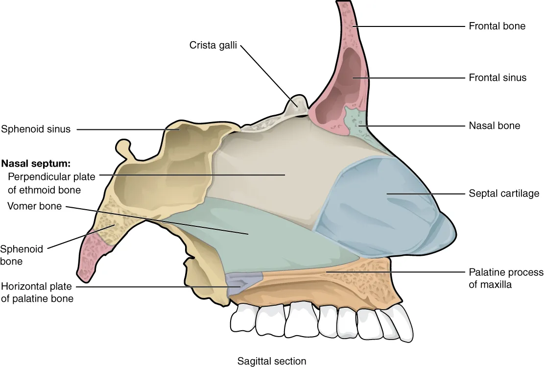
Divides the nasal cavity in two; it consists of the septal cartilage in front, the perpendicular plate of the ethmoid above and behind, and the vomer below and behind. Kiesselbach’s plexus (Little’s area) at the anteroinferior part, where five arteries anastomose, is the source of about 90% of nosebleeds.
- 03
Nasal conchae and meatuses
Conchae nasales; Meatus nasi
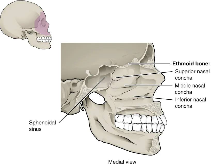
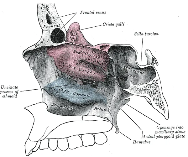
The lateral wall bears three conchae (superior, middle, inferior) that enlarge the mucosal surface for warming, humidifying and filtering air. The inferior meatus receives the nasolacrimal duct; the middle meatus the frontal, maxillary and anterior ethmoidal sinuses via the ostiomeatal complex; the superior meatus the posterior ethmoidal cells; the sphenoid sinus drains into the sphenoethmoidal recess.
- 04
Respiratory mucosa
Tunica mucosa nasi; Regio respiratoria
Ciliated pseudostratified columnar epithelium with goblet cells over a lamina propria rich in glands and erectile venous plexuses (especially on the inferior concha) that swell in the nasal cycle. Cilia sweep the mucus blanket back to the pharynx at about 6 mm a minute.
- 05
Olfactory region
Regio olfactoria
A small patch of about 2–5 cm² in the roof of the nasal cavity, on the superior concha and upper septum, containing olfactory receptor cells (bipolar neurons renewed throughout life), supporting cells and Bowman glands.
- 06
Paranasal sinuses
Sinus paranasales
Four paired air spaces in the frontal, ethmoid, sphenoid and maxillary bones, lined by mucosa continuous with the nose; they lighten the skull and add resonance to the voice. The maxillary sinus is the largest and drains poorly because its ostium is high on the medial wall; upper molar roots may project into its floor.
Position and relations
The cavity opens anteriorly at the nostrils and posteriorly into the nasopharynx through the choanae. Its roof is the cribriform plate, its floor the hard palate; the lateral wall bears three conchae (superior, middle, inferior) that define the meatuses. The maxillary, frontal and anterior ethmoidal sinuses drain into the middle meatus; the nasolacrimal duct opens into the inferior meatus.
Form and structure
The septum consists of septal cartilage in front and the perpendicular plate of the ethmoid and the vomer behind. The vestibule is lined by hair-bearing skin; the rest is respiratory mucosa (ciliated pseudostratified epithelium with goblet cells) with dense venous plexuses over the conchae; a small olfactory area in the roof holds the olfactory neurons.
Vessels and nerves
Blood comes mainly from the sphenopalatine artery (maxillary) and the anterior and posterior ethmoidal arteries (ophthalmic). On the anterior septum their branches anastomose in Kiesselbach’s plexus, the commonest site of epistaxis. The olfactory nerve (I) carries smell; general sensation comes from branches of V1 and V2.
Nasal cavity, nasal valves and sinuses
The nasal cavity warms and humidifies inspired air and removes fine particles before it reaches the lower airways. Its ciliated columnar epithelium secretes mucus that traps particles, which the cilia then sweep away (mucociliary clearance). The hard palate forms its floor. The lateral wall carries scroll-shaped turbinates that enlarge the surface exposed to airflow, together with the sinus drainage openings. Odorants are carried to the olfactory recess in the upper cavity, medial to the superior turbinate.[1]
The internal nasal valve is the narrowest part of the nasal airway. It is classically bounded by the septum medially, the caudal margin of the upper lateral cartilage laterally and the inferior turbinate below, about 1.3 cm into the cavity. In white populations the angle between the upper lateral cartilage and the septum usually measures 10–15°. Computational airflow models show the valve to be a three-dimensional, collapsible and heterogeneous structure rather than a flat cross-section, calling for a new framework in rhinoplasty planning.[2]
Four air-filled paranasal sinuses surround the nasal cavity, each named after its bone: maxillary, ethmoid, sphenoid and frontal. Their mucosa also secretes mucus, which the cilia move towards thin-walled ostia draining into the nose. Terminologia Anatomica currently lists only the ethmoidal cells and the frontal, sphenoidal and maxillary sinuses, whereas clinicians use many more terms for important structures; extensions to the terminology have been proposed to bring anatomical and clinical language together.[3][1][4]
Full-text reading (StatPearls)
The StatPearls chapters on this topic, reproduced verbatim. Their CC BY-NC-ND 4.0 licence allows unmodified copies only, so they are not translated or shortened.
Anatomy, Head and Neck, Nasal CavitySobiesk JL, Munakomi S · StatPearls 2023 · NBK544232 · 27k characters
Loading…
Sobiesk JL, Munakomi S. Anatomy, Head and Neck, Nasal Cavity. StatPearls [Internet]. Treasure Island (FL): StatPearls Publishing; 2023. NBK544232 · PMID 31334952 · CC BY-NC-ND 4.0. Reproduced verbatim, unmodified.
Anatomy, Head and Neck, Nose Paranasal SinusesCappello ZJ, Minutello K, Dublin AB · StatPearls 2023 · NBK499826 · 21k characters
Loading…
Cappello ZJ, Minutello K, Dublin AB. Anatomy, Head and Neck, Nose Paranasal Sinuses. StatPearls [Internet]. Treasure Island (FL): StatPearls Publishing; 2023. NBK499826 · PMID 29763001 · CC BY-NC-ND 4.0. Reproduced verbatim, unmodified.
Anatomy, Head and Neck, Nose BonesAlJulaih GH, Sharma P, Lasrado S · StatPearls 2023 · NBK541117 · 14k characters
Loading…
AlJulaih GH, Sharma P, Lasrado S. Anatomy, Head and Neck, Nose Bones. StatPearls [Internet]. Treasure Island (FL): StatPearls Publishing; 2023. NBK541117 · PMID 31082161 · CC BY-NC-ND 4.0. Reproduced verbatim, unmodified.
Anatomy, Head and Neck, Nose SinusesHenson B, Drake TM, Edens MA · StatPearls 2023 · NBK513272 · 9k characters
Loading…
Henson B, Drake TM, Edens MA. Anatomy, Head and Neck, Nose Sinuses. StatPearls [Internet]. Treasure Island (FL): StatPearls Publishing; 2023. NBK513272 · PMID 30020644 · CC BY-NC-ND 4.0. Reproduced verbatim, unmodified.
Anatomy, Head and Neck, NoseGalarza-Paez L, Marston G, Downs BW · StatPearls 2023 · NBK532870 · 8k characters
Loading…
Galarza-Paez L, Marston G, Downs BW. Anatomy, Head and Neck, Nose. StatPearls [Internet]. Treasure Island (FL): StatPearls Publishing; 2023. NBK532870 · PMID 30422465 · CC BY-NC-ND 4.0. Reproduced verbatim, unmodified.
Organs in this system
References
Sources for the values and descriptions on this page, indexed in PubMed (US National Library of Medicine).
- [1]Anatomy, Head and Neck, Nasal CavitySobiesk JL et al., StatPearls 2023PMID 31334952
- [2]Anatomy and Physiology of the Nasal ValvesLiu DH et al., Otolaryngologic clinics of North America 2025PMID 39426874
- [3]Anatomy, Head and Neck, Nose SinusesHenson B et al., StatPearls 2023PMID 30020644
- [4]Clinical anatomy of the paranasal sinuses and its terminologyChmielewski PP, Anatomical science international 2024PMID 37807010
- [5]The normal human lung: ultrastructure and morphometric estimation of diffusion capacityGehr P, Bachofen M, Weibel ER, 1978PMID 644146Supports: Alveolar surface area
- [6]Physiology, LungStatPearls (NCBI Bookshelf)PMID 31424761Supports: Respiratory rate