Thymus
Model
Layers
Z-Anatomy anatomical model based on BodyParts3D, licensed CC BY-SA 4.0.
Description
A bilobed primary lymphoid organ in the superior and anterior mediastinum, behind the manubrium of the sternum. T lymphocytes differentiate and are selected here; it is relatively largest in early childhood and involutes after puberty, being replaced by fat.
Parts
Each part has its own card; press “Show on the model” to fly to it and select it in the 3D model above.
- 01
Right and left lobes
Lobus dexter et lobus sinister thymi
The thymus has two asymmetrical lobes joined in the midline by connective tissue, lying in the superior and anterior mediastinum behind the manubrium and in front of the pericardium and great vessels. It is relatively largest at birth and absolutely largest around puberty (about 30–40 g), then involutes into fat.
- 02
Lobules
Lobuli thymi
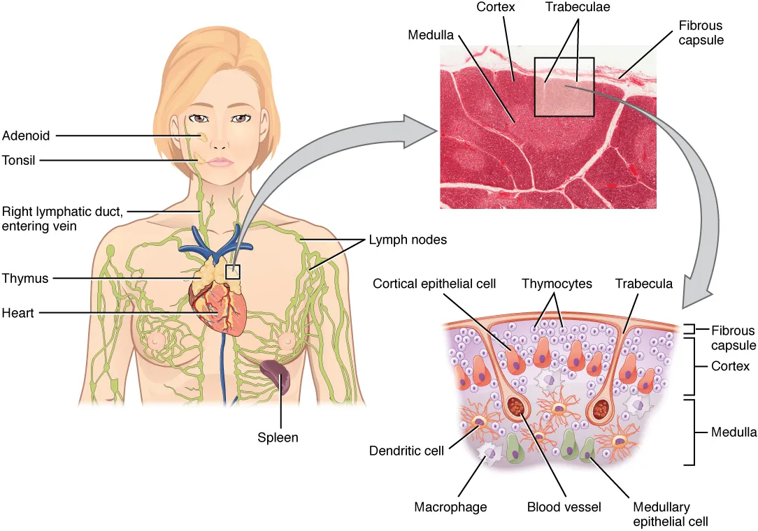
A thin capsule sends septa inwards that divide each lobe into lobules 0.5–2 mm across, each with an outer cortex and inner medulla; the medulla is continuous between lobules. The framework is not reticular fibres as in lymph nodes but a meshwork of thymic epithelial cells.
- 03
Cortex
Cortex thymi
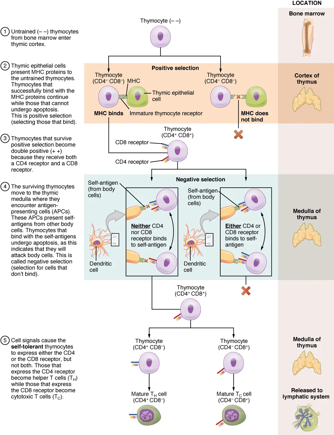
The dark zone packed with proliferating thymocytes (T-cell precursors). Here positive selection takes place: only cells able to recognise MHC molecules on cortical epithelial cells survive. A blood–thymus barrier around cortical capillaries keeps foreign antigen out.
- 04
Medulla
Medulla thymi
The paler, less cellular zone. Medullary epithelial cells express antigens from tissues throughout the body (driven by AIRE) so that self-reactive T cells are deleted: negative selection. Only about 2–5% of thymocytes survive both rounds and leave through postcapillary venules at the corticomedullary junction.
- 05
Thymic (Hassall’s) corpuscles
Corpuscula thymica
Concentric whorls of epithelial cells, keratinised at the centre, found only in the medulla and diagnostic of the thymus on a slide. They secrete TSLP, which drives dendritic cells to generate regulatory T cells (Tregs) that maintain immune tolerance.
- 06
Blood supply
Vasa thymi
Arteries come from branches of the internal thoracic and inferior thyroid arteries. Veins drain mainly into the left brachiocephalic vein, which must be respected during thymectomy for myasthenia gravis or thymoma.
Position and relations
The thymus lies in the superior mediastinum and extends into the anterior mediastinum, behind the manubrium and upper costal cartilages and in front of the pericardium, the aortic arch and the left brachiocephalic vein. In young children it may reach the neck near the thyroid; in adults it is largely replaced by fat.
Form and structure
Each lobe is divided into lobules, each with a densely packed cortex of thymocytes (T-cell precursors) and a paler medulla containing Hassall’s corpuscles. The framework is formed by epithelial reticular cells; cortical epithelial cells mediate positive selection, while medullary epithelial cells display tissue antigens (via AIRE) to delete self-reactive T clones. A blood–thymus barrier protects the cortex.
Vessels and nerves
Arteries come from branches of the internal thoracic and inferior thyroid arteries; the veins drain mainly into the left brachiocephalic vein. The thymus has no afferent lymphatics, only efferent vessels to the mediastinal nodes. Thymoma is associated with myasthenia gravis.
Physiology
Bone-marrow progenitors migrate to the thymus (via CCR7 and CCR9) and are steered to the T lineage by NOTCH signalling. Developing T cells are positively selected in the cortex (keeping cells whose receptors recognise self-peptide–MHC complexes) and further selected in the medulla to remove self-reactive cells, using machinery unique to thymic epithelial cells.[1][2]
The thymus involutes with age, a change conserved across species; declining function leaves older people with weaker responses to new pathogens, tumours and vaccines. Inflammation and endocrine signals can also cause acute thymic atrophy.[3][1]
Thymic architecture: capsule, cortex, corticomedullary junction and medulla
The thymus and parathyroids share a common primordium in the third pharyngeal pouch. Organogenesis involves fate determination, detachment from the parathyroid, migration into the thorax and colonisation by haematopoietic progenitors. In humans the thymus reaches its final position at 8–10 weeks of gestation, as haematopoietic precursors begin to colonise it; it keeps growing after birth, peaks in cellularity a few weeks postnatally and begins to decline before puberty. FOXN1 is the earliest thymus-specific marker.[1]
The thymus has two lobes separated by an interlobular septum, wrapped in a two-layer connective tissue capsule of collagen and reticular fibres. The inner layer folds inwards as septa carrying capillaries deep into the lobe; about 5% of developing thymocytes sit in the subcapsular zone. The cortex is the most cell-dense region, with about 70–80% of thymic cells, and the main site of positive selection by cortical thymic epithelial cells (cTECs); mitotic activity decreases inwards, mirroring the maturation stages of pre-T cells.[1]
The corticomedullary junction is rich in blood and lymphatic vessels. Arteries enter there and branch into capillaries for medulla and cortex; cortical capillaries are less fenestrated, limiting antigen access, whereas medullary capillaries are fenestrated. Progenitors enter through venules at the junction and mature T cells leave through post-capillary venules. The thymus has only efferent lymphatics, with no afferent lymph, so unlike lymph nodes it does not swell during infection. Nerve fibres follow the vessels in the capsule and septa.[1]
The medulla is less dense and holds about 10–15% of thymocytes, mainly single-positive CD4⁺ or CD8⁺ cells undergoing negative selection. Medullary thymic epithelial cells (mTECs), dendritic cells, macrophages and B cells present self-peptides on MHC class I and II, and thymocytes whose T-cell receptors bind self too strongly are removed by programmed cell death. Hassall’s corpuscles in the medulla probably help clear dying thymocytes and are a source of TSLP, which instructs dendritic cells and supports regulatory T-cell induction.[1]
Full-text reading (StatPearls)
The StatPearls chapters on this topic, reproduced verbatim. Their CC BY-NC-ND 4.0 licence allows unmodified copies only, so they are not translated or shortened.
Anatomy, Head and Neck: Blood Thymus BarrierBruss DM, Ely S · StatPearls 2023 · NBK546601 · 11k characters
Loading…
Bruss DM, Ely S. Anatomy, Head and Neck: Blood Thymus Barrier. StatPearls [Internet]. Treasure Island (FL): StatPearls Publishing; 2023. NBK546601 · PMID 31536208 · CC BY-NC-ND 4.0. Reproduced verbatim, unmodified.
Anatomy, Head and Neck, ThymusRemien K, Jozsa F, Jan A · StatPearls 2025 · NBK539748 · 11k characters
Loading…
Remien K, Jozsa F, Jan A. Anatomy, Head and Neck, Thymus. StatPearls [Internet]. Treasure Island (FL): StatPearls Publishing; 2025. NBK539748 · PMID 30969570 · CC BY-NC-ND 4.0. Reproduced verbatim, unmodified.
Organs in this system
References
Sources for the values and descriptions on this page, indexed in PubMed (US National Library of Medicine).
- [1]The thymus road to a T cell: migration, selection, and atrophyRuiz Pérez M et al., Frontiers in immunology 2024PMID 39257583
- [2]Thymus machinery for T-cell selectionKondo K et al., International immunology 2019PMID 30476234
- [3]Age-Related Thymic InvolutionLins MP et al., Advances in experimental medicine and biology 2025PMID 40067592