Skull
Model
Layers
Z-Anatomy anatomical model based on BodyParts3D, licensed CC BY-SA 4.0.
Description
Composed of 22 bones (8 neurocranial and 14 facial, excluding the auditory ossicles), it encloses and protects the brain and the special sense organs.
Parts
Each part has its own card; press “Show on the model” to fly to it and select it in the 3D model above.
- 01
Cranial vault (calvaria)
Calvaria
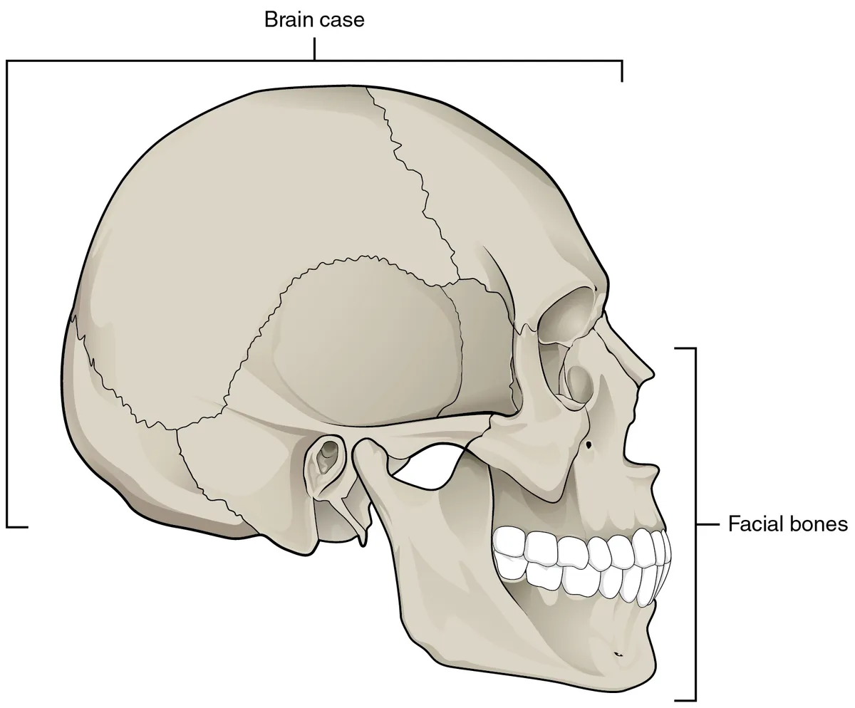
The roof of the skull is formed by the frontal bone, the two parietal bones and the squamous occipital, joined by the coronal, sagittal and lambdoid sutures. Each bone has two compact tables enclosing spongy diploë. In the newborn the anterior and posterior fontanelles stay open until about 18 months and 2–3 months respectively.
- 02
Cranial base
Basis cranii
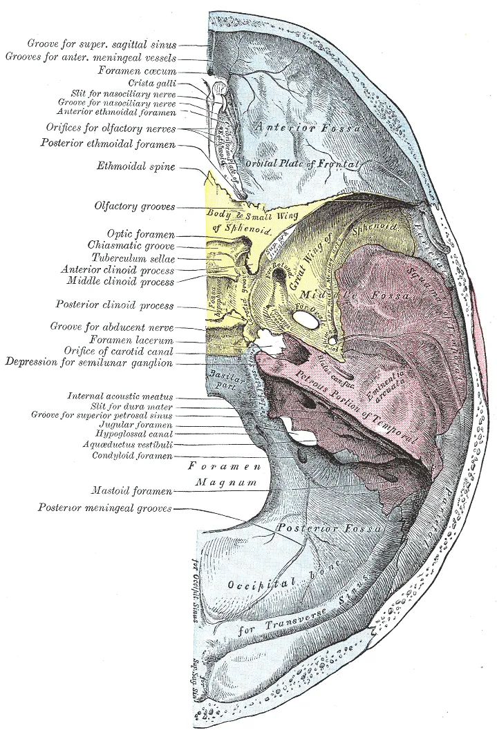
The floor of the cranial cavity is divided into three fossae: anterior (frontal lobes, cribriform plate of the ethmoid), middle (temporal lobes, sella turcica of the sphenoid holding the pituitary) and posterior (cerebellum, brainstem, foramen magnum). Its many foramina transmit cranial nerves and vessels, so basal fractures can cause CSF leak from the nose or ear and cranial nerve palsies.
- 03
Temporal bone
Os temporale
Made of the squamous, tympanic, styloid and petrous parts. The petrous part, the densest bone in the body, houses the middle and inner ear and the internal acoustic meatus for nerves VII and VIII. The mastoid process contains air cells continuous with the middle ear. The squama meets the sphenoid and parietal at the pterion, a thin spot overlying the middle meningeal artery.
- 04
Orbit
Orbita
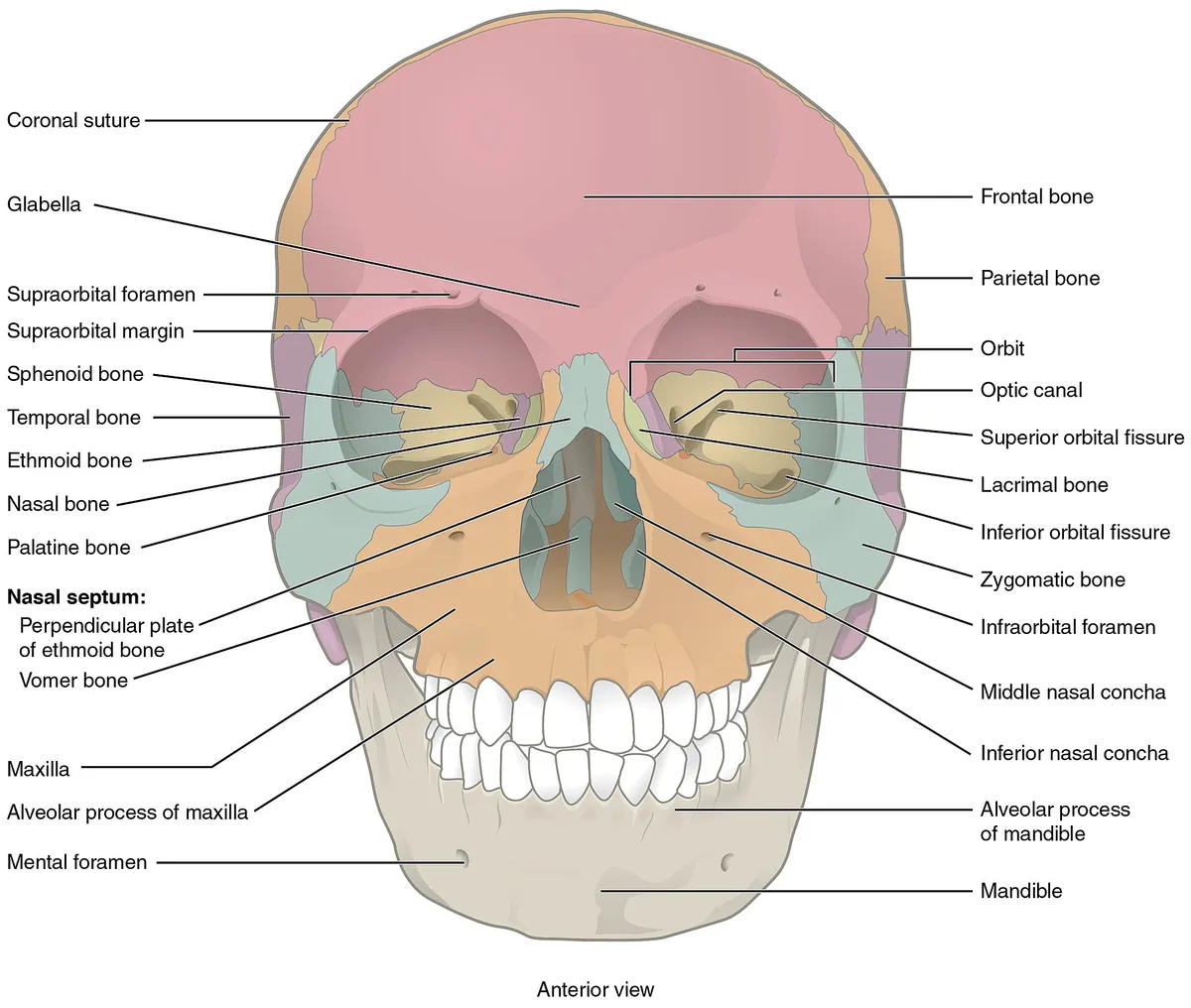
A pyramidal cavity formed by seven bones: frontal, zygomatic, maxilla, lacrimal, ethmoid, palatine and sphenoid. The medial wall (lamina papyracea of the ethmoid) and the floor (maxilla) are the thinnest and break under a direct blow to the eye (blow-out fracture). The optic canal and superior orbital fissure open at the apex.
- 05
Nasal skeleton
Skeleton nasi
The nasal cavity is bounded by the nasal bones, maxillae, ethmoid (perpendicular plate, superior and middle conchae), inferior nasal conchae, vomer and palatine bones. The nasal septum is formed by the perpendicular plate of the ethmoid and the vomer behind and the septal cartilage in front.
- 06
Paranasal sinuses
Sinus paranasales
Four pairs of air spaces lined by respiratory mucosa within the frontal, ethmoid, sphenoid and maxillary bones, all draining into the nasal cavity. The maxillary sinus is the largest; its ostium lies high on the medial wall, which hampers drainage, and the roots of the upper molars may reach its floor. The sinuses lighten the skull and give resonance to the voice.
- 07
Maxilla
Maxilla
The two maxillae form the middle of the face: they carry the upper dental arch (alveolar process) and form the orbital floor, the lateral wall of the nasal cavity and the front of the hard palate. Midface fractures are classified as Le Fort I, II or III according to how the fracture lines cross the maxilla.
- 08
Mandible
Mandibula
The only movable bone of the skull: a horseshoe-shaped body carrying the lower teeth and two rami, each ending in a condylar process (temporomandibular joint) and a coronoid process (temporalis insertion). The mandibular canal carries the inferior alveolar nerve, which emerges at the mental foramen as the mental nerve.
- 09
Hyoid bone
Os hyoideum
A U-shaped bone in the neck at the level of C3, articulating with no other bone and slung by muscles and ligaments. It anchors the suprahyoid and infrahyoid muscles that raise and lower the larynx in swallowing and speech. A fractured hyoid suggests manual strangulation.
Form and structure
The skull has 22 bones (excluding the ossicles), divided into the neurocranium (8 bones: frontal, two parietal, two temporal, occipital, sphenoid, ethmoid) enclosing the brain, and the viscerocranium (14 bones) forming the face, orbits, nasal and oral cavities. The cranial base has anterior, middle and posterior fossae, pierced by many foramina for vessels and cranial nerves.[1]
Position and relations
In the newborn, the vault bones are not yet fused and are joined by fibrous membranes at the fontanelles (the anterior closes at about 18–24 months) and at the sutures, allowing moulding during birth and growth of the skull with the brain. Premature suture closure causes skull deformity.[2]
Clinical correlations
The bones of the cranial vault are joined by sutures, which allow the skull to grow with the brain. Craniosynostosis is the premature closure of one or more sutures, causing skull deformity and potentially impairing brain, visual and respiratory development; early recognition and specialist surgery are essential.[2]
The skull base
The skull base packs many neurovascular structures into a small space, and understanding it begins with the sphenoid bone. The cavernous sinus contains the internal carotid artery and some of its branches with cranial nerves III, IV, VI and V1, and drains venous blood from many sources. The anterior skull base extends to the frontal sinus and matters for sinus surgery and sinonasal tumours; the clivus shields the brainstem and posterior fossa. This knowledge enables endoscopic endonasal approaches.[3]
The temporal bone is dense bone housing many minute structures: the external auditory canal, the middle ear with its ossicles, the inner ear (cochlea, vestibule, semicircular canals), the facial canal and numerous foramina, canals and clefts. High-resolution MRI shows cranial nerves traversing the skull base, while CT assesses the bony foramina; in a cranial neuropathy the whole course from brainstem nucleus to periphery must be examined.[4][5][6]
The orbit is a pyramidal bony box containing the globe, extraocular muscles, orbital fat, nerves and vessels; it communicates with the skull base through the optic canal and superior orbital fissure, which allows transorbital approaches to some intracranial regions. Shape anomalies such as orbital hypertelorism mark many craniofacial malformations.[7][8][9]
Full-text reading (StatPearls)
The StatPearls chapters on this topic, reproduced verbatim. Their CC BY-NC-ND 4.0 licence allows unmodified copies only, so they are not translated or shortened.
Anatomy, Head and Neck, SkullAnderson BW, Kortz MW, Black AC, Al Kharazi KA · StatPearls 2023 · NBK499834 · 19k characters
Loading…
Anderson BW, Kortz MW, Black AC, Al Kharazi KA. Anatomy, Head and Neck, Skull. StatPearls [Internet]. Treasure Island (FL): StatPearls Publishing; 2023. NBK499834 · PMID 29763009 · CC BY-NC-ND 4.0. Reproduced verbatim, unmodified.
Anatomy, Head and Neck, Skull ForamenLipsett BJ, Alsayouri K · StatPearls 2023 · NBK546621 · 9k characters
Loading…
Lipsett BJ, Alsayouri K. Anatomy, Head and Neck, Skull Foramen. StatPearls [Internet]. Treasure Island (FL): StatPearls Publishing; 2023. NBK546621 · PMID 31536228 · CC BY-NC-ND 4.0. Reproduced verbatim, unmodified.
Organs in this system
References
Sources for the values and descriptions on this page, indexed in PubMed (US National Library of Medicine).
- [1]Cranial Nerve AnatomyTraylor KS et al., Neuroimaging clinics of North America 2022PMID 35843663
- [2]Craniosynostosis - Recognition, clinical characteristics, and treatmentKajdic N et al., Bosnian journal of basic medical sciences 2018PMID 28623672
- [3]Skull Base AnatomyPatel CR et al., Otolaryngologic clinics of North America 2016PMID 26614826
- [4]Temporal Bone AnatomyBenson JC et al., Neuroimaging clinics of North America 2022PMID 36244722
- [5]Clinical anatomy and imaging of the cranial nerves and skull baseJha RM et al., Seminars in neurology 2012PMID 23361480
- [6]Skull base, orbits, temporal bone, and cranial nerves: anatomy on MR imagingMorani AC et al., Magnetic resonance imaging clinics of North America 2011PMID 21816324
- [7]Anatomy of the OrbitReinshagen KL et al., Neuroimaging clinics of North America 2022PMID 36244718
- [8]Transorbital Route to Intracranial SpaceDi Somma A et al., Advances and technical standards in neurosurgery 2024PMID 39017795
- [9]Orbital HypertelorismTashima A et al., Clinics in plastic surgery 2025PMID 39986883
- [10]Anatomy, BonesStatPearls (NCBI Bookshelf)PMID 30725884Supports: Number of bones
- [11]Calcium metabolism in health and diseasePeacock M, 2010PMID 20089499Supports: Skeletal calcium store