Meninges and ventricles
Model
Layers
Z-Anatomy anatomical model based on BodyParts3D, licensed CC BY-SA 4.0.
Description
Three membranes (dura, arachnoid and pia mater) wrap the brain and spinal cord; dural folds form the falx cerebri and tentorium cerebelli. The ventricular system (two lateral ventricles, the third ventricle, the cerebral aqueduct and the fourth ventricle) holds about 150 ml of cerebrospinal fluid secreted by the choroid plexus.
Parts
Each part has its own card; press “Show on the model” to fly to it and select it in the 3D model above.
- 01
Dura mater
Dura mater
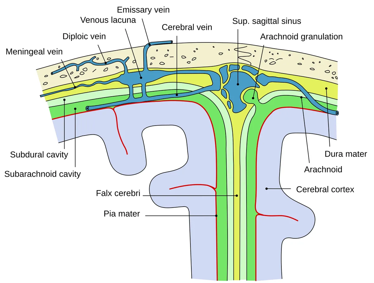
The thick outer fibrous membrane. In the skull it has periosteal and meningeal layers that separate to enclose the dural venous sinuses; in the vertebral canal it is a single layer separated from bone by the epidural space with fat and a venous plexus.
- 02
Falx cerebri and tentorium cerebelli
Falx cerebri; Tentorium cerebelli
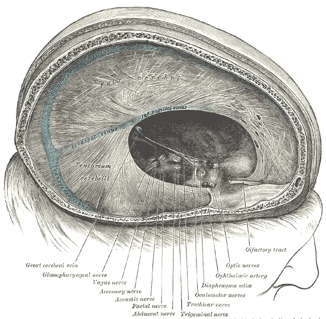
Folds of the meningeal layer: the falx cerebri lies in the longitudinal fissure between the hemispheres, and the tentorium cerebelli separates cerebrum from cerebellum, leaving the tentorial notch for the brainstem. Its free edge is where uncal herniation compresses the oculomotor nerve.
- 03
Arachnoid mater and subarachnoid space
Arachnoidea mater; Spatium subarachnoideum
A thin avascular membrane applied to the inner dura; arachnoid trabeculae cross the subarachnoid space, which holds cerebrospinal fluid and the major cerebral arteries. The space widens into cisterns (cerebellomedullary, chiasmatic, lumbar), and arachnoid granulations drain fluid into the venous sinuses.
- 04
Pia mater
Pia mater
The innermost, delicate and vascular layer, adherent to the surface of the brain and cord and following vessels into the parenchyma. Along the cord it forms the denticulate ligaments anchoring it to the dura, and the filum terminale.
- 05
Lateral ventricles
Ventriculus lateralis; Septum pellucidum
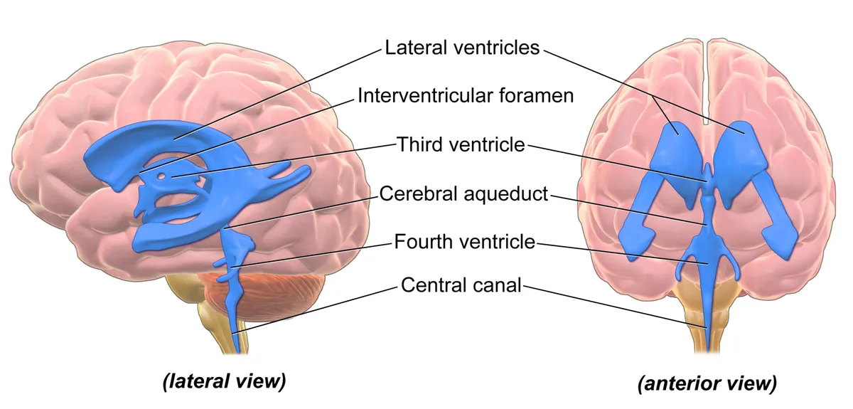
Two C-shaped cavities in the hemispheres, each with a frontal horn, body, atrium (trigone), occipital horn and temporal horn; the frontal horns are separated by the septum pellucidum. Each opens into the third ventricle through the interventricular foramen (of Monro).
- 06
Third ventricle
Ventriculus tertius
A narrow midline slit between the two thalami and the hypothalamus, often crossed by the interthalamic adhesion. Its anterior wall is the lamina terminalis; the floor bears the optic and infundibular recesses, and the pineal recess lies behind.
- 07
Cerebral aqueduct
Aqueductus mesencephali
A channel about 15 mm long through the midbrain, linking the third and fourth ventricles and surrounded by periaqueductal grey. As the narrowest part of the ventricular system, aqueductal stenosis is a common cause of obstructive hydrocephalus.
- 08
Fourth ventricle
Ventriculus quartus
A tent-shaped cavity between the pons and medulla in front and the cerebellum behind; its floor, the rhomboid fossa, carries the facial colliculus and cranial nerve nuclei. Fluid leaves for the subarachnoid space through the median aperture (Magendie) and two lateral apertures (Luschka).
- 09
Choroid plexus
Plexus choroideus
Tufts of fenestrated capillaries covered by specialised ependymal epithelium in the lateral, third and fourth ventricles. Tight junctions between the epithelial cells form the blood–CSF barrier, and the plexus secretes about 500 mL of fluid a day.
The three meninges
The dura mater is the tough outermost layer. In the skull it has a periosteal layer, adherent to the inner table of the bones, and a meningeal layer; the dural venous sinuses run between them. In the vertebral canal the dura has a single layer, separated from the canal wall by the epidural space, which contains fat and a venous plexus and is the target of epidural anaesthesia.
The meningeal layer folds inwards to form partitions: the falx cerebri between the hemispheres, the tentorium cerebelli separating cerebrum from cerebellum, the falx cerebelli and the diaphragma sellae over the pituitary fossa. They stabilise the brain, but they are also the edges against which it herniates when intracranial pressure rises.
The arachnoid mater is thin and avascular and lies against the dura; beneath it the subarachnoid space contains cerebrospinal fluid and the major cerebral arteries. The space widens into cisterns (the cerebellomedullary cistern or cisterna magna, the chiasmatic and interpeduncular cisterns) and, below L2, into the lumbar cistern. The pia mater adheres to the surface of the brain and cord, following every sulcus; along the cord it forms the denticulate ligaments and the filum terminale.
Ventricular system
Each hemisphere contains a C-shaped lateral ventricle with a frontal horn, body, atrium, occipital horn and temporal horn. The lateral ventricles open into the third ventricle through the interventricular foramina (of Monro). The third ventricle is a narrow cleft between the thalami, connected to the fourth ventricle by the cerebral aqueduct (of Sylvius) in the midbrain.
The fourth ventricle lies between the pons and medulla in front and the cerebellum behind; its floor, the rhomboid fossa, overlies the cranial nerve nuclei. Cerebrospinal fluid leaves it for the subarachnoid space through the median aperture (of Magendie) and two lateral apertures (of Luschka), and continues into the central canal of the cord.
Cerebrospinal fluid
Cerebrospinal fluid is secreted by the choroid plexuses of the ventricles at about 500 ml a day, while the circulating volume is only about 150 ml, so it is renewed several times daily. It flows from the lateral ventricles through the third ventricle, aqueduct and fourth ventricle into the subarachnoid space and is absorbed into the dural venous sinuses through the arachnoid granulations. It buoys and cushions the brain and takes part in metabolic exchange with brain tissue.
Clinical correlations
An extradural haematoma usually follows a temporal fracture that tears the middle meningeal artery, and may show a lucid interval before rapid deterioration. A subdural haematoma comes from torn bridging veins, especially in older people and those with alcohol misuse. Spontaneous subarachnoid haemorrhage is mostly due to a ruptured aneurysm of the arterial circle. Obstruction of cerebrospinal fluid flow (for example aqueductal stenosis) causes hydrocephalus, dilating the ventricles above the block.
Cerebrospinal fluid
Cerebrospinal fluid fills the ventricles and the cranial and spinal subarachnoid spaces, with a mean volume of 150 ml (about 25 ml ventricular, 125 ml subarachnoid), renewed roughly four times a day. It is secreted mainly by the choroid plexuses, with possible contributions from interstitial fluid, ependyma and capillaries. Its flow is driven chiefly by the arterial pulse wave and modulated by respiration, posture, jugular venous pressure and effort.[1]
Arachnoid villi were long considered the main site of CSF absorption into the venous sinuses. Experimental data show that a substantial share also drains along cranial and spinal nerve sheaths, through the cribriform plate and the adventitia of cerebral arteries into the lymphatic system.[1]
The choroid plexus does more than provide a cushion and a waste sink: factors it secretes into the CSF regulate neural stem cells, developmental critical periods and the overall health of the nervous system.[2]
The glymphatic system and perivascular spaces
The glymphatic system is the central nervous system’s analogue of the lymphatics: CSF flows from the subarachnoid space along periarterial spaces into the parenchyma, mixes with interstitial fluid and leaves along perivenous spaces, carrying metabolic waste. Astrocytes and vessels shape the perivascular space, and aquaporin-4 water channels on astrocytic endfeet facilitate the flow.[3][4]
Perivascular clearance is greatest during sleep. Enlarged perivascular spaces on MRI are associated with vascular risk factors, small vessel disease and cognitive decline, and glymphatic failure has been proposed as a common pathway to dementia, although parts of the model remain debated.[5][6][7]
Full-text reading (StatPearls)
The StatPearls chapters on this topic, reproduced verbatim. Their CC BY-NC-ND 4.0 licence allows unmodified copies only, so they are not translated or shortened.
Neuroanatomy, Ventricular SystemShenoy SS, Lui F · StatPearls 2023 · NBK532932 · 21k characters
Loading…
Shenoy SS, Lui F. Neuroanatomy, Ventricular System. StatPearls [Internet]. Treasure Island (FL): StatPearls Publishing; 2023. NBK532932 · PMID 30422527 · CC BY-NC-ND 4.0. Reproduced verbatim, unmodified.
Neuroanatomy, Cranial MeningesGhannam JY, Al Kharazi KA · StatPearls 2023 · NBK539882 · 13k characters
Loading…
Ghannam JY, Al Kharazi KA. Neuroanatomy, Cranial Meninges. StatPearls [Internet]. Treasure Island (FL): StatPearls Publishing; 2023. NBK539882 · PMID 30969704 · CC BY-NC-ND 4.0. Reproduced verbatim, unmodified.
Anatomy, Back, Spinal MeningesSehgal I, Das JM · StatPearls 2023 · NBK547755 · 9k characters
Loading…
Sehgal I, Das JM. Anatomy, Back, Spinal Meninges. StatPearls [Internet]. Treasure Island (FL): StatPearls Publishing; 2023. NBK547755 · PMID 31613535 · CC BY-NC-ND 4.0. Reproduced verbatim, unmodified.
Organs in this system
References
Sources for the values and descriptions on this page, indexed in PubMed (US National Library of Medicine).
- [1]Anatomy and physiology of cerebrospinal fluidSakka L et al., European annals of otorhinolaryngology, head and neck diseases 2011PMID 22100360
- [2]Development and functions of the choroid plexus-cerebrospinal fluid systemLun MP et al., Nature reviews. Neuroscience 2015PMID 26174708
- [3]The Glymphatic System: A Novel Component of Fundamental NeurobiologyHablitz LM et al., The Journal of neuroscience 2021PMID 34526407
- [4]Aquaporin-4 in glymphatic system, and its implication for central nervous system disordersPeng S et al., Neurobiology of disease 2023PMID 36796590
- [5]Perivascular spaces in the brain: anatomy, physiology and pathologyWardlaw JM et al., Nature reviews. Neurology 2020PMID 32094487
- [6]Glymphatic failure as a final common pathway to dementiaNedergaard M et al., Science 2020PMID 33004510
- [7]The Brain's Glymphatic System: Current ControversiesMestre H et al., Trends in neurosciences 2020PMID 32423764
- [8]The human brain in numbers: a linearly scaled-up primate brainHerculano-Houzel S, 2009PMID 19915731Supports: Neuron count
- [9]Determinants of conduction velocity in myelinated nerve fibersWaxman SG, 1980PMID 6245357Supports: Conduction velocity
- [10]Quantitative imaging of energy expenditure in human brainPMID 22487547Supports: Cerebral energy expenditure