Prostate
Model
Layers
Z-Anatomy anatomical model based on BodyParts3D, licensed CC BY-SA 4.0.
Description
A cone-shaped gland below the bladder that surrounds the prostatic urethra and weighs about 20 g in young men. It has a peripheral zone (where most carcinomas arise), a central zone and a transition zone (the site of benign hyperplasia); its secretion contains PSA and forms part of the seminal fluid.
Parts
Each part has its own card; press “Show on the model” to fly to it and select it in the 3D model above.
- 01
Peripheral zone
Zona peripherica prostatae
About 70% of glandular tissue, posterior and lateral, against the rectum and palpable on rectal examination. About 70% of prostate cancers arise here, and it is the zone sampled by systematic biopsy.
- 02
Transition zone
Zona transitionalis prostatae
Only about 5% of the gland in young men, two small lobes around the proximal prostatic urethra. It is where benign prostatic hyperplasia arises, so its enlargement compresses the urethra, causing hesitancy and nocturia.
- 03
Central zone
Zona centralis prostatae
About 25% of the gland, a cone surrounding the ejaculatory ducts from the base to the seminal colliculus; embryologically related to the Wolffian duct. It rarely gives rise to cancer or hyperplasia.
- 04
Anterior fibromuscular stroma
Stroma fibromusculare anterius
The non-glandular anterior part of smooth muscle and fibrous tissue, continuous with the internal sphincter at the bladder neck and the external sphincter at the apex. In front lies Santorini’s plexus (the dorsal venous complex), which must be controlled during prostatectomy.
- 05
Capsule, base and apex
Capsula prostatica; Basis et apex prostatae
A chestnut-shaped gland of about 20 g, its base against the bladder neck and apex on the urogenital diaphragm. The thin fibromuscular capsule is not a true capsule; the neurovascular bundles for erection run posterolateral to it and are preserved in nerve-sparing prostatectomy.
- 06
Ejaculatory ducts and seminal colliculus
Ductus ejaculatorii; Colliculus seminalis
Each ejaculatory duct, about 2 cm long, is formed by the ampulla of the ductus deferens and the duct of the seminal gland and runs obliquely through the central zone to open on the colliculus beside the utricle. The prostate contributes about 30% of semen volume (PSA, zinc, citric acid).
Position and relations
The prostate lies in the lesser pelvis below the bladder neck and above the urogenital diaphragm; anteriorly it is separated from the pubis by the retropubic space (of Retzius), and posteriorly it faces the rectal ampulla across the rectoprostatic fascia (Denonvilliers’ fascia). Its posterior surface can therefore be palpated on rectal examination.
Form and structure
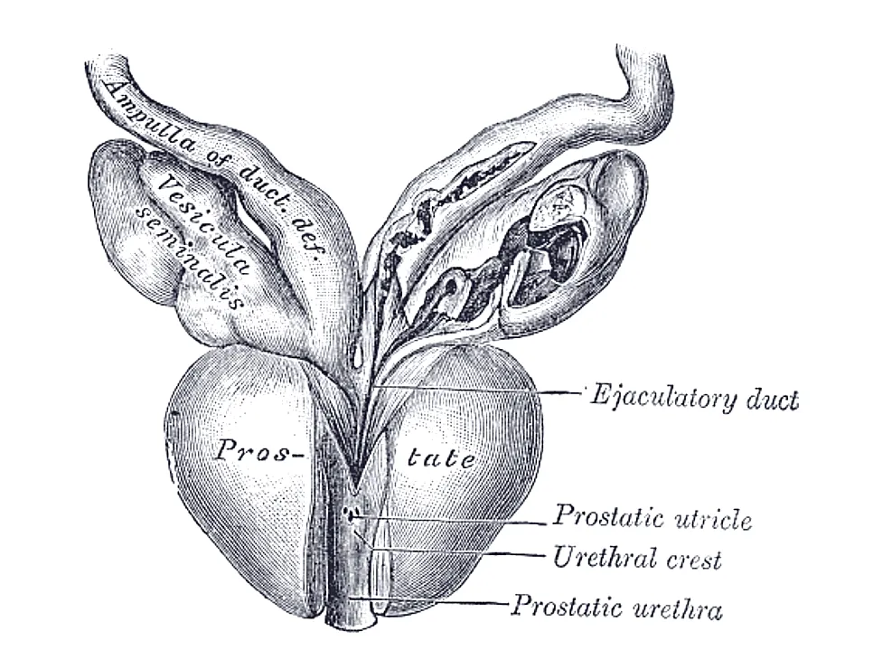
The gland has a base above and an apex below and is traversed by the prostatic urethra; the two ejaculatory ducts pass obliquely through it to open on the seminal colliculus. Following McNeal, the glandular tissue is divided into a peripheral zone (about 70% of glandular tissue), a central zone surrounding the ejaculatory ducts and a periurethral transition zone; anteriorly lies the non-glandular fibromuscular stroma.
Vessels and nerves
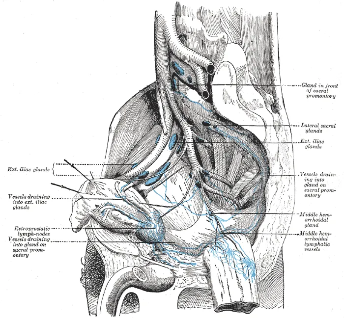
Arterial supply comes mainly from the inferior vesical artery with branches of the middle rectal and internal pudendal arteries (from the internal iliac). The veins form the prostatic plexus, which communicates with the vertebral venous plexus, one route for vertebral metastases of prostate cancer. Nerves come from the inferior hypogastric plexus; the neurovascular bundles for erection run along its posterolateral surface.
Development
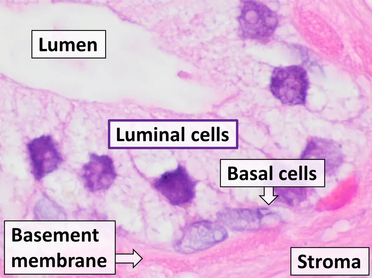
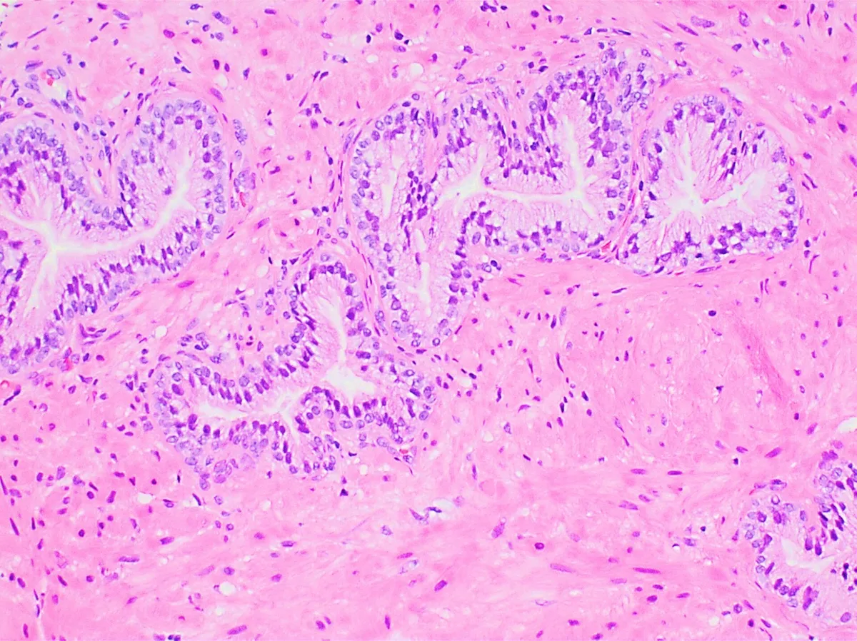
The prostate develops from epithelial buds growing out of the urogenital sinus, passing through organ specification, budding, branching morphogenesis, canalisation and cytodifferentiation, all driven by androgens. Many developmental genes and pathways are reactivated in prostate cancer, which is why antiandrogen therapy is first-line treatment.[1]
Unlike the lobular rodent prostate, whose lobes radiate away from the urethra, the human prostate has a compact zonal anatomy immediately around the urethra below the bladder, and it is the site of benign hyperplasia, cancer and prostatitis; animal models therefore reproduce human disease only in part.[2]
Relations, lobes, zones and function
The prostate is a dense fibromuscular gland shaped like an inverted cone, its base above surrounding the bladder neck and its apex below resting on the external urethral sphincter, wrapped around the proximal urethra in the lesser pelvis. A fibrous capsule containing nerves and a vascular plexus encloses it, surrounded in turn by visceral pelvic fascia. Anteriorly it lies behind the pubic symphysis, separated by retropubic fat and the prostatic venous plexus; posteriorly it is close to the rectum, separated by Denonvilliers’ fascia; laterally it is related to levator ani covered by endopelvic fascia; and below lies the external urethral sphincter, which helps control urinary and ejaculatory flow. Classical textbooks describe five lobes (anterior, posterior, two lateral and a median lobe), whereas clinicians describe two lateral lobes and a median lobe. Histologically there are three zones: the central zone at the base around the ejaculatory ducts; the peripheral zone, the largest (70%), surrounding most of the central zone and part of the distal prostatic urethra; and the small transition zone around the urethra between the bladder and the verumontanum. Anteriorly lies the non-glandular fibromuscular stroma.[3]
The gland’s main function is to secrete an alkaline fluid that protects sperm in the acidic vagina and prolongs their survival; it also contains nourishing proteins and enzymes, and its added volume makes semen easier to propel through the urethra. The two ejaculatory ducts enter the prostate as they leave the seminal vesicles, run obliquely from posterolateral to medial and inferior, and converge on the seminal colliculus of the prostatic urethra. The gland develops from epithelial buds growing laterally from the wall of the urogenital sinus, which branch into solid cords and then canalise into ducts and acini; the surrounding urogenital-sinus mesenchyme forms its fibroblasts and smooth muscle. Androgens act on androgen receptors in the mesenchyme, and androgen from Leydig cells is the chief regulator of growth. Blood comes mainly from the inferior vesical artery, with branches of the middle rectal and internal pudendal arteries; the veins form the prostatic plexus draining to the internal iliac veins, and lymph passes to internal iliac and sacral nodes. Autonomic nerves come from the inferior hypogastric plexus, sympathetic through the hypogastric nerve and parasympathetic through the pelvic nerve; both carry sensation.[3]
Peripheral zone
- Position
- Posterolateral, around the distal urethra
- Share of gland
- About 70%
- Typical disease
- About 75% of carcinomas
Central zone
- Position
- Base, around the ejaculatory ducts
- Share of gland
- —
- Typical disease
- —
Transition zone
- Position
- Around the urethra from bladder to verumontanum
- Share of gland
- Small
- Typical disease
- Benign prostatic hyperplasia
Anterior fibromuscular stroma
- Position
- Anterior surface, down to the apex
- Share of gland
- Non-glandular
- Typical disease
- —
| Zone | Position | Share of gland | Typical disease |
|---|---|---|---|
| Peripheral zone | Posterolateral, around the distal urethra | About 70% | About 75% of carcinomas |
| Central zone | Base, around the ejaculatory ducts | — | — |
| Transition zone | Around the urethra from bladder to verumontanum | Small | Benign prostatic hyperplasia |
| Anterior fibromuscular stroma | Anterior surface, down to the apex | Non-glandular | — |
Benign hyperplasia, cancer and prostatitis
The three notable diseases of the gland are benign hyperplasia, cancer and prostatitis. Benign prostatic hyperplasia is nodular rather than diffuse enlargement, usually of the transition zone, which narrows the urethra or protrudes into the bladder and obstructs urine flow, with frequency, nocturia and a weak stream. Most cases are treated conservatively: alpha-1 blockers lower smooth-muscle tone in the bladder neck and prostatic urethra; 5-alpha-reductase inhibitors block conversion of testosterone to dihydrotestosterone so that the gland shrinks; antimuscarinic drugs inhibit detrusor contraction to reduce urgency. Severe cases that fail medical treatment can undergo transurethral resection of the prostate (TURP), in which an instrument passed retrograde from the tip of the penis trims glandular tissue to widen the channel. Early complications include haematuria, a fall in haemoglobin, transfusion, clot retention and urinary infection; late ones include urethral stricture, bladder-neck contracture, stress incontinence, reoperation, retrograde ejaculation and erectile dysfunction.[3]
Prostate cancer is usually adenocarcinoma, about 75% arising in the peripheral zone, which is why the posterior surface is palpable on rectal examination. Associated factors include family history, high BMI, smoking and ionising or ultraviolet radiation. Symptoms can resemble benign hyperplasia, but low back pain suggests metastasis, since the cancer spreads through venous channels communicating with those of the lower lumbar vertebrae. Prostate-specific antigen (PSA) is a controversial marker: useful for detecting recurrence but not on its own for initial diagnosis, because benign hyperplasia also raises it. 5-alpha-reductase inhibitors are associated with more high-grade cancers but reduce the overall cancer burden. Prostatitis is caused by the bacteria that commonly cause urinary infection and presents mainly with pain and urinary symptoms; it is hard to diagnose because bacteria are seldom isolated, and is treated with antibiotics covering Gram-negative organisms.[3]
Full-text reading (StatPearls)
The StatPearls chapters on this topic, reproduced verbatim. Their CC BY-NC-ND 4.0 licence allows unmodified copies only, so they are not translated or shortened.
Anatomy, Abdomen and Pelvis, ProstateSingh O, Bolla SR · StatPearls 2023 · NBK540987 · 8k characters
Loading…
Singh O, Bolla SR. Anatomy, Abdomen and Pelvis, Prostate. StatPearls [Internet]. Treasure Island (FL): StatPearls Publishing; 2023. NBK540987 · PMID 31082031 · CC BY-NC-ND 4.0. Reproduced verbatim, unmodified.
Organs in this system
References
Sources for the values and descriptions on this page, indexed in PubMed (US National Library of Medicine).
- [1]Prostate OrganogenesisFrancis JC et al., Cold Spring Harbor perspectives in medicine 2018PMID 29229667
- [2]Review of Prostate Anatomy and Embryology and the Etiology of Benign Prostatic HyperplasiaAaron L et al., The Urologic clinics of North America 2016PMID 27476121
- [3]Anatomy, Abdomen and Pelvis, ProstateSingh O et al., StatPearls 2023PMID 31082031