Blood and blood cells
Model
Hover or tap to see names, click to read a description; click a name in the legend to fly to it. Procedurally built to histological proportions; colours follow illustration conventions.
Description
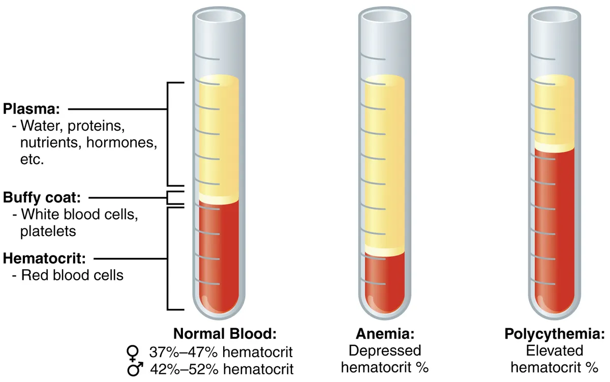
A fluid connective tissue, about 5 litres in an adult, made of plasma (about 55%) and formed elements: red cells that carry oxygen, five kinds of white cells that defend the body and platelets that stop bleeding. All arise from haematopoietic stem cells in the bone marrow.
Red blood cells
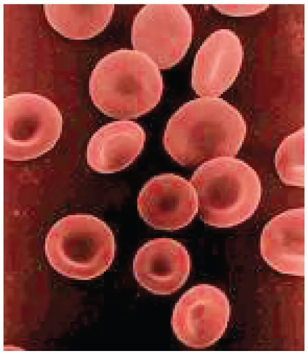
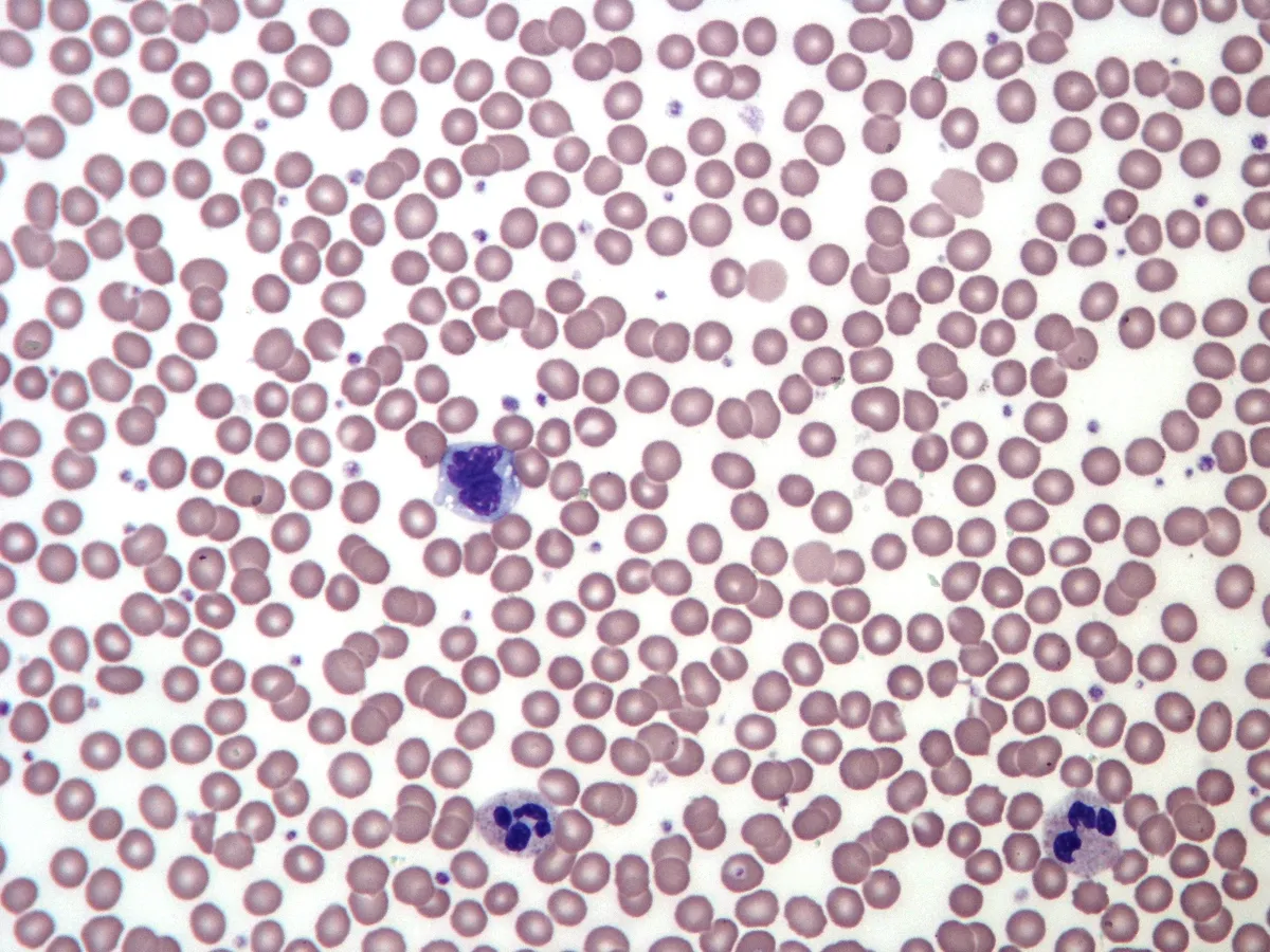
Blood is a fluid connective tissue making up about 7% of body mass; plasma occupies over half its volume, the rest being red cells, white cells and platelets. The mature red cell is a biconcave disc about 7.5 µm across and 2 µm thick, with no nucleus or organelles and a cytoplasm holding some 270 million haemoglobin molecules. The biconcave shape gives a large surface-to-volume ratio for gas exchange and lets the cell fold to pass capillaries narrower than itself. A person has about 25 trillion red cells, each living about 120 days before macrophages of the spleen and liver remove it.[1][2]
The red cell’s strength and pliability come from a membrane skeleton: a hexagonal spectrin network joined to short actin filaments and tethered to the membrane by ankyrin and band 3, which is also the Cl⁻/HCO₃⁻ exchanger for CO₂ transport. Mutations in these proteins make red cells spherical or elliptical, fragile in the spleen, and cause haemolytic anaemia. Red cell production is driven by erythropoietin from the kidney: when tissue oxygen falls, the kidney releases more of this hormone and the marrow raises output within days. Erythroblasts expel their nucleus at the last stage, producing reticulocytes that still carry some RNA and mature into red cells after about a day in the blood.[3][4][5][6]
White blood cells
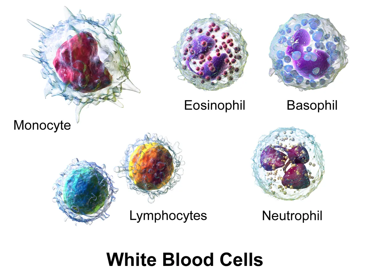
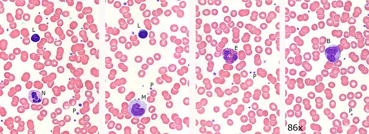
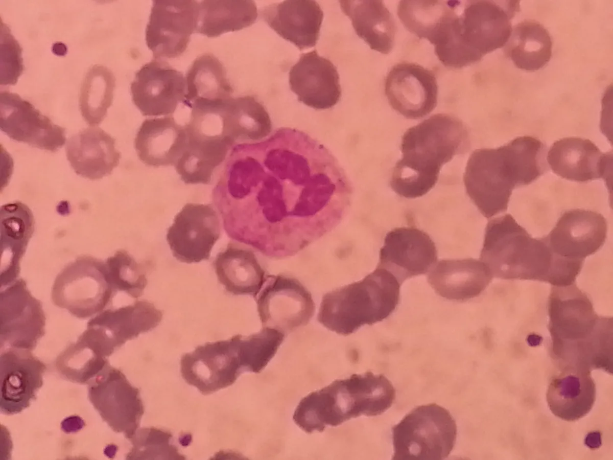
White cells are only in the blood in transit to the tissues where they actually work. Granulocytes have lobed nuclei and specific granules. Neutrophils are the majority; with 3–5 nuclear lobes they are first to reach a bacterial focus, engulfing and killing bacteria with oxidants, the enzymes of azurophil granules such as myeloperoxidase, and extracellular DNA traps; they live only a few days and are the main component of pus. Eosinophils have bilobed nuclei and orange-red granules with a crystalline core of major basic protein; they fight parasites and take part in asthma. Basophils, the rarest, have dark purple granules containing histamine and bind IgE like mast cells.[7][8][9]
Agranulocytes are lymphocytes and monocytes. Lymphocytes are small, their dense round nucleus filling almost the whole cell; B cells mature in the marrow and in lymph-node germinal centres differentiate into antibody-secreting plasma cells, T cells mature and are selected in the thymus, and NK cells kill virus-infected and tumour cells without antigen recognition. Monocytes are the largest blood cells, with a bean-shaped nucleus and grey-blue vacuolated cytoplasm; after 1–3 days in the blood they enter tissues and become macrophages or dendritic cells. Persistent eosinophilia calls for a search for parasites, drug allergy or blood disease.[10][11][12][13]
Neutrophil
- Share
- 50–70%
- Morphology
- 3–5 lobes, fine granules
- Main role
- Kill bacteria, acute inflammation
Lymphocyte
- Share
- 20–40%
- Morphology
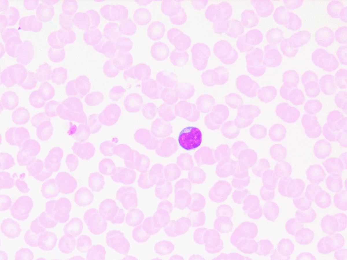
- Small, dense round nucleus
- Main role
- Adaptive immunity (B, T), NK
Monocyte
- Share
- 2–8%
- Morphology
- Large, bean-shaped nucleus
- Main role
- Becomes macrophage, dendritic cell
Eosinophil
- Share
- 1–4%
- Morphology
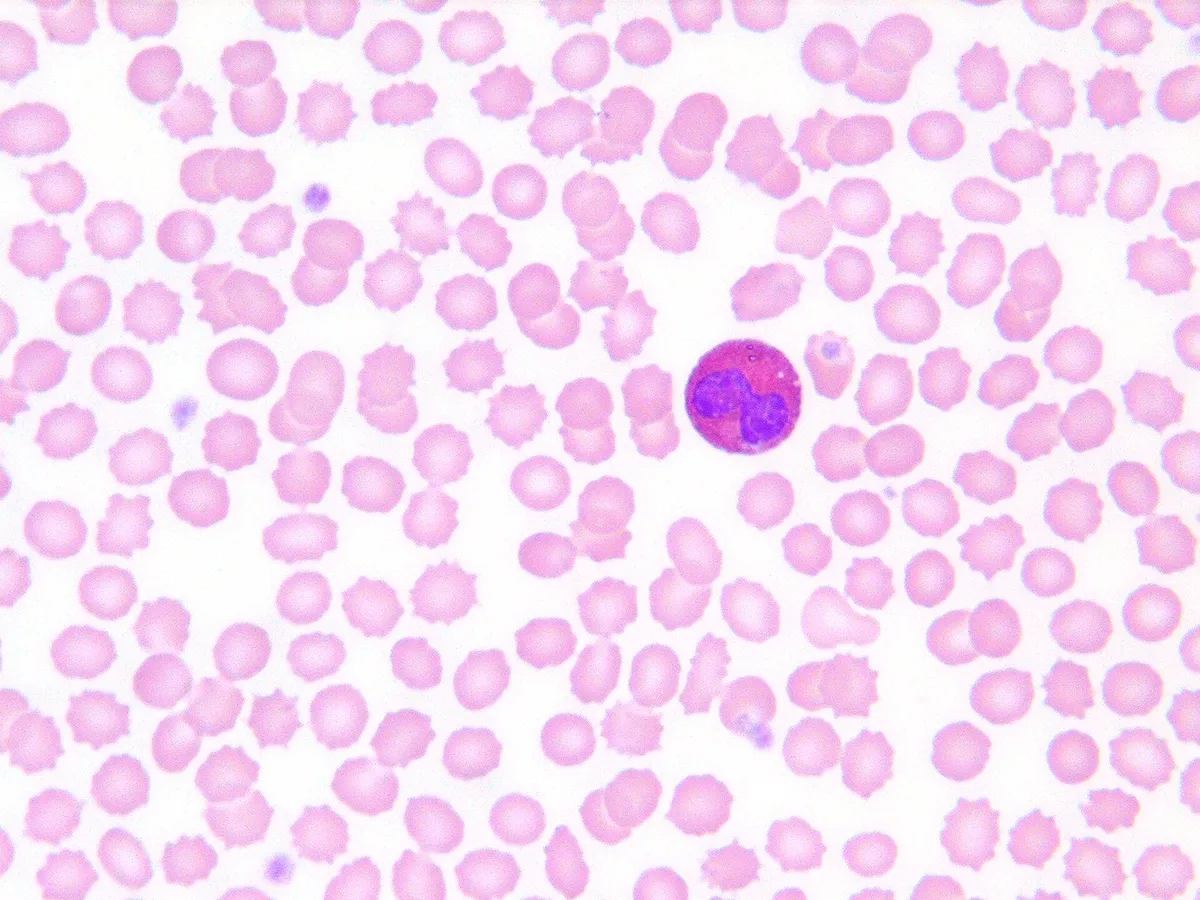
- Bilobed, orange-red granules
- Main role
- Parasites, allergy
Basophil
- Share
- < 1%
- Morphology
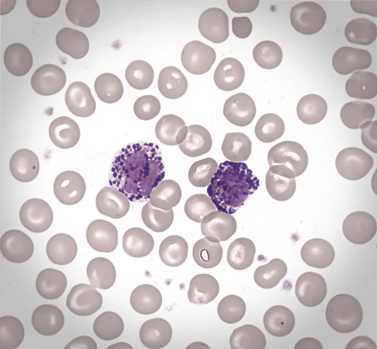
- Dark granules hide nucleus
- Main role
- Histamine, IgE reactions
| Type | Share | Morphology | Main role |
|---|---|---|---|
| Neutrophil | 50–70% | 3–5 lobes, fine granules | Kill bacteria, acute inflammation |
| Lymphocyte | 20–40% | Small, dense round nucleus | Adaptive immunity (B, T), NK |
| Monocyte | 2–8% | Large, bean-shaped nucleus | Becomes macrophage, dendritic cell |
| Eosinophil | 1–4% | Bilobed, orange-red granules | Parasites, allergy |
| Basophil | < 1% | Dark granules hide nucleus | Histamine, IgE reactions |
Platelets and haemostasis
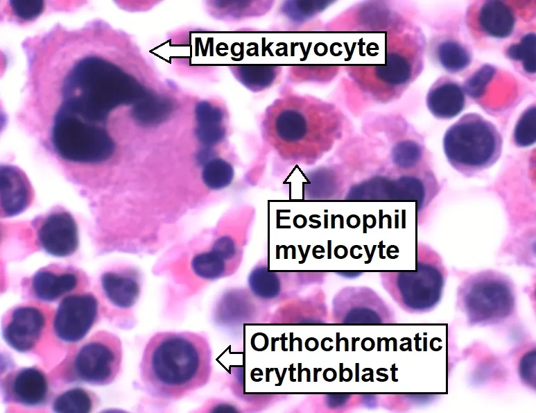
Platelets are anucleate cytoplasmic fragments 2–3 µm across that live 7–10 days, numbering 150–400 thousand per microlitre. They arise from megakaryocytes, giant polyploid marrow cells that extend long proplatelets into the marrow sinusoids, where blood flow sheds them as platelets; one megakaryocyte yields thousands of platelets under the control of thrombopoietin from the liver. Inside, a marginal band of microtubules keeps the disc shape, an open canalicular system connects to the surface, α-granules hold fibrinogen, von Willebrand factor and growth factors, and dense granules hold ADP, serotonin and Ca²⁺.[14][15][16][17]
When a vessel wall is injured, platelets adhere to exposed collagen through von Willebrand factor and the GPIb receptor and become activated: they change from discs to spiky spheres, release their granules and generate thromboxane A₂. ADP and thromboxane recruit more platelets; the GPIIb/IIIa receptor switches to a fibrinogen-binding state, cross-linking platelets into the primary haemostatic plug. Activated platelet membranes expose phosphatidylserine as a surface for the coagulation cascade, and thrombin cleaves fibrinogen into a fibrin mesh that secures the plug. Aspirin blocks thromboxane irreversibly for the platelet’s lifetime, while clopidogrel blocks the ADP receptor.[18][14]
Haematopoiesis
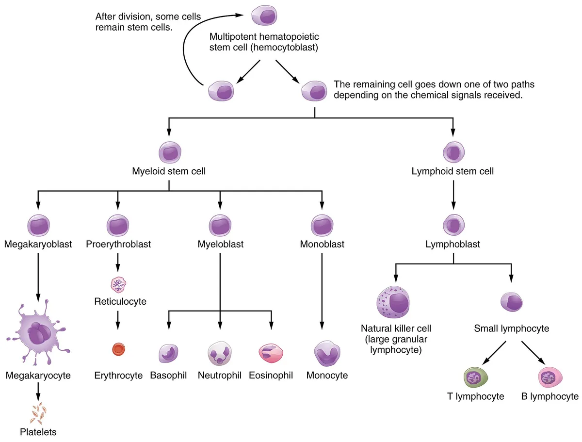
In adults haematopoiesis takes place in the red marrow of flat bones, vertebrae and the ends of long bones; in the fetus it moves from yolk sac to liver and spleen before settling in the marrow. Marrow is a reticular tissue with sinusoids lined by discontinuous endothelium, interspersed with adipocytes, macrophages and maturing blood cell lines. Haematopoietic stem cells, very rare and mostly quiescent, sit in perivascular niches where endothelial and reticular cells provide signals such as CXCL12 and stem cell factor; their self-renewal is what makes marrow transplantation curative for blood diseases.[19][20]
The classical branching tree, in which the stem cell splits into myeloid and lymphoid progenitors that narrow step by step, has given way to a more continuous picture: single-cell sequencing shows early progenitors already biased towards one lineage, and the megakaryocyte line can branch directly from the stem cell. Growth factors set the output of each line: erythropoietin for red cells, thrombopoietin for platelets, G-CSF for neutrophils. The marrow makes some 200 billion red cells, 100 billion granulocytes and 100 billion platelets a day; recombinant G-CSF is used to restore neutrophils after chemotherapy and to mobilise stem cells for transplantation.[21][19][2]
Full-text reading (StatPearls)
The StatPearls chapters on this topic, reproduced verbatim. Their CC BY-NC-ND 4.0 licence allows unmodified copies only, so they are not translated or shortened.
Biochemistry of Platelet Activating FactorAshraf MA, Nookala V · StatPearls 2023 · NBK557392 · 28k characters
Loading…
Ashraf MA, Nookala V. Biochemistry of Platelet Activating Factor. StatPearls [Internet]. Treasure Island (FL): StatPearls Publishing; 2023. NBK557392 · PMID 32491324 · CC BY-NC-ND 4.0. Reproduced verbatim, unmodified.
Histology, B-Cell LymphocyteAlthwaiqeb SA, Fakoya AO, Bordoni B · StatPearls 2024 · NBK560905 · 27k characters
Loading…
Althwaiqeb SA, Fakoya AO, Bordoni B. Histology, B-Cell Lymphocyte. StatPearls [Internet]. Treasure Island (FL): StatPearls Publishing; 2024. NBK560905 · PMID 32809740 · CC BY-NC-ND 4.0. Reproduced verbatim, unmodified.
Histology, T-Cell LymphocyteSauls RS, McCausland C, Taylor BN · StatPearls 2023 · NBK535433 · 14k characters
Loading…
Sauls RS, McCausland C, Taylor BN. Histology, T-Cell Lymphocyte. StatPearls [Internet]. Treasure Island (FL): StatPearls Publishing; 2023. NBK535433 · PMID 30571054 · CC BY-NC-ND 4.0. Reproduced verbatim, unmodified.
Histology, PlateletsWilliams O, Sergent SR · StatPearls 2022 · NBK557800 · 13k characters
Loading…
Williams O, Sergent SR. Histology, Platelets. StatPearls [Internet]. Treasure Island (FL): StatPearls Publishing; 2022. NBK557800 · PMID 32491732 · CC BY-NC-ND 4.0. Reproduced verbatim, unmodified.
Physiology, PlateletDenault D, Launico MV · StatPearls 2026 · NBK470328 · 12k characters
Loading…
Denault D, Launico MV. Physiology, Platelet. StatPearls [Internet]. Treasure Island (FL): StatPearls Publishing; 2026. NBK470328 · PMID 29262002 · CC BY-NC-ND 4.0. Reproduced verbatim, unmodified.
Histology, Red Blood CellBarbalato L, Pillarisetty LS · StatPearls 2022 · NBK539702 · 10k characters
Loading…
Barbalato L, Pillarisetty LS. Histology, Red Blood Cell. StatPearls [Internet]. Treasure Island (FL): StatPearls Publishing; 2022. NBK539702 · PMID 30969524 · CC BY-NC-ND 4.0. Reproduced verbatim, unmodified.
Histology, HematopoiesisChapman J, Zhang Y · StatPearls 2023 · NBK534246 · 10k characters
Loading…
Chapman J, Zhang Y. Histology, Hematopoiesis. StatPearls [Internet]. Treasure Island (FL): StatPearls Publishing; 2023. NBK534246 · PMID 30480979 · CC BY-NC-ND 4.0. Reproduced verbatim, unmodified.
Histology, White Blood CellTigner A, Ibrahim SA, Murray IV · StatPearls 2026 · NBK563148 · 10k characters
Loading…
Tigner A, Ibrahim SA, Murray IV. Histology, White Blood Cell. StatPearls [Internet]. Treasure Island (FL): StatPearls Publishing; 2026. NBK563148 · PMID 33085295 · CC BY-NC-ND 4.0. Reproduced verbatim, unmodified.
Histology, MonocytesEspinoza VE, Emmady PD · StatPearls 2023 · NBK557618 · 7k characters
Loading…
Espinoza VE, Emmady PD. Histology, Monocytes. StatPearls [Internet]. Treasure Island (FL): StatPearls Publishing; 2023. NBK557618 · PMID 32491550 · CC BY-NC-ND 4.0. Reproduced verbatim, unmodified.
Organs in this system
References
Sources for the values and descriptions on this page, indexed in PubMed (US National Library of Medicine).
- [1]Histology, Red Blood CellBarbalato L et al., StatPearls 2022PMID 30969524
- [2]Hematopoiesis in numbersCosgrove J et al., Trends in immunology 2021PMID 34742656
- [3]Red cell membrane: past, present, and futureMohandas N et al., Blood 2008PMID 18988878
- [4]ErythropoietinJelkmann W, Frontiers of hormone research 2016PMID 27348128
- [5]Erythropoiesis: development and differentiationDzierzak E et al., Cold Spring Harbor perspectives in medicine 2013PMID 23545573
- [6]Cell fate decision in erythropoiesis: Insights from multiomics studiesTur S et al., Experimental hematology 2024PMID 38262486
- [7]Histology, White Blood CellTigner A et al., StatPearls 2026PMID 33085295
- [8]Spatial adaptation of eosinophils and their emerging roles in homeostasis, infection and diseaseArnold IC et al., Nature reviews. Immunology 2024PMID 38982311
- [9]IgE, mast cells, basophils, and eosinophilsStone KD et al., The Journal of allergy and clinical immunology 2010PMID 20176269
- [10]Histology, MonocytesEspinoza VE et al., StatPearls 2023PMID 32491550
- [11]The unique biology of germinal center B cellsYoung C et al., Immunity 2021PMID 34380063
- [12]The thymus road to a T cell: migration, selection, and atrophyRuiz Pérez M et al., Frontiers in immunology 2024PMID 39257583
- [13]Approach to Patients with EosinophiliaKuang FL, The Medical clinics of North America 2020PMID 31757229
- [14]Histology, PlateletsWilliams O et al., StatPearls 2022PMID 32491732
- [15]The incredible journey: From megakaryocyte development to platelet formationMachlus KR et al., The Journal of cell biology 2013PMID 23751492
- [16]Platelet generation in vivo and in vitroFurniss JA et al., Blood 2024PMID 39357055
- [17]MegakaryopoiesisGeddis AE, Seminars in hematology 2010PMID 20620431
- [18]Physiology, PlateletDenault D et al., StatPearls 2026PMID 29262002
- [19]Histology, HematopoiesisChapman J et al., StatPearls 2023PMID 30480979
- [20]HematopoiesisRieger MA et al., Cold Spring Harbor perspectives in biology 2012PMID 23209149
- [21]New paradigms on hematopoietic stem cell differentiationCheng H et al., Protein & cell 2020PMID 31201709