Heart
Model
Hover or tap to see names, click to read a description; click a name in the legend to fly to it. Procedurally built to histological proportions; colours follow illustration conventions.
Description
A hollow muscular organ with four chambers (two atria, two ventricles) lying in the middle mediastinum, with about two-thirds of its mass to the left of the midline. Resting stroke volume is about 70 mL.
Parts
Each part has its own card; press “Show on the model” to fly to it and select it in the 3D model above.
- 01
Right atrium
Atrium dextrum
Receives venous blood from the superior and inferior venae cavae and the coronary sinus. Its smooth sinus venarum is separated from the pectinate-muscled part and auricle by the crista terminalis; the septum bears the fossa ovalis, remnant of the fetal foramen ovale. The sinoatrial node lies at the entry of the superior vena cava.
- 02
Right ventricle
Ventriculus dexter
A crescent-shaped chamber wrapped round the left ventricle, with a wall 3–5 mm thick, pumping into the low-pressure pulmonary circulation. Its inlet has coarse trabeculae and three papillary muscles; the moderator band carries the right bundle branch; the smooth outflow tract (infundibulum) leads to the pulmonary valve.
- 03
Left atrium
Atrium sinistrum
The most posterior chamber, receiving the four pulmonary veins; its wall is mostly smooth, with pectinate muscles only in the left auricle. It lies directly in front of the oesophagus, so an enlarged atrium can indent it and transoesophageal echo images it well; the left auricle is the usual site of thrombus in atrial fibrillation.
- 04
Left ventricle
Ventriculus sinister
A conical chamber forming the apex, with a wall 8–12 mm thick (about three times the right) to pump into the systemic circulation. Fine trabeculae and two large papillary muscles (anterolateral and posteromedial) anchor the mitral leaflets; the outflow tract below the aortic valve is smooth-walled.
- 05
Interatrial and interventricular septa
Septum interatriale; Septum interventriculare
The thin interatrial septum bears the fossa ovalis; a patent foramen ovale persists in about a quarter of adults. The interventricular septum is mostly thick muscle, with a small membranous part just below the aortic valve that is the commonest site of ventricular septal defects.
- 06
Atrioventricular valves
Valva tricuspidalis; Valva mitralis
The tricuspid (anterior, posterior, septal leaflets) on the right and the mitral (anterior, posterior) on the left close at the start of systole, producing the first heart sound. Chordae tendineae tie each leaflet to papillary muscles so it cannot evert into the atrium; rupture causes acute regurgitation.
- 07
Semilunar valves
Valva aortae; Valva trunci pulmonalis
Each has three pocket-like cusps without chordae and closes at the start of diastole, producing the second heart sound. Behind the aortic cusps are the three sinuses of Valsalva; the right and left coronary arteries arise from the right and left sinuses.
- 08
Papillary muscles and chordae tendineae
Musculi papillares; Chordae tendineae
Papillary muscles are muscular columns projecting from the ventricular wall that contract with the ventricle to tension the chordae. The posteromedial papillary muscle of the left ventricle usually has a single blood supply (often the posterior interventricular artery) and is prone to rupture in myocardial infarction, causing acute mitral regurgitation.
- 09
Cardiac conduction system
Systema conducente cordis
The sinoatrial node (Keith–Flack) fires 60–100 times a minute; the impulse spreads through atrial muscle to the atrioventricular node (Aschoff–Tawara) in the triangle of Koch, is delayed about 0.1 s, then passes down the bundle of His, the bundle branches and the Purkinje network so the ventricles contract from apex to base.
- 10
Coronary arteries
Arteriae coronariae
The left coronary artery divides into the anterior interventricular (LAD, supplying the anterior two-thirds of the septum and the anterior wall) and circumflex branches; the right coronary runs in the right coronary sulcus and supplies the SA node in about 60% and the AV node in about 80% of people. The artery giving the posterior interventricular branch defines dominance (usually right).
- 11
Cardiac veins and coronary sinus
Venae cordis; Sinus coronarius
The great (with the LAD), middle (with the posterior interventricular artery) and small cardiac veins drain into the coronary sinus in the posterior coronary sulcus, which opens into the right atrium guarded by the Thebesian valve. The coronary sinus is the route for left-ventricular leads in resynchronisation pacing.
- 12
Heart wall and pericardium
Endocardium; Myocardium; Epicardium; Pericardium
The wall comprises endocardium, myocardium and epicardium (the visceral serous pericardium, carrying fat and the coronary vessels). Outside lie the fibrous pericardium and the parietal serous layer; the pericardial cavity holds 15–50 mL of fluid, and rapid effusion can cause tamponade.
- 13
Fibrous skeleton of the heart
Skeleton fibrosum cordis
Four fibrous rings round the valve orifices joined by fibrous trigones; they anchor valves and muscle and electrically insulate atria from ventricles. The bundle of His is the only normal conducting path through it; an accessory pathway (bundle of Kent) causes Wolff–Parkinson–White syndrome.
Position and relations
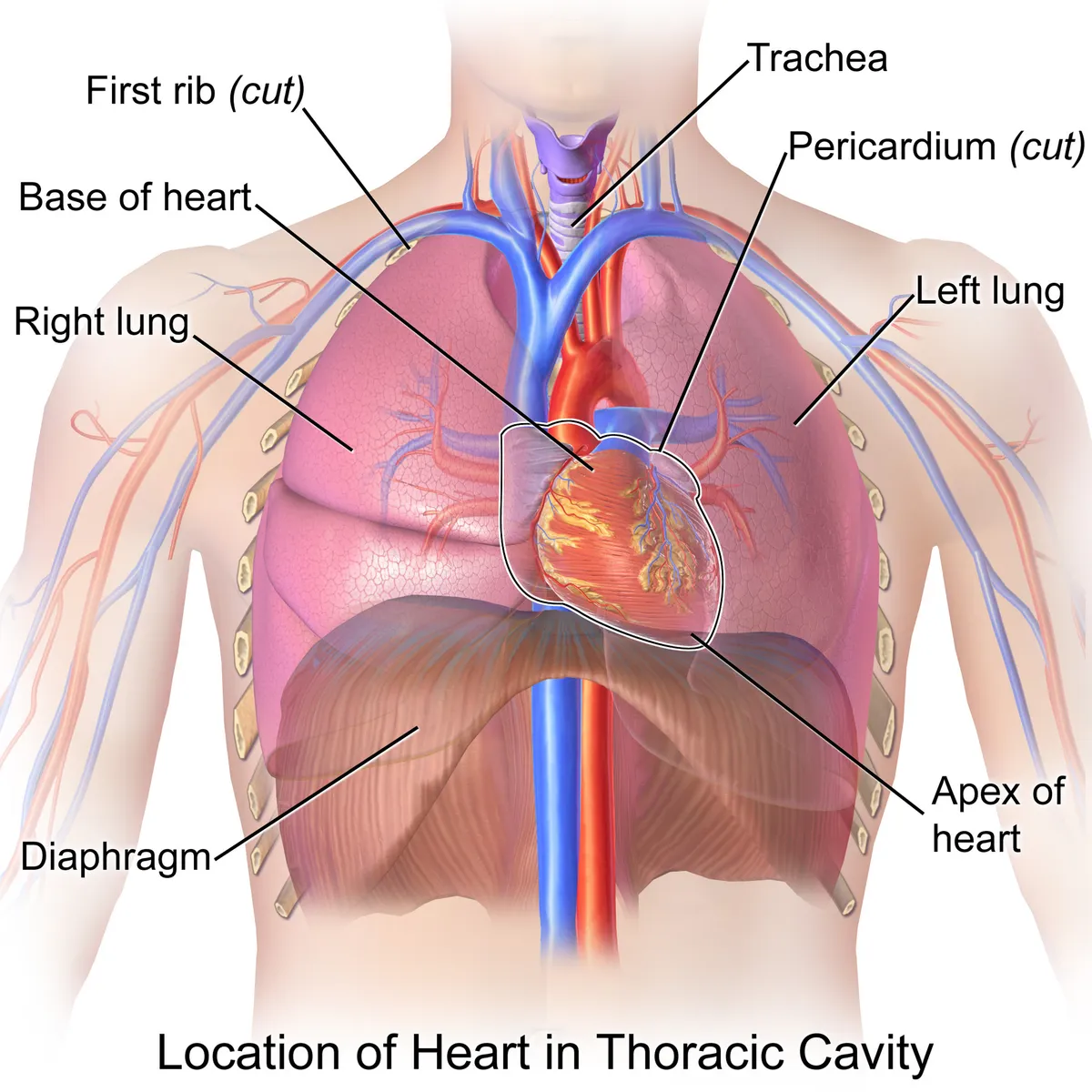
The heart lies in the middle mediastinum, behind the sternum, above the diaphragm and between the lungs; about two-thirds of it lies left of the midline. Its base faces posteriorly and to the right, its apex anteriorly, inferiorly and to the left, usually in the fifth intercostal space on the left midclavicular line. It is enclosed in the pericardial sac.[1]
Form and structure
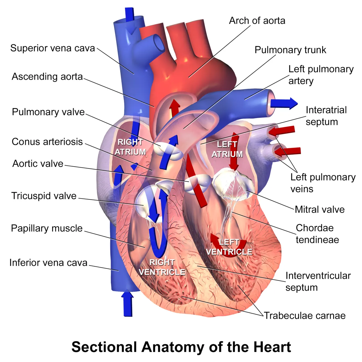
The heart has four chambers: two atria that receive blood and two ventricles that pump it out, separated by the interatrial and interventricular septa. Four valves keep flow one-way: the tricuspid and mitral (atrioventricular) valves, and the pulmonary and aortic (semilunar) valves. The left ventricular wall is about three times thicker than the right, as it must overcome systemic pressure.
Vessels and nerves
The myocardium is supplied by the right and left coronary arteries, arising from the aortic sinuses just above the aortic valve; venous blood drains into the coronary sinus and then the right atrium. Sympathetic nerves increase rate and contractility, while parasympathetic (vagal) fibres slow the rate and atrioventricular conduction.[2]
Tricuspid valve and right heart
The tricuspid valve (valva tricuspidalis) comprises three leaflets (anterior, posterior, septal), chordae tendineae and papillary muscles attached to a saddle-shaped annulus. Its anatomy varies considerably between individuals; understanding the normal structure is the basis for explaining secondary tricuspid regurgitation, in which right ventricular and annular dilatation prevent leaflet coaptation.[3]
The subendocardial Purkinje network distributes the impulse to the whole ventricular myocardium within a few tens of milliseconds. Purkinje cells are large, poor in myofibrils and electrophysiologically distinct, so they conduct fast but can also become ectopic foci.[4]
Conduction system and coronary arteries
All cardiomyocytes can conduct the impulse, but a specialised population both generates it and carries it from atria to ventricles: the conduction system (sinoatrial node, atrioventricular node, His bundle and its branches, Purkinje network). Its components cannot be distinguished from working myocardium by gross dissection; histological criteria proposed in 1910 require that conducting cells be histologically discrete, traceable from section to section and, for tracts, insulated by fibrous tissue.[5]
The atrioventricular conduction axis was described by Tawara over a century ago. Conduction system pacing (His bundle and, more recently, left bundle branch area pacing) is replacing chronic right ventricular pacing, which carries worse outcomes, and this experience has refined knowledge of the axis. In parallel, sinoatrial node pacemaker cells are the template for biological pacemakers.[6][7]
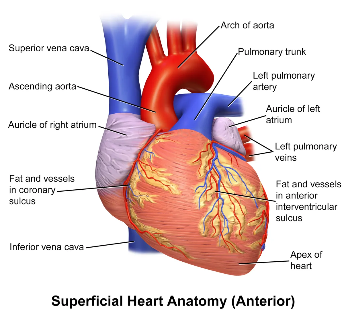
The right and left coronary arteries arise from their sinuses of Valsalva in the aortic root. Surgically the system is divided into the left main artery with its branches (left anterior descending and circumflex) and the right coronary artery; unrecognised variants of origin and course can lead to coronary injury, especially in redo surgery.[8]
As the embryonic heart wall thickens, passive diffusion gives way to a vascular plexus that remodels into the mature coronary tree. In adults, coronary microvascular dysfunction causes angina despite normal epicardial arteries, with poor quality of life despite a generally good prognosis.[9][10]
Applied coronary artery anatomy
The right coronary artery (RCA) arises from the right coronary sinus; the left main artery arises from the left sinus of Valsalva and divides into the circumflex and left anterior descending (LAD) arteries. Dominance is set by the vessel giving the posterior descending artery, which supplies the posterior septum and usually the atrioventricular nodal branch: about 70–80% of people are right dominant, 5–10% left dominant and 10–20% codominant. Patients with bicuspid aortic valves more often have a short left main and left dominance.[8]
The conduction system is supplied mainly by the RCA. The sinoatrial nodal artery arises from the RCA in 60–70% of people and otherwise from the circumflex; it also supplies Bachmann’s bundle, the crista terminalis and the right atrial free wall, running along the anterior interatrial groove to the superior cavoatrial junction. It may pass in front of, behind or around the superior vena cava, so right atrial incisions near the cavoatrial junction, sinus venosus defect repair or Cox-Maze ablation can injure it.[8]
In about 90% of people the atrioventricular nodal artery arises from the U-shaped segment of the RCA at the crux cordis, and in 10% (left dominance) from the circumflex. It runs towards the right fibrous trigone near the mitral annulus and ends at the apex of the triangle of Koch, making it an angiographic landmark for the AV node and a predictor of AV block during ablation of AV nodal re-entrant tachycardia. Its closeness to the attachment of the posterior mitral leaflet exposes it during valve surgery.[8]
The LAD, almost a continuation of the left main, runs between the pulmonary trunk root and the left auricle and then down the anterior interventricular groove to the apex; it is divided into thirds by the first septal perforator and the last diagonal. Diagonals leave at acute angles to the anterolateral left ventricle. About 9 septal perforators (6–14) leave at right angles to supply the anterior two-thirds of the muscular septum; they supply the moderator band, which carries the right bundle branch, so their occlusion can cause right bundle branch block or complete heart block. Injecting 1–4 ml of 96% ethanol into a septal perforator is used to thin the septum in hypertrophic obstructive cardiomyopathy.[8]
A myocardial bridge is a segment of coronary artery that dips into the myocardium instead of running epicardially. The LAD accounts for 67–98% of cases, mostly its proximal and middle parts. Most people are asymptomatic because coronary flow is mainly diastolic, but ischaemia, infarction and sudden death have been reported; angiography shows systolic narrowing of the lumen.[8]
Full-text reading (StatPearls)
The StatPearls chapters on this topic, reproduced verbatim. Their CC BY-NC-ND 4.0 licence allows unmodified copies only, so they are not translated or shortened.
Anatomy, Thorax, Heart Left Anterior Descending (LAD) ArteryJindal H, Fakoya AO · StatPearls 2026 · NBK482375 · 18k characters
Loading…
Jindal H, Fakoya AO. Anatomy, Thorax, Heart Left Anterior Descending (LAD) Artery. StatPearls [Internet]. Treasure Island (FL): StatPearls Publishing; 2026. NBK482375 · PMID 29493997 · CC BY-NC-ND 4.0. Reproduced verbatim, unmodified.
Anatomy, Thorax, Heart Great VesselsBamalan OA, Jozsa F, Soos MP · StatPearls 2023 · NBK547680 · 15k characters
Loading…
Bamalan OA, Jozsa F, Soos MP. Anatomy, Thorax, Heart Great Vessels. StatPearls [Internet]. Treasure Island (FL): StatPearls Publishing; 2023. NBK547680 · PMID 31613460 · CC BY-NC-ND 4.0. Reproduced verbatim, unmodified.
Histology, HeartArackal A, Alsayouri K · StatPearls 2023 · NBK545143 · 14k characters
Loading…
Arackal A, Alsayouri K. Histology, Heart. StatPearls [Internet]. Treasure Island (FL): StatPearls Publishing; 2023. NBK545143 · PMID 31424727 · CC BY-NC-ND 4.0. Reproduced verbatim, unmodified.
Anatomy, Thorax, Heart Pulmonic ValveSundjaja JH, Bordoni B · StatPearls 2023 · NBK547706 · 14k characters
Loading…
Sundjaja JH, Bordoni B. Anatomy, Thorax, Heart Pulmonic Valve. StatPearls [Internet]. Treasure Island (FL): StatPearls Publishing; 2023. NBK547706 · PMID 31613486 · CC BY-NC-ND 4.0. Reproduced verbatim, unmodified.
Anatomy, Thorax, Heart Foramen OvaleYee K, Lui F · StatPearls 2023 · NBK545203 · 12k characters
Loading…
Yee K, Lui F. Anatomy, Thorax, Heart Foramen Ovale. StatPearls [Internet]. Treasure Island (FL): StatPearls Publishing; 2023. NBK545203 · PMID 31424787 · CC BY-NC-ND 4.0. Reproduced verbatim, unmodified.
Anatomy, Thorax, Heart Left Atrial AppendageArora Y, Jozsa F, Soos MP · StatPearls 2023 · NBK553218 · 12k characters
Loading…
Arora Y, Jozsa F, Soos MP. Anatomy, Thorax, Heart Left Atrial Appendage. StatPearls [Internet]. Treasure Island (FL): StatPearls Publishing; 2023. NBK553218 · PMID 31985999 · CC BY-NC-ND 4.0. Reproduced verbatim, unmodified.
Anatomy, Thorax, Heart Anomalous Left Coronary ArteryCarter YM, Wehrle CJ, Mahajan K · StatPearls 2023 · NBK537186 · 11k characters
Loading…
Carter YM, Wehrle CJ, Mahajan K. Anatomy, Thorax, Heart Anomalous Left Coronary Artery. StatPearls [Internet]. Treasure Island (FL): StatPearls Publishing; 2023. NBK537186 · PMID 30725871 · CC BY-NC-ND 4.0. Reproduced verbatim, unmodified.
Anatomy, Thorax, Heart ArteriesChaudhry R, Rahman S, Law MA · StatPearls 2023 · NBK470522 · 11k characters
Loading…
Chaudhry R, Rahman S, Law MA. Anatomy, Thorax, Heart Arteries. StatPearls [Internet]. Treasure Island (FL): StatPearls Publishing; 2023. NBK470522 · PMID 29261906 · CC BY-NC-ND 4.0. Reproduced verbatim, unmodified.
Anatomy, Thorax, Heart VeinsMiao JH, Makaryus AN · StatPearls 2023 · NBK549786 · 10k characters
Loading…
Miao JH, Makaryus AN. Anatomy, Thorax, Heart Veins. StatPearls [Internet]. Treasure Island (FL): StatPearls Publishing; 2023. NBK549786 · PMID 31747193 · CC BY-NC-ND 4.0. Reproduced verbatim, unmodified.
Anatomy, Thorax, Heart Anomalous Left Anterior Descending (LAD) ArteryWeber C, Brown KN, Borger J · StatPearls 2023 · NBK538162 · 10k characters
Loading…
Weber C, Brown KN, Borger J. Anatomy, Thorax, Heart Anomalous Left Anterior Descending (LAD) Artery. StatPearls [Internet]. Treasure Island (FL): StatPearls Publishing; 2023. NBK538162 · PMID 30844189 · CC BY-NC-ND 4.0. Reproduced verbatim, unmodified.
Anatomy, Thorax, Heart Fossa OvalisOduah MTA, Sharma P, Brown KN · StatPearls 2023 · NBK538432 · 10k characters
Loading…
Oduah MTA, Sharma P, Brown KN. Anatomy, Thorax, Heart Fossa Ovalis. StatPearls [Internet]. Treasure Island (FL): StatPearls Publishing; 2023. NBK538432 · PMID 30860703 · CC BY-NC-ND 4.0. Reproduced verbatim, unmodified.
Anatomy, Thorax, Heart AortaShahoud JS, Miao JH, Bolla SR · StatPearls 2023 · NBK538140 · 8k characters
Loading…
Shahoud JS, Miao JH, Bolla SR. Anatomy, Thorax, Heart Aorta. StatPearls [Internet]. Treasure Island (FL): StatPearls Publishing; 2023. NBK538140 · PMID 30844167 · CC BY-NC-ND 4.0. Reproduced verbatim, unmodified.
Anatomy, Thorax, Heart Ductus ArteriosusHuff T, Chaudhry R, Arora Y, Mahajan K · StatPearls 2023 · NBK470160 · 8k characters
Loading…
Huff T, Chaudhry R, Arora Y, Mahajan K. Anatomy, Thorax, Heart Ductus Arteriosus. StatPearls [Internet]. Treasure Island (FL): StatPearls Publishing; 2023. NBK470160 · PMID 29262217 · CC BY-NC-ND 4.0. Reproduced verbatim, unmodified.
Anatomy, Thorax, HeartRehman I, Rehman A · StatPearls 2023 · NBK470256 · 7k characters
Loading…
Rehman I, Rehman A. Anatomy, Thorax, Heart. StatPearls [Internet]. Treasure Island (FL): StatPearls Publishing; 2023. NBK470256 · PMID 29262022 · CC BY-NC-ND 4.0. Reproduced verbatim, unmodified.
Anatomy, Thorax, Heart Thebesian VeinsNordick K, Weber C, Singh P · StatPearls 2023 · NBK541040 · 7k characters
Loading…
Nordick K, Weber C, Singh P. Anatomy, Thorax, Heart Thebesian Veins. StatPearls [Internet]. Treasure Island (FL): StatPearls Publishing; 2023. NBK541040 · PMID 31082084 · CC BY-NC-ND 4.0. Reproduced verbatim, unmodified.
Organs in this system
References
Sources for the values and descriptions on this page, indexed in PubMed (US National Library of Medicine).
- [1]Imaging of Pericardial DiseaseKligerman S, Radiologic clinics of North America 2019PMID 30454812
- [2]Vagally mediated atrioventricular block: pathophysiology and diagnosisAlboni P et al., Heart 2013PMID 23286970
- [3]Anatomy and Physiology of the Tricuspid ValveDahou A et al., JACC. Cardiovascular imaging 2019PMID 30846121
- [4]Cardiac Purkinje cellsBoyden PA et al., Heart rhythm 2010PMID 19939742
- [5]The anatomy of the cardiac conduction systemAnderson RH et al., Clinical anatomy 2009PMID 18773472
- [6]Anatomy of the cardiac conduction systemPadala SK et al., Pacing and clinical electrophysiology 2021PMID 33118629
- [7]New Approaches to Biological Pacemakers: Links to Sinoatrial Node DevelopmentVedantham V, Trends in molecular medicine 2015PMID 26611337
- [8]Comprehensive Review of Coronary Artery Anatomy Relevant to Cardiac SurgeryKesieme EB et al., Current cardiology reviews 2025PMID 39484768
- [9]Vascularizing the heartRiley PR et al., Cardiovascular research 2011PMID 21282300
- [10]Coronary microvascular dysfunctionSucato V et al., Minerva cardioangiologica 2020PMID 32083426
- [11]Physiology, Cardiac OutputStatPearls (NCBI Bookshelf)PMID 29262215Supports: Heart rate, cardiac output
- [12]Physiology, CardiovascularStatPearls (NCBI Bookshelf)PMID 29630249