Spinal nerves and plexuses
Model
Layers
Z-Anatomy anatomical model based on BodyParts3D, licensed CC BY-SA 4.0.
Description
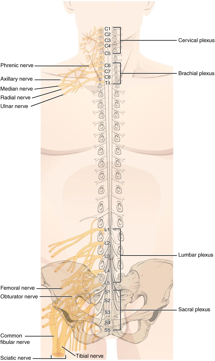
Thirty-one pairs of spinal nerves (8 cervical, 12 thoracic, 5 lumbar, 5 sacral, 1 coccygeal), each formed by an anterior (motor) and a posterior (sensory, with the spinal ganglion) root. The anterior rami of the cervical, lumbar and sacral nerves interweave into the cervical, brachial, lumbar and sacral plexuses that supply the neck and limbs.
Parts
Each part has its own card; press “Show on the model” to fly to it and select it in the 3D model above.
- 01
Roots and spinal ganglion
Radix anterior et posterior; Ganglion sensorium nervi spinalis
Each cord segment gives a posterior (sensory) root bearing the spinal ganglion of pseudounipolar neurons, and an anterior (motor) root. They join at the intervertebral foramen into a mixed spinal nerve about 1 cm long, which divides into anterior and posterior rami. There are 31 pairs: 8 cervical, 12 thoracic, 5 lumbar, 5 sacral and 1 coccygeal.
- 02
Brachial plexus
Plexus brachialis
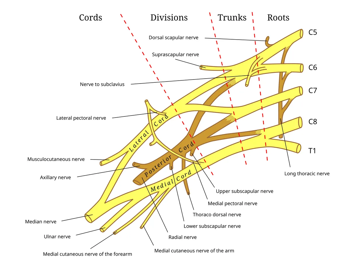
Formed by the anterior rami of C5–T1 and organised into roots, trunks (upper, middle, lower), anterior and posterior divisions and cords (lateral, posterior, medial) around the axillary artery. Upper-trunk injury (Erb, C5–C6) gives the “waiter’s tip” posture; lower-trunk injury (Klumpke, C8–T1) weakens the intrinsic hand muscles.
- 03
Musculocutaneous and axillary nerves
Nervus musculocutaneus; Nervus axillaris
The musculocutaneous nerve (lateral cord, C5–C7) pierces coracobrachialis, supplies biceps and brachialis and ends as the lateral antebrachial cutaneous nerve. The axillary nerve (posterior cord, C5–C6) winds round the surgical neck of the humerus through the quadrangular space to supply deltoid and teres minor; it is at risk in surgical-neck fractures and shoulder dislocation.
- 04
Median nerve
Nervus medianus
Formed by roots from the lateral and medial cords (C6–T1), it accompanies the brachial artery, passes between the heads of pronator teres and through the carpal tunnel. It supplies most forearm flexors, the thenar muscles and the lateral two lumbricals, and the palmar skin of the lateral three and a half digits; carpal tunnel compression is the commonest entrapment neuropathy.
- 05
Ulnar nerve
Nervus ulnaris
The terminal branch of the medial cord (C8–T1), passing behind the medial epicondyle (the “funny bone”) and through Guyon’s canal at the wrist. It supplies flexor carpi ulnaris, the medial half of flexor digitorum profundus and most intrinsic hand muscles; injury causes ulnar claw hand and numbness of the little finger.
- 06
Radial nerve
Nervus radialis
The largest branch of the posterior cord (C5–T1), running in the radial groove on the back of the humerus and dividing into superficial (sensory) and deep (posterior interosseous) branches. It supplies all extensors of elbow, wrist and fingers; a humeral shaft fracture can cause wrist drop.
- 07
Intercostal nerves
Nervi intercostales
The anterior rami of T1–T11 do not form plexuses; each runs in the costal groove below its vein and artery (vein–artery–nerve from above), between the internal and innermost intercostal muscles. They supply the intercostal and abdominal wall muscles and segmental skin; T12 is the subcostal nerve.
- 08
Lumbar plexus
Plexus lumbalis
Formed by the anterior rami of L1–L4 within psoas major. It gives the iliohypogastric, ilio-inguinal, genitofemoral and lateral femoral cutaneous nerves, the obturator nerve (adductors) and the femoral nerve (quadriceps, ending as the saphenous nerve).
- 09
Sacral plexus and sciatic nerve
Plexus sacralis; Nervus ischiadicus
Formed by the lumbosacral trunk (L4–L5) and the anterior rami of S1–S4 in front of piriformis. Its largest branch is the sciatic nerve (L4–S3), the largest nerve in the body, which leaves through the greater sciatic foramen below piriformis and divides into the tibial and common fibular nerves; the plexus also gives the superior and inferior gluteal and pudendal nerves.
- 10
Cauda equina
Cauda equina
The bundle of lumbar, sacral and coccygeal roots descending in the dural sac below the conus (from L1–L2) to reach their intervertebral foramina. Compression by a large disc herniation causes bilateral radicular pain, saddle anaesthesia and urinary retention, a surgical emergency.
Form and structure
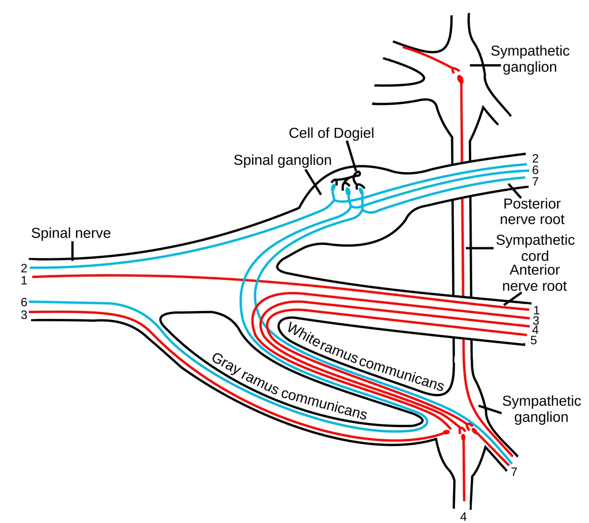
Each spinal nerve is formed by an anterior root (motor fibres from the anterior horn and, from T1 to L2, preganglionic sympathetic fibres) and a posterior root (sensory fibres whose cell bodies lie in the spinal ganglion). The roots unite in the intervertebral foramen into a short mixed nerve that immediately divides into a posterior ramus (muscles and skin of the back) and an anterior ramus (trunk and limbs). White and grey rami communicantes connect it with the sympathetic trunk.
In the thoracic region the anterior rami remain separate as the eleven pairs of intercostal nerves and the subcostal nerve, preserving the segmental pattern. In the cervical and lumbosacral regions they interweave into plexuses, so each peripheral nerve carries fibres from several cord segments and each segment contributes to several nerves.
Plexuses
The brachial plexus is described in five successive parts: roots (C5–T1), trunks (upper, middle, lower), divisions (anterior, posterior), cords (lateral, medial, posterior, named for their position around the axillary artery) and terminal branches. The lumbar plexus lies within psoas major; the sacral plexus lies on the anterior surface of piriformis and gives the sciatic nerve, the largest nerve in the body.
Cervical
- Roots
- C1–C4
- Main branches
- Phrenic nerve (C3–C5), ansa cervicalis, cutaneous nerves of the neck
- Supplies
- Diaphragm, infrahyoid muscles, skin of the neck
Brachial
- Roots
- C5–T1
- Main branches
- Musculocutaneous, axillary, radial, median, ulnar
- Supplies
- Shoulder and upper limb
Lumbar
- Roots
- L1–L4
- Main branches
- Iliohypogastric, ilioinguinal, genitofemoral, lateral femoral cutaneous, femoral, obturator
- Supplies
- Lower abdominal wall, front and medial thigh
Sacral
- Roots
- L4–S4
- Main branches
- Sciatic (tibial, common fibular), superior and inferior gluteal, pudendal
- Supplies
- Buttock, back of thigh, leg, foot, perineum
| Plexus | Roots | Main branches | Supplies |
|---|---|---|---|
| Cervical | C1–C4 | Phrenic nerve (C3–C5), ansa cervicalis, cutaneous nerves of the neck | Diaphragm, infrahyoid muscles, skin of the neck |
| Brachial | C5–T1 | Musculocutaneous, axillary, radial, median, ulnar | Shoulder and upper limb |
| Lumbar | L1–L4 | Iliohypogastric, ilioinguinal, genitofemoral, lateral femoral cutaneous, femoral, obturator | Lower abdominal wall, front and medial thigh |
| Sacral | L4–S4 | Sciatic (tibial, common fibular), superior and inferior gluteal, pudendal | Buttock, back of thigh, leg, foot, perineum |
Dermatomes and myotomes
The area of skin supplied by one posterior root is a dermatome; useful landmarks are C6 at the thumb, T4 at the nipple, T10 at the umbilicus, L4 on the medial leg and S1 along the lateral foot. Tendon reflexes test individual segments: biceps (C5–C6), triceps (C7), knee (L3–L4), ankle (S1). Together with muscle strength, they localise a lesion to a root or a peripheral nerve.
Clinical correlations
A lumbar disc herniation usually compresses the root that descends to the level below (an L4–L5 disc compresses the L5 root), causing pain along that dermatome. Carpal tunnel syndrome is the most common entrapment of the median nerve. In diabetes, radiculoplexus neuropathies differ from the usual polyneuropathy: they are subacute, painful and often monophasic with incomplete recovery, caused by immune-mediated ischaemic injury (microvasculitis), and may respond to immune therapy.[1]
Functional organisation
A dermatome is the skin area supplied by a single dorsal spinal root, a fundamental concept in neurological examination. A systematic review found that many textbook dermatome maps rest on flawed studies; the evidence-based map stresses that dermatomes overlap and vary between individuals.[2]
The brachial plexus is formed by the ventral rami of C5–T1, which successively form trunks, divisions, cords and terminal branches (musculocutaneous, axillary, radial, median, ulnar). Variants are common and affect plexus blocks; ultrasound and MRI assess each segment in trauma, tumours and plexitis.[3][4]
The median nerve is mixed: the nerve of pronation, of thumb, index, middle finger and wrist flexion, of thumb antepulsion and opposition, and of sensation over the palmar first three and a half digits. Along its course it passes several narrow passages where it can be compressed, notably the carpal tunnel and between the heads of pronator teres, and it may communicate with the ulnar nerve in the forearm.[5]
Clinical correlations
Entrapment neuropathies are compressions of peripheral nerves in anatomical tunnels: carpal tunnel syndrome (median nerve), the ulnar nerve at the elbow, the fibular nerve at the fibular neck and others. Diagnosis rests on topographic anatomy, examination, electrophysiology and neuromuscular ultrasound.[6]
The lumbosacral plexus (L1–S4) gives the femoral, obturator, sciatic and pudendal nerves. Plexopathy produces pain, weakness and sensory loss not confined to one root or nerve; causes include compression by retroperitoneal haematoma or tumour, diabetic and non-diabetic radiculoplexus neuropathy (ischaemic injury from perivascular inflammation and microvasculitis, which may respond to immunotherapy).[7][1]
Full-text reading (StatPearls)
The StatPearls chapters on this topic, reproduced verbatim. Their CC BY-NC-ND 4.0 licence allows unmodified copies only, so they are not translated or shortened.
Anatomy, Shoulder and Upper Limb, Brachial PlexusBayot ML, Nassereddin A, Varacallo MA · StatPearls 2023 · NBK500016 · 14k characters
Loading…
Bayot ML, Nassereddin A, Varacallo MA. Anatomy, Shoulder and Upper Limb, Brachial Plexus. StatPearls [Internet]. Treasure Island (FL): StatPearls Publishing; 2023. NBK500016 · PMID 29763192 · CC BY-NC-ND 4.0. Reproduced verbatim, unmodified.
Anatomy, Head and Neck: Brachial PlexusPolcaro L, Charlick M, Daly DT · StatPearls 2023 · NBK531473 · 10k characters
Loading…
Polcaro L, Charlick M, Daly DT. Anatomy, Head and Neck: Brachial Plexus. StatPearls [Internet]. Treasure Island (FL): StatPearls Publishing; 2023. NBK531473 · PMID 30285368 · CC BY-NC-ND 4.0. Reproduced verbatim, unmodified.
Organs in this system
References
Sources for the values and descriptions on this page, indexed in PubMed (US National Library of Medicine).
- [1]Diabetic radiculoplexus neuropathiesLaughlin RS et al., Handbook of clinical neurology 2014PMID 25410213
- [2]An evidence-based approach to human dermatomesLee MW et al., Clinical anatomy 2008PMID 18470936
- [3]Brachial plexus anatomy: normal and variantOrebaugh SL et al., TheScientificWorldJournal 2009PMID 19412559
- [4]Anatomy, Imaging, and Pathologic Conditions of the Brachial PlexusGilcrease-Garcia BM et al., Radiographics 2020PMID 33001787
- [5]Anatomy of the median nerve and its clinical applicationsSoubeyrand M et al., Hand surgery & rehabilitation 2020PMID 31816428
- [6]Common Entrapment NeuropathiesMalek E et al., Seminars in neurology 2019PMID 31639838
- [7]Lumbosacral plexopathyDyck PJ et al., Continuum 2014PMID 25299286
- [8]The human brain in numbers: a linearly scaled-up primate brainHerculano-Houzel S, 2009PMID 19915731Supports: Neuron count
- [9]Determinants of conduction velocity in myelinated nerve fibersWaxman SG, 1980PMID 6245357Supports: Conduction velocity
- [10]Quantitative imaging of energy expenditure in human brainPMID 22487547Supports: Cerebral energy expenditure