Triceps surae
Model
Layers
Z-Anatomy anatomical model based on BodyParts3D, licensed CC BY-SA 4.0.
Description
The calf muscle, made of gastrocnemius (two heads) and soleus, inserting on the calcaneus through the calcaneal (Achilles) tendon, the largest and strongest tendon in the body. It is the main plantarflexor, propelling the body in walking, running and jumping.
Parts
Each part has its own card; press “Show on the model” to fly to it and select it in the 3D model above.
- 01
Medial head of gastrocnemius
Caput mediale musculi gastrocnemii
Arises from the popliteal surface of the femur above the medial condyle and is larger and reaches lower than the lateral head. Gastrocnemius crosses both knee and ankle, flexing the knee and plantarflexing the foot, and is most effective with the knee straight (running, jumping). The medial head often tears in a sudden push-off (“tennis leg”).
- 02
Lateral head of gastrocnemius
Caput laterale musculi gastrocnemii
Arises from the lateral surface of the lateral femoral condyle; its tendon may contain a small sesamoid bone (fabella). The two heads bound the lower popliteal fossa and unite in the middle of the calf.
- 03
Soleus
Musculus soleus
A broad flat muscle deep to gastrocnemius, arising from the head and shaft of the fibula, the soleal line of the tibia and the tendinous soleal arch. Rich in fatigue-resistant slow fibres, it keeps us balanced when standing. Its intramuscular veins form the “calf muscle pump” returning blood to the heart; stasis there is where deep vein thrombosis usually begins.
- 04
Calcaneal (Achilles) tendon
Tendo calcaneus
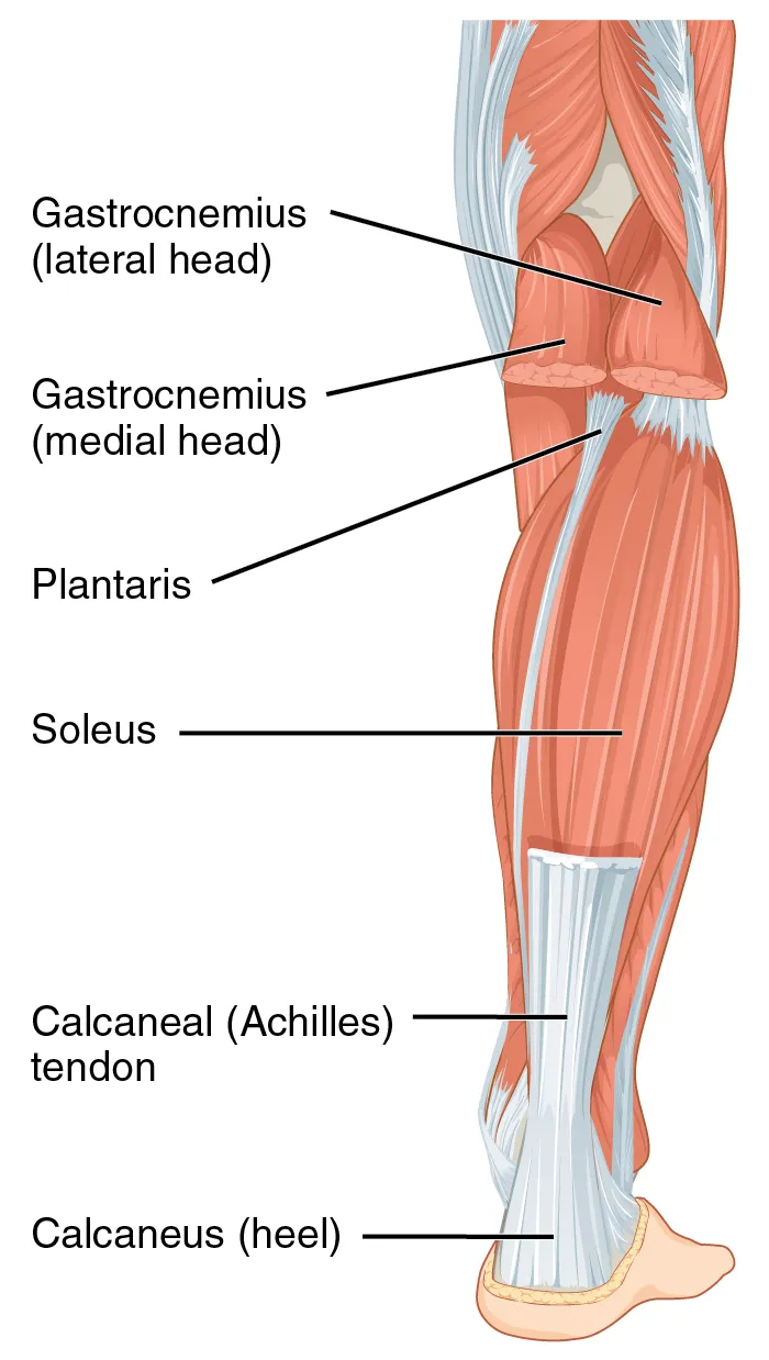
The thickest and strongest tendon in the body, about 15 cm long, inserting into the middle of the posterior surface of the calcaneus. Its fibres spiral about 90° as they descend. The zone 2–6 cm above the insertion is the most poorly vascularised and the usual site of rupture. Tapping it tests the ankle jerk (S1–S2).
- 05
Plantaris
Musculus plantaris
A small belly above the lateral femoral condyle with a very long, slender tendon running between gastrocnemius and soleus to insert beside the calcaneal tendon. It is absent in about 7–20% of people; its tendon is often harvested for grafts.
- 06
Nerve supply
Innervatio
The tibial nerve (S1–S2), a branch of the sciatic nerve, runs down the middle of the popliteal fossa and supplies both heads of gastrocnemius, soleus and plantaris. Injury abolishes standing on tiptoe.
Position and relations
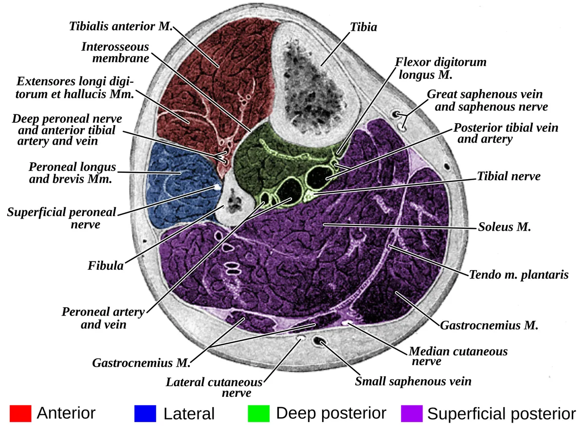
The two heads of gastrocnemius arise from the medial and lateral femoral condyles, so it crosses both knee and ankle; soleus lies deeper, arising from the upper tibia and fibula, and crosses only the ankle. The calcaneal tendon forms around mid-calf, twists slightly as it descends and inserts on the calcaneal tuberosity.
Form and structure
Gastrocnemius is rich in fast fibres and strongest with the knee extended (jumping, sprinting); soleus is rich in slow fibres and works continuously in standing to stop the body falling forward. Soleus contains large venous sinuses whose compression pumps blood back to the heart (the musculovenous pump or “second heart”), so prolonged immobility predisposes to deep vein thrombosis.
Vessels and nerves
The tibial nerve (S1–S2) supplies all three heads; the ankle jerk tests the S1 root. Blood comes from the sural arteries (popliteal) and branches of the posterior tibial and fibular arteries. The tendon 2–6 cm above its insertion is poorly vascularised and is the usual site of rupture.
Gastrocnemius, soleus and calcaneal tendon
The gastrocnemius is a superficial, biarticular muscle of the posterior leg that forms the triceps surae with the soleus. It arises from the medial and lateral femoral condyles and inserts into the calcaneus via the calcaneal tendon, plantarflexing the ankle and assisting knee flexion in gait, posture and propulsion. It is supplied by the sural branches of the popliteal artery and innervated by the tibial nerve.[1]
Variants include accessory heads, such as the gastrocnemius tertius, and altered origin or morphology. Common disorders are calf tears, calcaneal tendon disease and entrapment of the tibial nerve or popliteal vessels. Surgeons often use the medial head as a rotational or free flap, and operate on the muscle to treat equinus deformity.[1]
Skeletal muscles are length- and velocity-sensitive force generators built from vast numbers of sarcomeres; each muscle’s size, shape, fibre architecture and moment arms are tuned to its task, and muscles may have a preferred sarcomere-length operating range. In the leg, the gastrocnemius crosses two joints whereas the soleus crosses only the ankle, so their contribution to plantarflexion varies with knee position.[2]
Full-text reading (StatPearls)
The StatPearls chapters on this topic, reproduced verbatim. Their CC BY-NC-ND 4.0 licence allows unmodified copies only, so they are not translated or shortened.
Anatomy, Bony Pelvis and Lower Limb: Gastrocnemius MuscleBordoni B, Varacallo MA · StatPearls 2026 · NBK532946 · 29k characters
Loading…
Bordoni B, Varacallo MA. Anatomy, Bony Pelvis and Lower Limb: Gastrocnemius Muscle. StatPearls [Internet]. Treasure Island (FL): StatPearls Publishing; 2026. NBK532946 · PMID 30422541 · CC BY-NC-ND 4.0. Reproduced verbatim, unmodified.
Organs in this system
References
Sources for the values and descriptions on this page, indexed in PubMed (US National Library of Medicine).
- [1]Anatomy, Bony Pelvis and Lower Limb: Gastrocnemius MuscleBordoni B et al., StatPearls 2026PMID 30422541
- [2]Skeletal muscle design to meet functional demandsLieber RL et al., Philosophical transactions of the Royal Society of London. Series B, Biological sciences 2011PMID 21502118
- [3]Anatomy, Bony Pelvis and Lower Limb, Gluteus Maximus MuscleStatPearls (NCBI Bookshelf)PMID 30855781Supports: Gluteus maximus