Lymphatic system
Model
Z-Anatomy anatomical model based on BodyParts3D, licensed CC BY-SA 4.0.
Overview
The lymphatic system comprises the lymphatic vessels, the lymph and the lymphoid organs. The primary lymphoid organs (bone marrow, thymus) are where lymphocytes mature; the secondary lymphoid organs (lymph nodes, spleen, tonsils, mucosa-associated lymphoid tissue) are where they meet antigen.
Functions
- 01Return of interstitial fluid and leaked plasma proteins to the venous circulation
- 02Absorption of dietary fat from the small intestine through the lacteals
- 03Immune surveillance: filtering lymph and blood and initiating adaptive immune responses
Lymph circulation
Lymphatic vessels form the body’s secondary circulatory system, absorbing and transporting lymph and orchestrating immune surveillance and responses. The network forms mainly by lymphangiogenesis, the sprouting of new vessels from pre-existing lymphatics or embryonic veins; abnormal lymphangiogenesis accompanies many diseases.[1]
At the blood capillaries, part of the fluid filtered into the interstitium is not reabsorbed into the venules. Lymphatic capillaries, blind-ended and lined by overlapping endothelial cells that act as valves, take up this fluid together with proteins and immune cells. Lymph then flows through valved collecting vessels whose smooth muscle contracts rhythmically, passes through successive node stations and reaches the thoracic duct and right lymphatic duct, which return it to the veins at the root of the neck; several litres of lymph return to the circulation each day.
When lymph flow is obstructed, for example after axillary node dissection or radiotherapy, protein-rich fluid accumulates in the tissues and causes lymphoedema. In the small intestine, the lacteals of the villi absorb fat as chylomicrons, giving intestinal lymph its milky appearance (chyle).
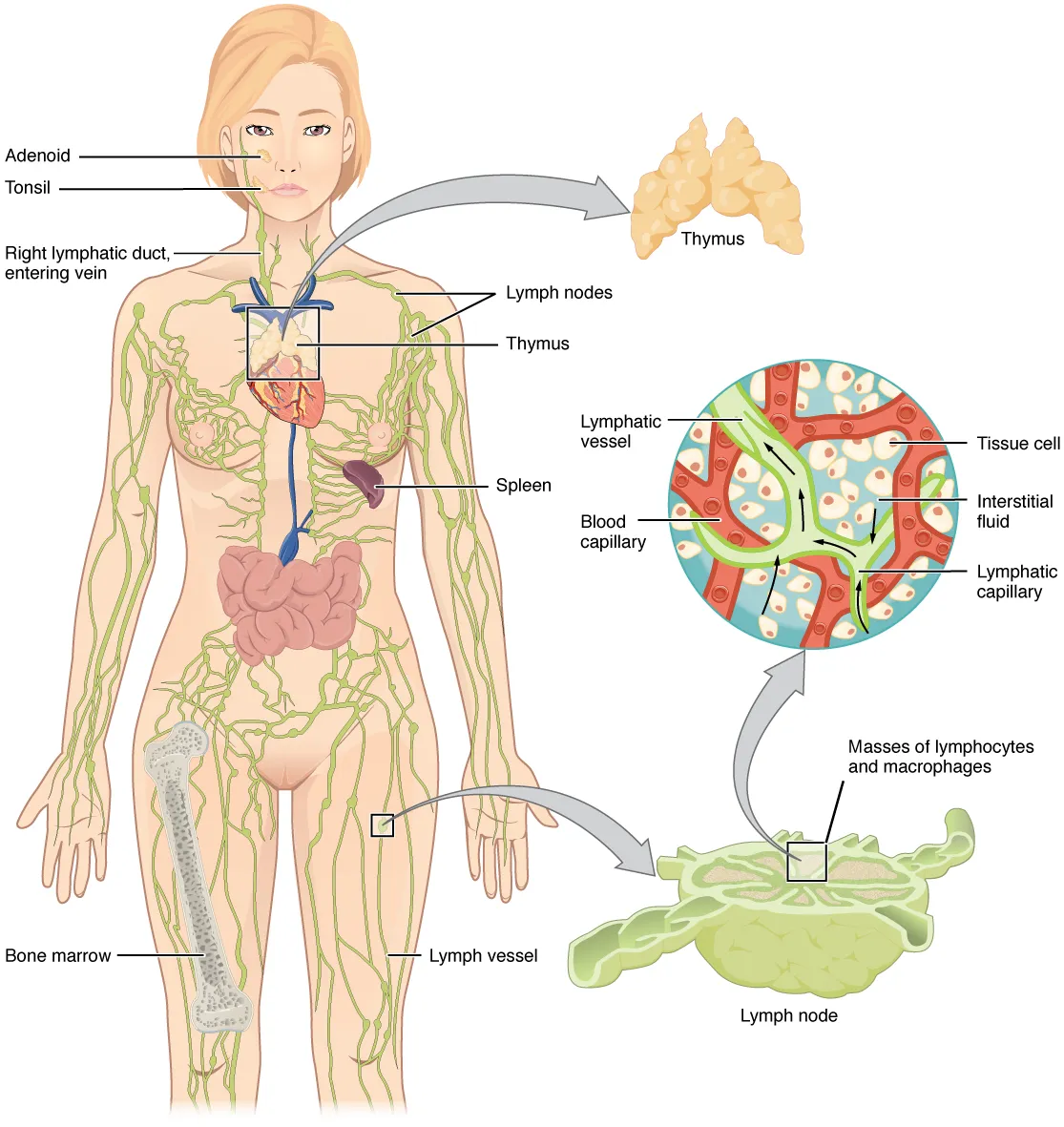
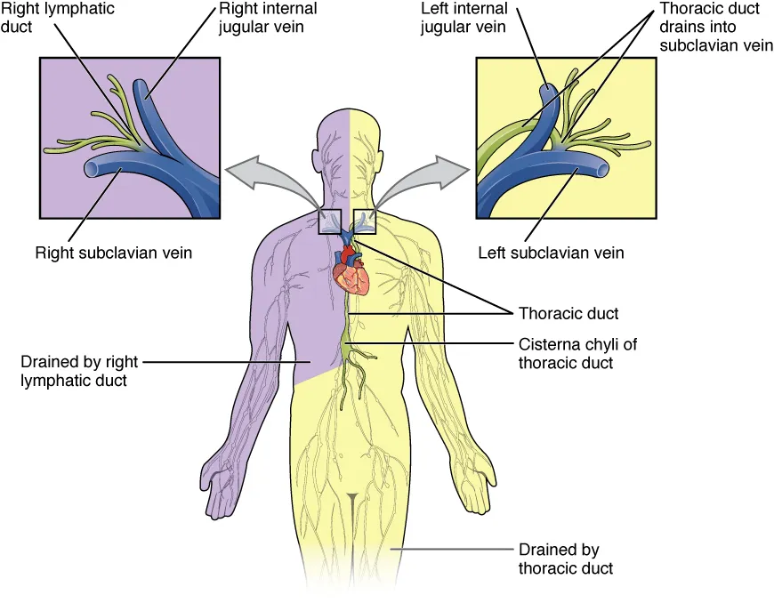
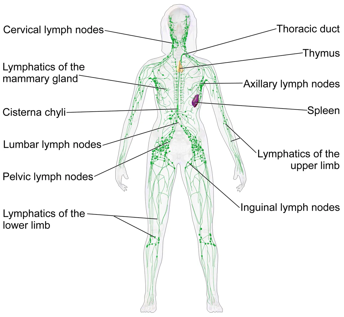
Lymphatic pathways: from capillaries to the two great ducts
By the Starling law, fluid is filtered into the interstitium at the arteriolar end of capillaries and reabsorbed at the venular end; depending on the tissue, filtration exceeds reabsorption by about 10%, and this excess is carried away by the lymphatic system. Once interstitial fluid enters a lymphatic capillary it is called lymph. Capillaries drain into collecting vessels, which converge into the large lymphatic trunks; on the way lymph passes regional nodes and then collecting nodes (which receive several regional nodes), where it is filtered and screened by the immune system.[2]
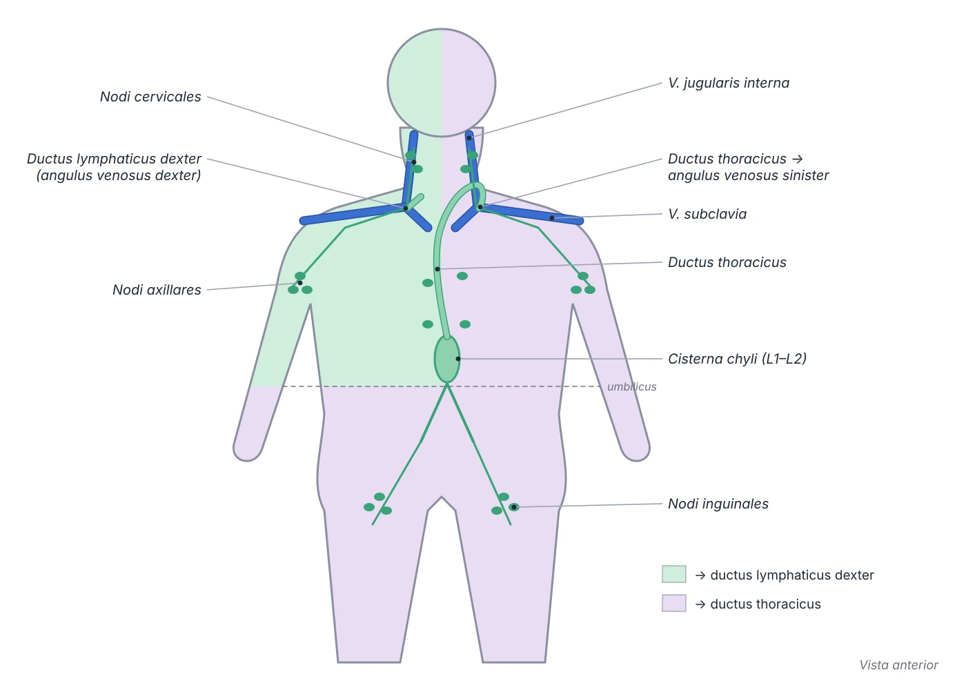
Drainage is asymmetric. The right lymphatic duct receives lymph only from the upper right quadrant of the body through three trunks: the right subclavian trunk (right upper limb and right body wall down to the umbilicus), the right bronchomediastinal trunk (thoracic cavity) and the right jugular trunk (right half of the head and neck), and empties at the right jugulovenous angle. The thoracic duct receives the corresponding left trunks plus the cisterna chyli, and so drains the abdominal viscera below the diaphragm, the abdominal wall below the umbilicus and both lower limbs, emptying at the left venous angle. In the lower limb, popliteal nodes drain to the inguinal nodes; these form a vertical chain along the great saphenous vein and a horizontal group below the inguinal ligament, also receive the lower abdominal wall and lower back up to the umbilicus, and drain to the external iliac nodes.[2]
Architecture of a lymph node and cell traffic
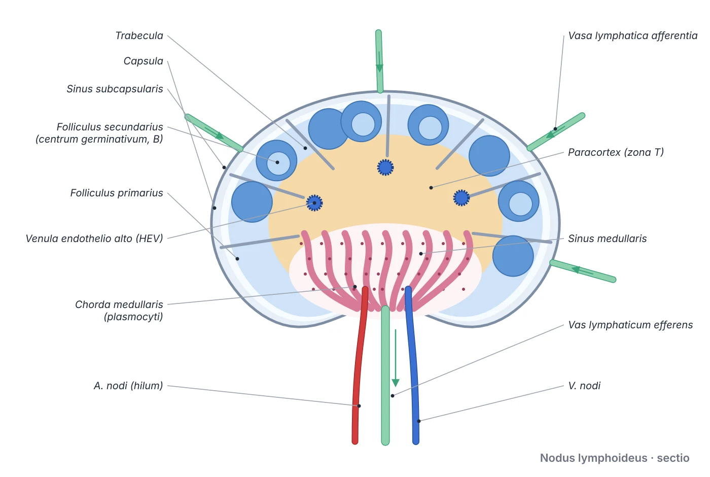
A node is bean-shaped and physiologically 1–20 mm in size depending on its site. The convex surface receives several afferent lymphatics; the hilum on the concave surface is where efferent lymphatics leave and blood vessels enter. The fibrous capsule sends trabeculae inward. A meshwork of reticular cells making type III collagen supports three zones: the superficial cortex holds B cells with follicular dendritic cells in primary follicles, which form germinal centres and become secondary follicles when B cells are activated; the deep cortex (paracortex) holds T cells with dendritic cells in deep cortical units, where T cells proliferate without forming germinal centres; the medulla consists of medullary cords containing plasma cells. Lymph enters the subcapsular sinus, runs through trabecular sinuses to the medullary sinuses and leaves by the efferent vessel.[2][3]
Most lymphocytes enter a node not from lymph but through high endothelial venules in the paracortex, crossing the wall by means of endothelial adhesion molecules. Chemokine signals send naive B cells to the superficial cortex and naive T cells to the deep cortex; if they meet no antigen they leave by the efferent vessel. The meshwork in the sinuses traps antigens and bacteria that have breached the first barriers, and sinus macrophages phagocytose debris, red cells and carbon particles. Activated B cells become plasma cells, plasmablasts and memory B cells; plasma cells move to the medullary cords and release antibody into the lymph. Node enlargement may therefore reflect infection (necrotising granulomas in tuberculosis, fungal infection and cat-scratch disease; reactive hyperplasia in infectious mononucleosis), autoimmune disease, lymphoma or metastasis; the number, site and size of involved nodes are the most important prognostic factors in cancer staging. Loss of nodes after dissection causes lymphoedema.[2][3]
The thymus and T-cell selection
The thymus lies in the superior mediastinum behind the sternum and has two lobes 30–40 mm long and 25–35 mm wide; cervical extensions may reach the lower border of the thyroid (thyrothymic ligament), and posteriorly it is related to the great vessels, trachea and pericardium. It develops from the ventral part of the third pharyngeal pouch and descends into the mediastinum, enlarges until about 3 years of age, begins to involute at puberty (probably because of rising androgens) and is progressively replaced by fat, although it continues to produce T cells in adults at a much lower rate. Its arteries come from the inferior thyroid, internal thoracic and pericardiacophrenic arteries; its veins drain mainly to the left brachiocephalic vein.[4][5]
Each lobule has a darkly staining cortex (crowded with thymocytes, i.e. T lymphoblasts) and a paler medulla. The blood–thymus barrier of the cortex consists of capillary endothelium surrounded by pericytes, a basement membrane and a layer of joined squamous thymic epithelial cells, keeping blood-borne macromolecules away from maturing cells. Here T cells undergo positive selection (retaining cells whose receptor can recognise the body’s own MHC molecules) and then negative selection (removing cells that react too strongly to self-antigens), establishing central tolerance and limiting autoimmunity. The result is the CD4+ helper, CD8+ cytotoxic (killing targets through Fas/FasL or perforin and granzymes) and CD4+CD25+ regulatory lineages. Thymic hyperplasia commonly accompanies myasthenia gravis, and thymectomy is one of its treatments.[5][6][4]
Spleen, Waldeyer ring and mucosal lymphoid tissue
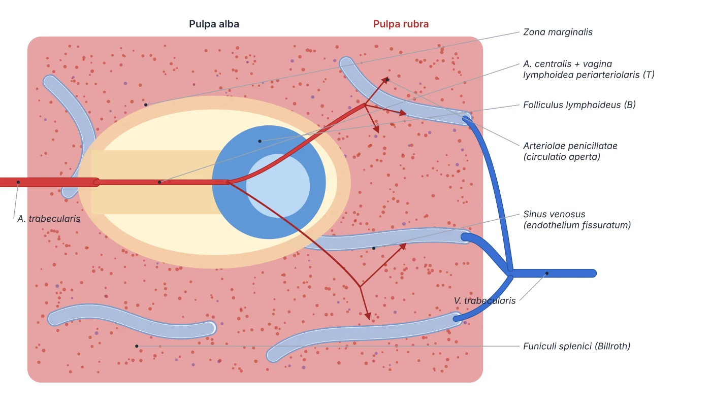
The spleen is the largest lymphatic organ, lying in the left hypochondrium between the gastric fundus and the diaphragm at the level of ribs 9–11; it is 10–12 cm long, weighs 150–200 g and is normally not palpable. The diaphragmatic surface is convex; the visceral surface is concave, with gastric, renal, colic and pancreatic impressions, and its hilum transmits the vessels and nerves and gives attachment to the splenorenal and gastrosplenic ligaments. It arises from mesenchyme between the layers of the dorsal mesogastrium in weeks 5–6 and is haemopoietic in fetal life. The splenic artery (from the coeliac trunk) divides into about five non-anastomosing branches, giving the spleen a vascular segmentation that permits partial splenectomy; the splenic vein runs behind the pancreas to help form the portal vein. The spleen has only efferent lymphatics.[7]
The white pulp around the central arterioles is where B and T cells respond to blood-borne antigens; the red pulp, most of the parenchyma, consists of the cords of Billroth (a reticular meshwork crowded with red cells, macrophages and platelets) alternating with venous sinusoids with fenestrated endothelium, where aged or deformed red cells are held back and destroyed; the marginal zone between them directs blood-borne pathogens to the white pulp. People without a spleen are prone to infection by encapsulated bacteria (pneumococcus, meningococcus, Haemophilus influenzae) and need vaccination. In the mucosa, the Waldeyer ring of palatine, tubal, pharyngeal and lingual tonsils surrounds the crossroads of the pharynx. The palatine tonsils develop from the second pharyngeal pouch and are mainly B-cell tissue; surface M cells capture antigen, B cells proliferate in germinal centres and secrete IgA, in a structure resembling the intestinal Peyer patches. Tonsillar activity peaks at 4–12 years of age and declines after puberty.[7][8]
Bone marrow
- Embryonic origin
- Mesenchyme
- Main role
- Primary lymphoid organ: production and maturation of B cells
Thymus
- Embryonic origin
- Third pharyngeal pouch (ventral part)
- Main role
- Primary lymphoid organ: T-cell selection
Lymph nodes
- Embryonic origin
- Mesenchymal condensations, lymph sacs
- Main role
- Filtering lymph, starting immune responses
Spleen
- Embryonic origin
- Mesenchyme of the dorsal mesogastrium
- Main role
- Filtering blood, removing aged red cells, responding to blood-borne antigen
Palatine tonsil
- Embryonic origin
- Second pharyngeal pouch
- Main role
- Mucosal immunity, IgA secretion
| Organ | Embryonic origin | Main role |
|---|---|---|
| Bone marrow | Mesenchyme | Primary lymphoid organ: production and maturation of B cells |
| Thymus | Third pharyngeal pouch (ventral part) | Primary lymphoid organ: T-cell selection |
| Lymph nodes | Mesenchymal condensations, lymph sacs | Filtering lymph, starting immune responses |
| Spleen | Mesenchyme of the dorsal mesogastrium | Filtering blood, removing aged red cells, responding to blood-borne antigen |
| Palatine tonsil | Second pharyngeal pouch | Mucosal immunity, IgA secretion |
Principal organs
The largest lymphoid organ, lying in the left hypochondrium behind ribs IX–XI, about 12 cm long and weighing about 150 g in adults. The red pulp filters blood and removes ageing red cells; the white pulp is lymphoid tissue sheathing the arterioles. It is supplied by the splenic artery, a branch of the coeliac trunk.
ThymusThymusA bilobed primary lymphoid organ in the superior and anterior mediastinum, behind the manubrium of the sternum. T lymphocytes differentiate and are selected here; it is relatively largest in early childhood and involutes after puberty, being replaced by fat.
Lymph nodesNodi lymphoideiSmall bean-shaped organs along the lymphatic vessels, clustered in the neck, axilla, groin, mediastinum and around the great abdominal vessels. The cortex holds B-cell follicles, the paracortex T cells, and the medulla the medullary cords and lymphatic sinuses.
Palatine tonsilsTonsilla palatinaPaired masses of lymphoid tissue in the tonsillar fossa between the palatoglossal and palatopharyngeal arches, forming part of the pharyngeal lymphoid ring (Waldeyer’s ring) with the pharyngeal, tubal and lingual tonsils. Surface crypts enlarge the area exposed to antigen.
Reference values
- The human body has some 500–700 lymph nodes, most numerous in the neck, axillae, groins and abdomen.
- The spleen holds about a third of the body’s platelets, which is why splenomegaly can cause thrombocytopenia.
References
Sources for the values and descriptions on this page, indexed in PubMed (US National Library of Medicine).
- [1]Lymphatic vessel: origin, heterogeneity, biological functions, and therapeutic targetsHu Z et al., Signal transduction and targeted therapy 2024PMID 38172098
- [2]Anatomy, Lymph NodesWięcław M et al., StatPearls 2026PMID 32491649
- [3]Histology, Lymph NodesMercadante AA et al., StatPearls 2023PMID 32644479
- [4]Anatomy, Head and Neck, ThymusRemien K et al., StatPearls 2025PMID 30969570
- [5]Anatomy, Head and Neck: Blood Thymus BarrierBruss DM et al., StatPearls 2023PMID 31536208
- [6]Histology, T-Cell LymphocyteSauls RS et al., StatPearls 2023PMID 30571054
- [7]Anatomy, Abdomen and Pelvis, SpleenOmole AE et al., StatPearls 2026PMID 29489170
- [8]Anatomy, Head and Neck: TonsilsMasters KG et al., StatPearls 2025PMID 30969614