Gluteus maximus
Model
Layers
Z-Anatomy anatomical model based on BodyParts3D, licensed CC BY-SA 4.0.
Description
The largest and heaviest muscle in the body, shaping the buttock. It extends and laterally rotates the hip and is essential for rising from sitting, climbing stairs, running and stabilising the trunk.
Parts
Each part has its own card; press “Show on the model” to fly to it and select it in the 3D model above.
- 01
Origin
Origo
The largest and heaviest muscle in the body, arising broadly from the ilium behind the posterior gluteal line, the back of the sacrum and coccyx, the sacrotuberous ligament and the thoracolumbar fascia. Its coarse fasciculi run obliquely down and laterally.
- 02
Insertion: iliotibial tract and gluteal tuberosity
Insertio
About three quarters of the fibres, the superficial ones, insert into the iliotibial tract, through which the muscle steadies the extended knee; the remaining deep fibres attach to the gluteal tuberosity of the femur. Bursae separate it from the greater trochanter and ischial tuberosity.
- 03
Actions
Functio
A powerful extensor and lateral rotator of the thigh, called on mainly for forceful movements: rising from a chair, climbing stairs, running and jumping. It is almost silent in level walking. It underpins upright posture by keeping the pelvis from tipping forwards.
- 04
Nerve and blood supply
Innervatio et vascularisatio
The inferior gluteal nerve (L5–S2) and artery leave the pelvis through the greater sciatic foramen below piriformis. Paralysis makes climbing stairs and rising from a seat difficult while level walking stays almost normal.
- 05
Relation to the sciatic nerve
Relationes cum nervo ischiadico
The sciatic nerve runs deep to gluteus maximus in the lower medial quadrant of the buttock. Intramuscular injections must therefore go into the upper outer quadrant (or the ventrogluteal site over gluteus medius) to avoid it.
Position and relations
It arises broadly from the ilium behind the posterior gluteal line, the back of the sacrum and coccyx, the thoracolumbar fascia and the sacrotuberous ligament. Most fibres insert into the iliotibial tract, the deep fibres into the gluteal tuberosity of the femur. Its lower border crosses the gluteal fold; it covers the ischial tuberosity when standing but uncovers it when sitting.
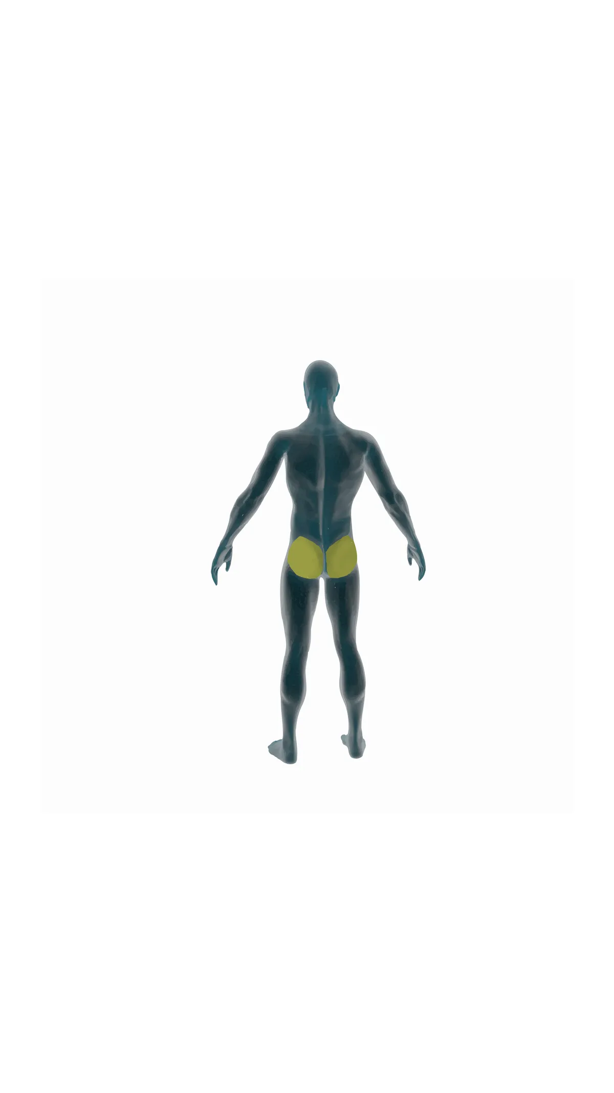
Form and structure
It is relatively quiet in level walking and works hard in loaded hip extension (rising, climbing, sprinting). Through the iliotibial tract it helps stabilise the extended knee. Beneath it lie bursae (trochanteric, ischial) and the deep lateral rotators (piriformis, gemelli, obturator internus, quadratus femoris).
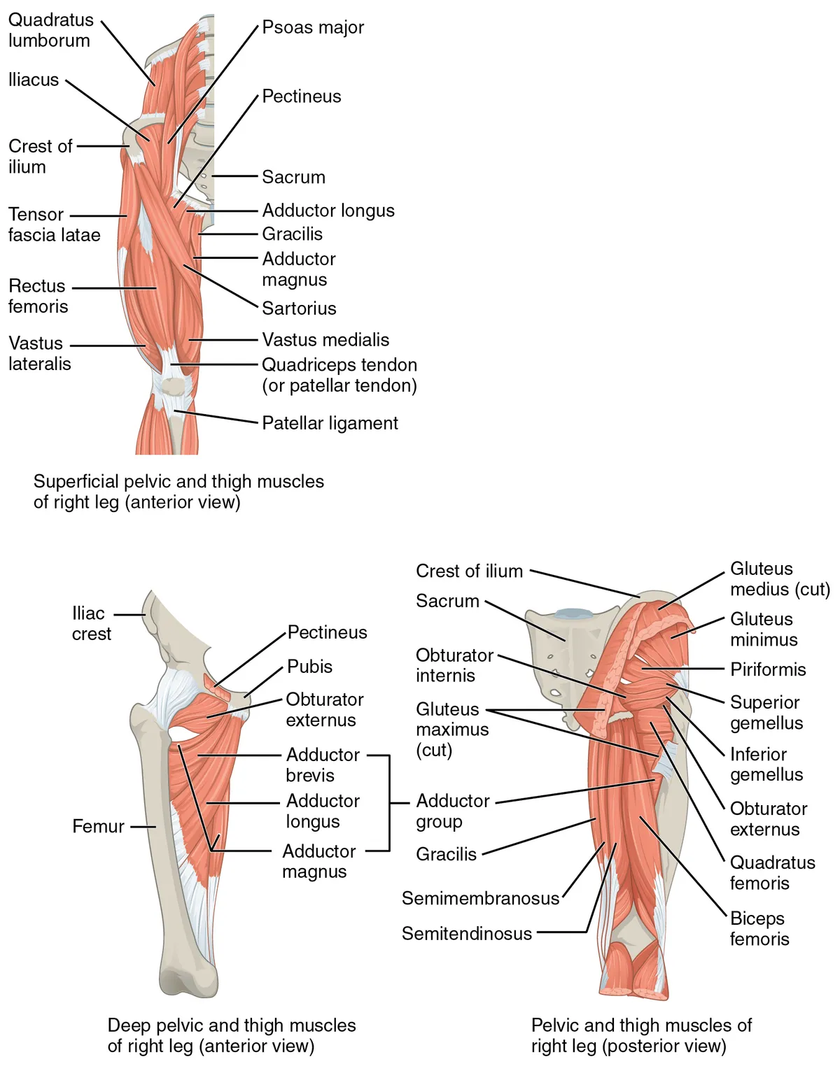
Vessels and nerves
It is supplied by the inferior gluteal nerve (L5–S2) and by the superior and inferior gluteal arteries (internal iliac). The sciatic nerve runs beneath it in the lower half of the buttock, which is why intramuscular injections are given in the upper outer quadrant.
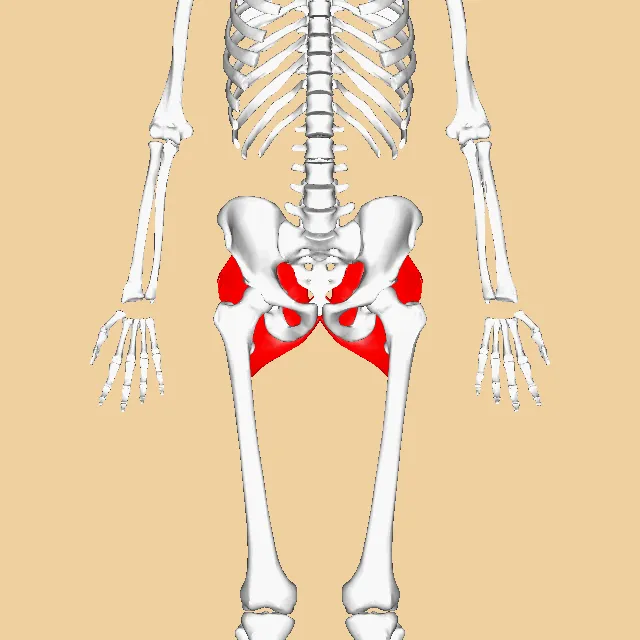
The gluteal region and hip extension
The gluteal region on the back of the pelvis contains muscles critical for movement and upright stability and is a conduit for several neurovascular structures to the lower limb. Of the gluteus maximus, medius and minimus, the maximus is the largest and most superficial, which makes it important in surgical approaches to the hip.[1]
The gluteus maximus is one of the main hip extensors. A systematic review of loaded exercises, expressing activation as a percentage of maximal voluntary isometric contraction, found the step-up and its variants produced the highest activation, followed by several exercises reaching very high levels (>60%), such as deadlifts, barbell hip thrusts, split squats and lunges.[2]
Lateral hip pain, formerly called trochanteric bursitis, is now thought to arise mainly from gluteal tendinopathy, a common condition, particularly in postmenopausal women, that markedly affects quality of life.[3]
Full-text reading (StatPearls)
The StatPearls chapters on this topic, reproduced verbatim. Their CC BY-NC-ND 4.0 licence allows unmodified copies only, so they are not translated or shortened.
Anatomy, Bony Pelvis and Lower Limb, Gluteus Maximus MuscleElzanie A, Borger J · StatPearls 2023 · NBK538193 · 11k characters
Loading…
Elzanie A, Borger J. Anatomy, Bony Pelvis and Lower Limb, Gluteus Maximus Muscle. StatPearls [Internet]. Treasure Island (FL): StatPearls Publishing; 2023. NBK538193 · PMID 30855781 · CC BY-NC-ND 4.0. Reproduced verbatim, unmodified.
Organs in this system
References
Sources for the values and descriptions on this page, indexed in PubMed (US National Library of Medicine).
- [1]Anatomy, Bony Pelvis and Lower Limb, Gluteus Maximus MuscleElzanie A et al., StatPearls 2023PMID 30855781
- [2]Gluteus Maximus Activation during Common Strength and Hypertrophy Exercises: A Systematic ReviewNeto WK et al., Journal of sports science & medicine 2020PMID 32132843
- [3]Gluteal Tendinopathy: Integrating Pathomechanics and Clinical Features in Its ManagementGrimaldi A et al., The Journal of orthopaedic and sports physical therapy 2015PMID 26381486