Liver
Model
Hover or tap to see names, click to read a description; click a name in the legend to fly to it. Procedurally built to histological proportions; colours follow illustration conventions.
Description
The largest solid organ (about 1.4–1.5 kg), situated in the right hypochondrium. It secretes bile, metabolises carbohydrates, lipids and proteins, synthesises plasma proteins and detoxifies xenobiotics.
Parts
Each part has its own card; press “Show on the model” to fly to it and select it in the 3D model above.
- 01
Right lobe
Lobus hepatis dexter
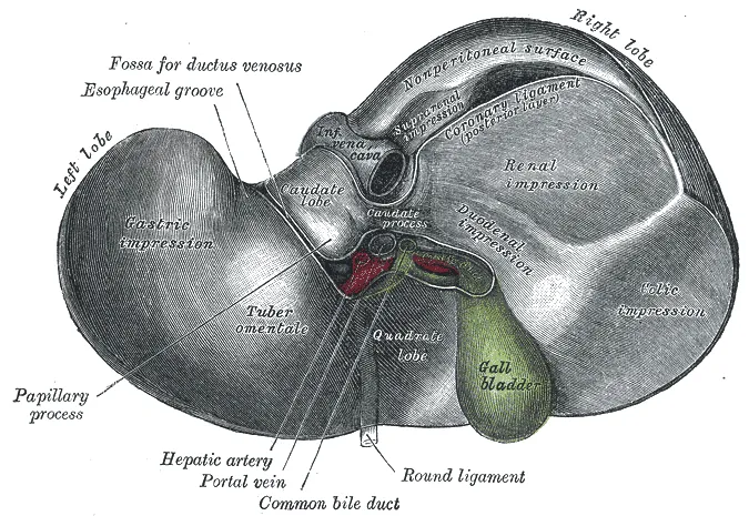
The largest lobe, anatomically to the right of the falciform ligament; functionally the right liver lies right of Cantlie’s line (from the gallbladder to the inferior vena cava) and comprises segments V–VIII. Its visceral surface bears impressions of the right kidney, colon and duodenum.
- 02
Left lobe
Lobus hepatis sinister
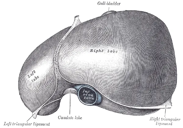
Lies left of the falciform ligament, thin and draped over the stomach, and comprises segments II and III. The functional left liver also includes segment IV (right of the falciform), the basis for left hepatectomy and living-donor grafts (the left lateral section for children).
- 03
Caudate and quadrate lobes
Lobus caudatus; Lobus quadratus
Two small lobes on the visceral surface on either side of the porta hepatis. The quadrate lobe (segment IVb) lies between the gallbladder and ligamentum teres; the caudate lobe (segment I) embraces the inferior vena cava, receives both portal branches and drains directly into the cava, which is why it enlarges in Budd–Chiari syndrome.
- 04
Couinaud segments
Segmenta hepatis
The three hepatic veins divide the liver vertically and the plane of the portal bifurcation horizontally, giving eight segments (I–VIII) numbered clockwise in frontal view. Each has its own portal pedicle (portal vein, hepatic artery, bile duct) and can be resected independently.
- 05
Porta hepatis
Porta hepatis
A transverse fissure about 5 cm long on the visceral surface where the portal vein (posterior), proper hepatic artery (anterior left) and hepatic duct (anterior right) enter or leave with nerves and lymphatics, wrapped in the free edge of the lesser omentum. The Pringle manoeuvre clamps this pedicle to limit bleeding in liver surgery.
- 06
Hepatic veins
Venae hepaticae
The right, middle and left hepatic veins run between segments, have no valves and drain directly into the inferior vena cava just below the diaphragm; the middle vein lies in Cantlie’s plane. Outflow obstruction here causes Budd–Chiari syndrome.
- 07
Ligaments and bare area
Ligamentum falciforme; Ligamentum teres hepatis; Area nuda
The falciform ligament attaches the liver to the anterior abdominal wall, its free edge carrying the ligamentum teres (the obliterated umbilical vein); the coronary and triangular ligaments outline the bare area on the posterior surface, where the liver contacts the diaphragm without peritoneum. The ligamentum venosum is the remnant of the ductus venosus.
- 08
Hepatic lobule
Lobulus hepatis
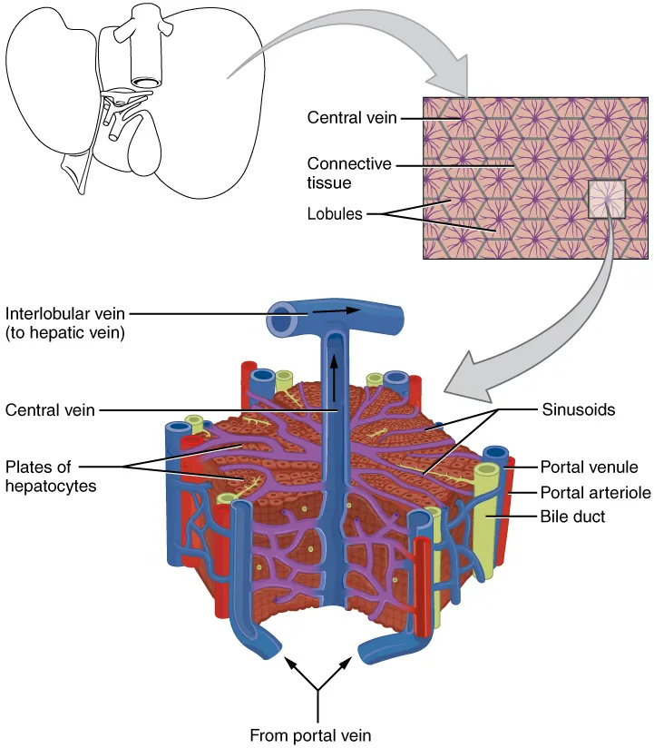
A hexagonal structural unit about 1–2 mm across, with a central vein at its centre and portal tracts (portal triads) at its corners. Radiating plates of hepatocytes alternate with sinusoids lined by Kupffer cells; bile flows in canaliculi opposite to blood towards the bile ductules of the portal tracts.
Position and relations
The liver lies in the right hypochondrium and epigastrium, under the right dome of the diaphragm, mostly covered by the ribs. Its smooth diaphragmatic surface faces the diaphragm; its visceral surface relates to the stomach, duodenum, hepatic flexure of the colon, right kidney and gallbladder. The porta hepatis transmits the portal vein, hepatic artery proper and hepatic ducts.[1]
Form and structure
The structural unit of the liver is the hexagonal hepatic lobule, with a central vein in the middle and portal triads (portal vein branch, hepatic artery, bile duct) at its corners. Hepatocytes form plates separated by sinusoids lined with Kupffer macrophages; the space of Disse contains vitamin A-storing stellate cells.[2][3]
Segments and vessels
Because the liver looks homogeneous, its anatomy is described by its vessels. In 1897 Cantlie first correctly divided right and left liver along a line from the gallbladder bed to the inferior vena cava; Couinaud later divided the liver into eight segments, each with its own portal pedicle (portal vein branch, hepatic artery, bile duct) and drained by hepatic veins lying between segments.[1]
This division underpins modern interventions such as transplantation, segmental resection, transarterial chemoembolisation and portal vein embolisation: none could be performed safely without a precise knowledge of hepatic vascular anatomy.[4]
Liver: metabolism, immunity and regeneration
The liver is a large organ for nutrient metabolism, detoxification and energy storage, and also an immune organ: it hosts B and T cells, dendritic cells and several resident macrophage populations (Kupffer cells), and produces acute-phase proteins in inflammation. The reason is its position in the circulation: it is the first major organ downstream of the gut, receiving nutrient-rich blood (with antigens and bacterial products) through the portal vein.[2][5]
Single-cell and spatial transcriptomics have built a liver cell atlas revealing zonation along the lobule from portal tract to central vein: hepatocytes in each zone express different metabolic programmes, and hepatocytes, sinusoidal endothelial cells, stellate cells and macrophages communicate in defined neighbourhoods.[6]
The liver regenerates remarkably after injury through coordinated action of many cell types and signalling pathways, linked to metabolic reprogramming of glucose, lipid and amino-acid metabolism. With ageing, oxidative stress, inflammaging, genomic instability and epigenetic dysregulation erode its structure and function and blunt regeneration.[7][8]
Metabolic dysfunction-associated steatotic liver disease (formerly non-alcoholic fatty liver disease) is the commonest chronic liver disease; hepatic stellate cells switch from quiescence to an activated fibrogenic state, driving progression from steatosis to steatohepatitis and cirrhosis.[9][10][11]
Full-text reading (StatPearls)
The StatPearls chapters on this topic, reproduced verbatim. Their CC BY-NC-ND 4.0 licence allows unmodified copies only, so they are not translated or shortened.
Anatomy, Abdomen and Pelvis: LiverVernon H, Wehrle CJ, Alia VSK, Kasi A · StatPearls 2022 · NBK500014 · 9k characters
Loading…
Vernon H, Wehrle CJ, Alia VSK, Kasi A. Anatomy, Abdomen and Pelvis: Liver. StatPearls [Internet]. Treasure Island (FL): StatPearls Publishing; 2022. NBK500014 · PMID 29763190 · CC BY-NC-ND 4.0. Reproduced verbatim, unmodified.
Organs in this system
References
Sources for the values and descriptions on this page, indexed in PubMed (US National Library of Medicine).
- [1]Clinical and surgical anatomy of the liver: a review for cliniciansJuza RM et al., Clinical anatomy 2014PMID 24453062
- [2]Liver macrophages: development, dynamics, and functionsNusse Y et al., Cellular & molecular immunology 2025PMID 40461700
- [3]Pathogenesis of liver fibrosisHernandez-Gea V et al., Annual review of pathology 2011PMID 21073339
- [4]Liver anatomyAbdel-Misih SR et al., The Surgical clinics of North America 2010PMID 20637938
- [5]Priming and Maintenance of Adaptive Immunity in the LiverKawashima K et al., Annual review of immunology 2024PMID 38360545
- [6]Single-cell and spatially resolved transcriptomics for liver biologyLin P et al., Hepatology 2024PMID 37002587
- [7]Molecular mechanisms in liver repair and regeneration: from physiology to therapeuticsMa X et al., Signal transduction and targeted therapy 2025PMID 39920130
- [8]Liver aging: underlying mechanisms and therapeutic strategiesZhong L et al., Molecular aspects of medicine 2025PMID 40886472
- [9]Pathophysiological underpinnings of metabolic dysfunction-associated steatotic liver diseaseKuchay MS et al., American journal of physiology. Cell physiology 2025PMID 40244183
- [10]Regulation of Hepatic Stellate Cell Phenotypes in Metabolic Dysfunction-Associated SteatohepatitisKisseleva T et al., Gastroenterology 2025PMID 40120772
- [11]MAFLD: A Consensus-Driven Proposed Nomenclature for Metabolic Associated Fatty Liver DiseaseEslam M et al., Gastroenterology 2020PMID 32044314
- [12]Gastrointestinal mean transit times in young and middle-aged healthy subjectsPMID 11318834Supports: Gastrointestinal transit time
- [13]Revised Estimates for the Number of Human and Bacteria Cells in the BodySender R, Fuchs S, Milo R, 2016PMID 27541692Supports: Colonic bacterial count