Hip bone
Model
Hover or tap to see names, click to read a description; click a name in the legend to fly to it. Procedurally built to histological proportions; colours follow illustration conventions.
Description
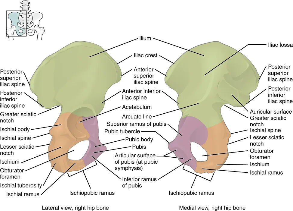
A large flat bone formed by three bones (ilium, ischium, pubis) that fuse at the acetabulum by adulthood. The two hip bones and the sacrum form the bony pelvis, which transmits body weight to the lower limbs and protects the pelvic organs.
Parts
Each part has its own card; press “Show on the model” to fly to it and select it in the 3D model above.
- 01
Ilium
Os ilium
The upper, widest part of the hip bone. The iliac crest is palpable under the skin at the level of L4 (the landmark for lumbar puncture) and ends at the anterior and posterior superior iliac spines. The inner concavity is the iliac fossa and the auricular surface behind articulates with the sacrum. The crest is a source of bone graft and marrow aspirates.
- 02
Ischium
Os ischii
The posteroinferior part, with a body and ramus. The ischial tuberosity bears weight in sitting and gives origin to the hamstrings. The ischial spine is the landmark for pudendal nerve block in obstetrics and separates the greater and lesser sciatic notches.
- 03
Pubis
Os pubis
The anterior part, with a body and superior and inferior rami. The two bodies meet at the pubic symphysis through a fibrocartilaginous disc; the pubic tubercle anchors the inguinal ligament. The rami and the ischium encircle the obturator foramen, crossed by the obturator neurovascular bundle.
- 04
Acetabulum
Acetabulum
A deep cup on the lateral surface where the ilium, ischium and pubis fuse through the triradiate cartilage (ossified at about 15–17 years). A C-shaped lunate articular surface surrounds the acetabular fossa, and the acetabular labrum deepens the socket. A shallow acetabulum is the basis of developmental dysplasia of the hip.
- 05
Obturator foramen
Foramen obturatum
The largest foramen of the skeleton, almost closed by the obturator membrane except for the obturator canal above, which passes the obturator artery, vein and nerve. A rare obturator hernia, typical of thin elderly women, can cause pain along the inner thigh.
- 06
Pelvic girdle and pelvis
Cingulum pelvicum
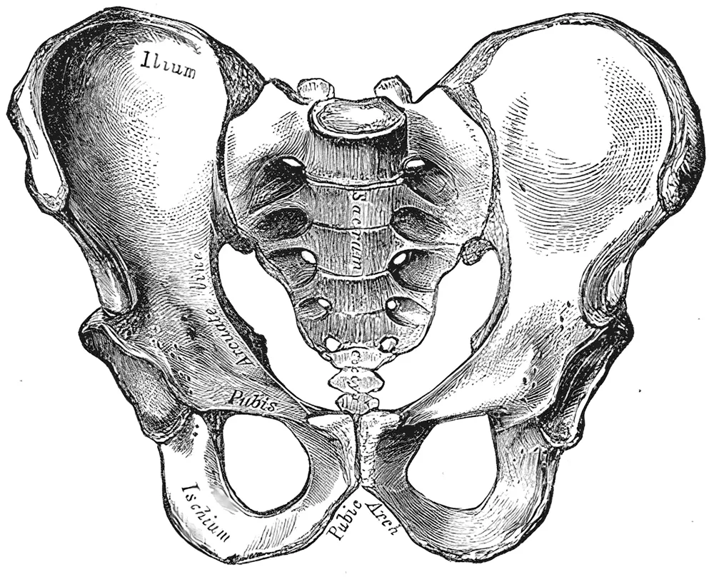
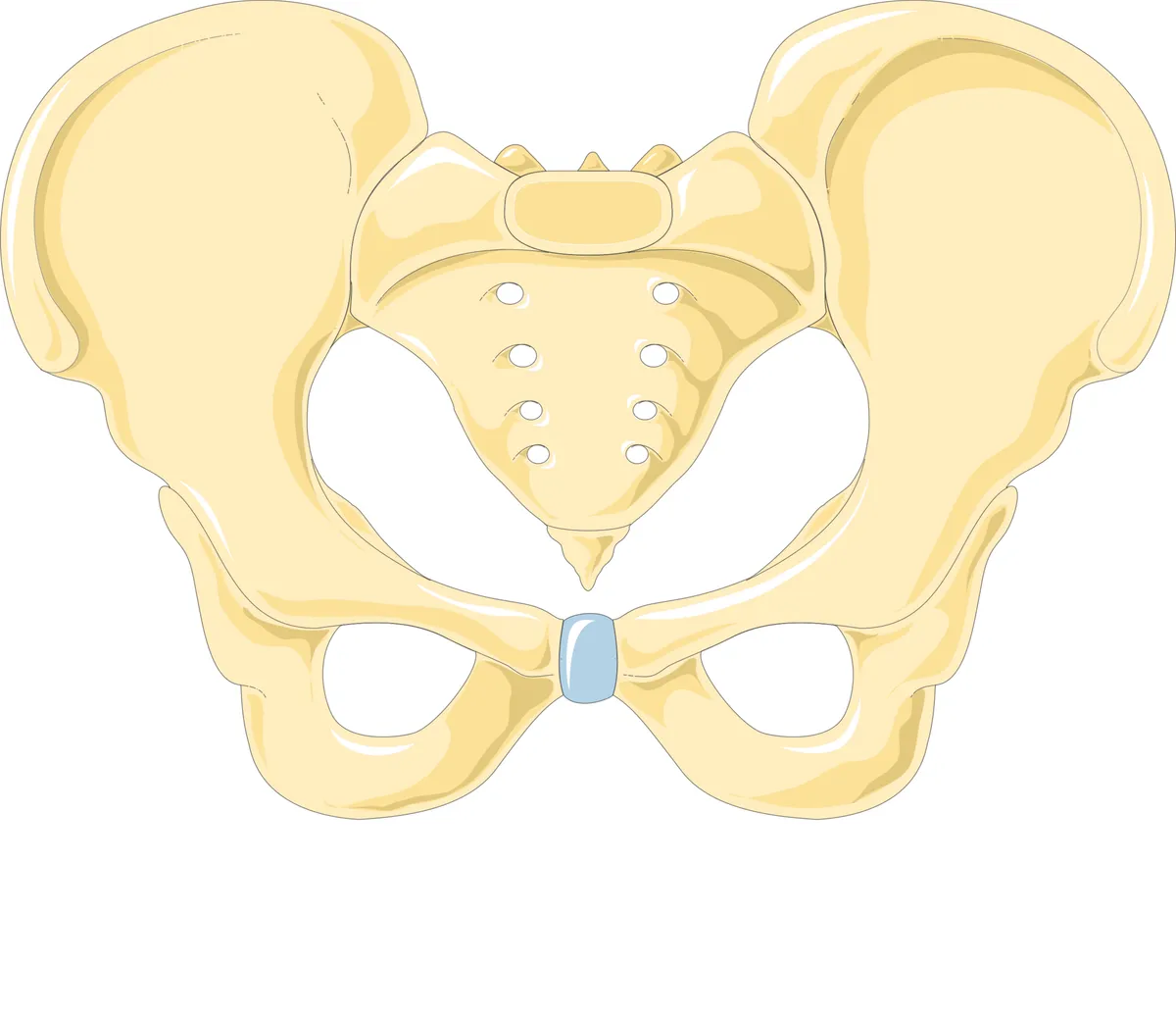
The two hip bones and the sacrum form a closed pelvic ring that transfers body weight from the spine to the lower limbs. The pelvic brim divides the greater pelvis (part of the abdomen) from the lesser pelvis (holding the pelvic viscera). The female pelvis is wider, with an oval inlet and a wider subpubic angle adapted to childbirth.
Position and relations
The hip bone articulates with the sacrum behind (sacroiliac joint), with its fellow in front (pubic symphysis) and with the femoral head at the acetabulum. The iliac crest is palpable; the anterior superior iliac spine is a measuring landmark and the attachment of the inguinal ligament. The arcuate line separates the greater pelvis above from the lesser pelvis below, whose inlet and outlet define the birth canal.
Form and structure
The cup-shaped acetabulum, to which all three bones contribute, has a cartilage-covered lunate surface and a central fossa. The obturator foramen is almost closed by the obturator membrane. The female pelvis is wider and shallower, with an oval inlet and a wider subpubic angle than the male, features used to sex skeletal remains.
Vessels and nerves
The bone is supplied by internal iliac branches (iliolumbar, gluteal, obturator) and the circumflex femoral arteries. Pelvic fractures can tear the presacral venous plexus and internal iliac branches, causing severe retroperitoneal bleeding. The sciatic nerve leaves the pelvis through the greater sciatic notch below piriformis.
Pelvic girdle, sacroiliac joint and acetabulum
The pelvic girdle consists of 3 bones (the two hip bones and the sacrum) and 3 joints (the two sacroiliac joints and the pubic symphysis). The pelvic floor muscles, mainly levator ani, receive somatic innervation from the lumbosacral plexus. The bony and muscular pelvis is closely linked to the hip and gluteal muscles, which together support the pelvic organs and core muscles. Pelvic floor physiology centres on bladder and bowel control, sexual function and pregnancy.[1]
The sacroiliac joints are essential for load transfer between the spine and the legs. Sacrum, pelvis and spine are functionally linked to the arms, legs and head through muscular, fascial and ligamentous connections. Joint stability is described by force-closure models, in which compression from muscles and ligaments may be insufficient or excessive, both with clinical implications. The pelvis and sacroiliac joint also show sexual dimorphism.[2]
The acetabulum of the hip bone receives the femoral head, and the acetabular labrum deepens it. Labral tears are a mechanical cause of hip pain, assessed with high-resolution MR arthrography. Femoroacetabular impingement, of cam or pincer type, causes hip pain, damages the acetabular cartilage and labrum and leads to degeneration; abnormal morphology of the acetabulum or proximal femur may be the primary predisposing factor.[3][4]
Full-text reading (StatPearls)
The StatPearls chapters on this topic, reproduced verbatim. Their CC BY-NC-ND 4.0 licence allows unmodified copies only, so they are not translated or shortened.
Anatomy, Bony Pelvis and Lower Limb: Foot and AnkleFicke J, Fakoya AO, Byerly DW · StatPearls 2026 · NBK546698 · 31k characters
Loading…
Ficke J, Fakoya AO, Byerly DW. Anatomy, Bony Pelvis and Lower Limb: Foot and Ankle. StatPearls [Internet]. Treasure Island (FL): StatPearls Publishing; 2026. NBK546698 · PMID 31536304 · CC BY-NC-ND 4.0. Reproduced verbatim, unmodified.
Anatomy, Bony Pelvis and Lower Limb: Tibial NerveDesai SS, La Rose J · StatPearls 2026 · NBK537028 · 25k characters
Loading…
Desai SS, La Rose J. Anatomy, Bony Pelvis and Lower Limb: Tibial Nerve. StatPearls [Internet]. Treasure Island (FL): StatPearls Publishing; 2026. NBK537028 · PMID 30725713 · CC BY-NC-ND 4.0. Reproduced verbatim, unmodified.
Anatomy, Bony Pelvis and Lower Limb: Iliopsoas MuscleBordoni B, Varacallo MA · StatPearls 2026 · NBK531508 · 20k characters
Loading…
Bordoni B, Varacallo MA. Anatomy, Bony Pelvis and Lower Limb: Iliopsoas Muscle. StatPearls [Internet]. Treasure Island (FL): StatPearls Publishing; 2026. NBK531508 · PMID 30285403 · CC BY-NC-ND 4.0. Reproduced verbatim, unmodified.
Anatomy, Bony Pelvis and Lower Limb, HipGlenister R, Sharma S · StatPearls 2023 · NBK526019 · 18k characters
Loading…
Glenister R, Sharma S. Anatomy, Bony Pelvis and Lower Limb, Hip. StatPearls [Internet]. Treasure Island (FL): StatPearls Publishing; 2023. NBK526019 · PMID 30252275 · CC BY-NC-ND 4.0. Reproduced verbatim, unmodified.
Anatomy, Bony Pelvis and Lower Limb, KneeGupton M, Imonugo O, Black AC, Launico MV, Terreberry RR · StatPearls 2023 · NBK500017 · 17k characters
Loading…
Gupton M, Imonugo O, Black AC, Launico MV, Terreberry RR. Anatomy, Bony Pelvis and Lower Limb, Knee. StatPearls [Internet]. Treasure Island (FL): StatPearls Publishing; 2023. NBK500017 · PMID 29763193 · CC BY-NC-ND 4.0. Reproduced verbatim, unmodified.
Anatomy, Bony Pelvis and Lower Limb: Metatarsal BonesLezak B, La Rose J, Massel DH · StatPearls 2026 · NBK549872 · 15k characters
Loading…
Lezak B, La Rose J, Massel DH. Anatomy, Bony Pelvis and Lower Limb: Metatarsal Bones. StatPearls [Internet]. Treasure Island (FL): StatPearls Publishing; 2026. NBK549872 · PMID 31751062 · CC BY-NC-ND 4.0. Reproduced verbatim, unmodified.
Anatomy, Bony Pelvis and Lower Limb: CalfBinstead JT, Varacallo MA · StatPearls 2026 · NBK459362 · 14k characters
Loading…
Binstead JT, Varacallo MA. Anatomy, Bony Pelvis and Lower Limb: Calf. StatPearls [Internet]. Treasure Island (FL): StatPearls Publishing; 2026. NBK459362 · PMID 29083629 · CC BY-NC-ND 4.0. Reproduced verbatim, unmodified.
Anatomy, Bony Pelvis and Lower Limb: Leg Anterior CompartmentLezak B, Launico MV · StatPearls 2026 · NBK539725 · 13k characters
Loading…
Lezak B, Launico MV. Anatomy, Bony Pelvis and Lower Limb: Leg Anterior Compartment. StatPearls [Internet]. Treasure Island (FL): StatPearls Publishing; 2026. NBK539725 · PMID 30969547 · CC BY-NC-ND 4.0. Reproduced verbatim, unmodified.
Anatomy, Bony Pelvis and Lower Limb: Posterior Tibial ArteryLezak B, Launico MV · StatPearls 2026 · NBK536981 · 12k characters
Loading…
Lezak B, Launico MV. Anatomy, Bony Pelvis and Lower Limb: Posterior Tibial Artery. StatPearls [Internet]. Treasure Island (FL): StatPearls Publishing; 2026. NBK536981 · PMID 30725666 · CC BY-NC-ND 4.0. Reproduced verbatim, unmodified.
Anatomy, Bony Pelvis and Lower Limb, Gemelli MusclesLezak B, Massel DH · StatPearls 2026 · NBK557420 · 9k characters
Loading…
Lezak B, Massel DH. Anatomy, Bony Pelvis and Lower Limb, Gemelli Muscles. StatPearls [Internet]. Treasure Island (FL): StatPearls Publishing; 2026. NBK557420 · PMID 32491352 · CC BY-NC-ND 4.0. Reproduced verbatim, unmodified.
Organs in this system
References
Sources for the values and descriptions on this page, indexed in PubMed (US National Library of Medicine).
- [1]Anatomy and Physiology of the Pelvic FloorEickmeyer SM, Physical medicine and rehabilitation clinics of North America 2017PMID 28676358
- [2]The sacroiliac joint: an overview of its anatomy, function and potential clinical implicationsVleeming A et al., Journal of anatomy 2012PMID 22994881
- [3]Acetabular labrumBlankenbaker DG et al., Magnetic resonance imaging clinics of North America 2013PMID 23168180
- [4]Femoroacetabular impingementKassarjian A et al., Topics in magnetic resonance imaging 2006PMID 17414995
- [5]Anatomy, BonesStatPearls (NCBI Bookshelf)PMID 30725884Supports: Number of bones
- [6]Calcium metabolism in health and diseasePeacock M, 2010PMID 20089499Supports: Skeletal calcium store