Shoulder and axilla
Model
Layers
Z-Anatomy anatomical model based on BodyParts3D, licensed CC BY-SA 4.0.
Overview
A regional model of the (right) shoulder and axilla: the scapula, clavicle and upper humerus, the glenohumeral joint with its capsule and the subacromial–subdeltoid bursae, the deltoid and rotator cuff, the walls of the axilla (pectoralis major and minor, subscapularis, latissimus dorsi, teres major, serratus anterior), the axillary artery and vein, the brachial plexus from roots to cords and terminal branches, and the axillary lymph nodes.
Boundaries and subdivisions
The axilla is a four-walled pyramidal space between the arm and the chest wall. Its apex, above, is the cervicoaxillary canal between the clavicle, the outer border of the first rib and the upper border of the scapula, through which vessels and nerves pass from the neck to the limb. The anterior wall is pectoralis major and minor; the posterior wall subscapularis, latissimus dorsi and teres major; the medial wall serratus anterior over the upper ribs; the lateral wall the intertubercular groove of the humerus. The base is skin and axillary fascia. The shoulder region includes the glenohumeral joint and the deltoid region, where the rotator cuff muscles (supraspinatus, infraspinatus, teres minor, subscapularis) wrap around the humeral head.[1][2][3]
Layers from superficial to deep
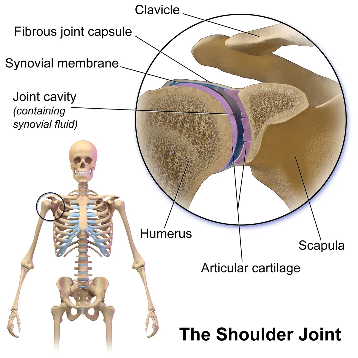
The most superficial muscle is deltoid, with clavicular, acromial and spinal parts inserting on the deltoid tuberosity; beneath it and under the acromion the subacromial–subdeltoid bursa lets the supraspinatus tendon glide. Deeper, the four rotator cuff muscles hold the humeral head in the shallow glenoid; the capsule is reinforced by the glenohumeral and coracohumeral ligaments. Behind, the quadrangular space (bounded by teres minor, teres major, the long head of triceps and the surgical neck of the humerus) transmits the axillary nerve and posterior circumflex humeral artery. In front, pectoralis minor divides the axillary artery into three parts.[2][4][5]
Vessels, nerves and lymphatics
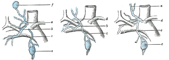
The axillary artery continues the subclavian from the outer border of the first rib to the lower border of teres major. Its first part (above pectoralis minor) gives the superior thoracic artery; the second part (behind it) the thoracoacromial and lateral thoracic arteries; the third part (below it) the subscapular and the anterior and posterior circumflex humeral arteries. The lateral, medial and posterior cords of the brachial plexus lie around the second part as their names indicate. Notable collateral branches are the long thoracic nerve (C5–C7) on the surface of serratus anterior, the thoracodorsal nerve to latissimus dorsi, and the axillary nerve (C5–C6) from the posterior cord. Most lymph from the breast drains to the five groups of axillary nodes: pectoral, subscapular, humeral, central and apical.[6][2][7][8]
Embryology
The pectoral girdle and the limb bud have different origins: the scapula and the muscles on its medial border come from paraxial somites, whereas most limb-bud structures arise from lateral plate mesoderm opposite somites 8 to 10, and the girdle connective tissue stays continuous with the axial skeleton. Growth factors such as SHH (zone of polarising activity), FGF and Hox genes position and pattern the limb. The clavicle is the first bone to ossify in the embryo, mainly from lateral mesoderm: its shaft ossifies in membrane while its two ends ossify in cartilage. The humerus is a typical endochondral bone, with a primary centre in the shaft and secondary centres at each end. The supraclavicular fossa is a surface depression shaped in weeks 4 to 8 as paraxial mesoderm forms the scalene muscles and the connective tissue of the lateral neck.[5][9][10][4][11]
Anatomical variants
The coracoclavicular ligament is sometimes replaced by a bony bridge (about 9% in a study of 2,724 people, more often in African American individuals and women), and the frequency of a coracoclavicular joint varies widely between populations. An unfused acromial apophysis (os acromiale) may cause pain and raises the risk of impingement and rotator cuff tears, while congenital elevation of the scapula (Sprengel deformity) limits girdle motion. Infraspinatus may fuse with teres minor, and subchondral cysts at the cuff insertions can be mistaken for disease. In the quadrangular space, fibrous bands or variant bordering muscles narrow the space during arm rotation. The supraclavicular fossa contains a variable number of lymph nodes (roughly 4 to 12), which should not be misread as pathology.[12][13][5][14][11]
Clinical correlations
Shoulder dislocation is usually anteroinferior and can stretch the axillary nerve, causing numbness over the lateral shoulder and deltoid weakness. Subacromial impingement and supraspinatus tears are common causes of pain on abduction. Injury to the long thoracic nerve during axillary dissection paralyses serratus anterior, so the medial border of the scapula lifts off the chest (winged scapula). A fracture of the surgical neck of the humerus endangers the axillary nerve, which winds around it in the quadrangular space.[2][6][7][15]
Surgical and procedural considerations
Proximal humeral fractures are common in older people who fall onto the shoulder or an outstretched arm; most are treated non-operatively, but multifragmentary fractures risk post-traumatic osteonecrosis of the head and may need arthroplasty. Acromioclavicular injuries (sprains and dislocations) are very common after direct impact to the lateral shoulder. Quadrangular space syndrome is rare and often missed; it affects people who repeat overhead motion (baseball pitchers, volleyball players, swimmers, electricians) and should be considered when shoulder pain and unexplained deltoid or teres minor weakness suggest compression of the axillary nerve and posterior circumflex humeral artery. In shoulder arthroplasty the scapula contributes the glenoid and labrum; a cystic mass in the supraclavicular fossa is assessed with ultrasound and CT, then fine-needle aspiration or core biopsy, before any operation.[4][12][14][13][11]
Key structures
Fossa axillaris
- Name
- Axillary fossa
- Position, role
- Four-walled pyramid; holds axillary vessels, brachial plexus and nodes
Spatium quadrangulare
- Name
- Quadrangular space
- Position, role
- Axillary nerve and posterior circumflex humeral artery
Cingulum musculotendineum rotatorum
- Name
- Rotator cuff
- Position, role
- Supraspinatus, infraspinatus, teres minor, subscapularis; stabilise the humeral head
Bursa subacromialis
- Name
- Subacromial bursa
- Position, role
- Between the acromion and the supraspinatus tendon
A. axillaris
- Name
- Axillary artery
- Position, role
- Three parts defined by pectoralis minor; six branches
N. thoracicus longus
- Name
- Long thoracic nerve
- Position, role
- C5–C7; serratus anterior; injury causes winged scapula
Nodi lymphoidei axillares
- Name
- Axillary lymph nodes
- Position, role
- Five groups; receive most breast lymph
| Terminologia Anatomica | Name | Position, role |
|---|---|---|
| Fossa axillaris | Axillary fossa | Four-walled pyramid; holds axillary vessels, brachial plexus and nodes |
| Spatium quadrangulare | Quadrangular space | Axillary nerve and posterior circumflex humeral artery |
| Cingulum musculotendineum rotatorum | Rotator cuff | Supraspinatus, infraspinatus, teres minor, subscapularis; stabilise the humeral head |
| Bursa subacromialis | Subacromial bursa | Between the acromion and the supraspinatus tendon |
| A. axillaris | Axillary artery | Three parts defined by pectoralis minor; six branches |
| N. thoracicus longus | Long thoracic nerve | C5–C7; serratus anterior; injury causes winged scapula |
| Nodi lymphoidei axillares | Axillary lymph nodes | Five groups; receive most breast lymph |
What each layer contains
SkinCutis17
- Cubital fossa
- Infraclavicular fossa
- Interscapular region
- Anterior region of arm
- Anterior region of elbow
- Deltoid region
- Infrascapular region
- Lateral region of thorax
- Mammary region
- Pectoral region
- Posterior region of arm
- Posterior region of elbow
- Scapular region
- Lateral bicipital groove
- Medial bicipital groove
- Triangle of auscultation
- Deltopectoral triangle
Muscles, fasciae and tendon sheathsMusculi, fasciae et vaginae tendinum69
- (Abdominal part of pectoralis major muscle)
- Subacromial bursa
- Subdeltoid bursa
- Subtendinous bursa of infraspinatus muscle
- Subtendinous bursa of teres major muscle
- Subtendinous bursa of trapezius muscle
- Subtendinous bursa of triceps brachii muscle
- Short head of biceps brachii
- Lateral head of triceps brachii
- Long head of biceps brachii
- Long head of triceps brachii
- Medial head of triceps brachii
- Coracobrachial bursa
- Depressor anguli oris
- Diaphragm
- Clavipectoral fascia
- Levator scapulae
- Levatores breves costarum
- Levatores longi costarum
- External intercostal muscles
- Internal intercostal muscles
- Innermost intercostal muscles
- Brachialis muscle
- Brachioradialis muscle
- Coracobrachialis muscle
- Iliocostalis colli muscle
- Iliocostalis lumborum muscle
- Iliocostalis thoracis muscle
- Infraspinatus muscle
- Latissimus dorsi muscle
- Longissimus capitis muscle
- Longissimus colli muscle
- Longissimus thoracis muscle
- External abdominal oblique muscle
- Internal abdominal oblique muscle
- Omohyoid muscle
- Pectoralis minor muscle
- Rhomboid major muscle
- Rhomboid minor muscle
- Scalenus anterior muscle
- Scalenus medius muscle
- Scalenus posterior muscle
- Serratus anterior muscle
- Serratus posterior inferior muscle
- Serratus posterior superior muscle
- Spinalis capitis muscle
- Splenius capitis muscle
- Splenius colli muscle
- Sternocleidomastoid muscle
- Subclavius muscle
- Subscapularis muscle
- Supraspinatus muscle
- Teres major muscle
- Teres minor muscle
- Transversus abdominis muscle
- Transversus thoracis muscle
- Acromial part of deltoid muscle
- Ascending part of trapezius muscle
- Clavicular part of deltoid muscle
- Clavicular head of pectoralis major muscle
- Descending part of trapezius muscle
- Scapular spinal part of deltoid muscle
- Sternocostal head of pectoralis major muscle
- Transverse part of trapezius muscle
- Platysma
- Lateral intermuscular septum of arm
- Medial intermuscular septum of arm
- Subcutaneous acromial bursa
- Intertubercular tendon sheath
Bones and cartilageOssa et cartilagines24
- Costal cartilage of eighth rib
- Costal cartilage of first rib
- Costal cartilage of fourth rib
- Costal cartilage of fifth rib
- Costal cartilage of second rib
- Costal cartilage of seventh rib
- Costal cartilage of sixth rib
- Costal cartilage of third rib
- Clavicle
- Costal cartilage of ninth rib
- First rib
- Second rib
- Tenth rib
- Twelfth rib
- Humerus
- Ninth rib
- Eighth rib
- Fourth rib
- Fifth rib
- Scapula
- Seventh rib
- Sixth rib
- Third rib
- Eleventh rib
Joints and ligamentsJuncturae et ligamenta23
- Articular capsule of acromioclavicular joint
- Articular capsule of glenohumeral joint
- Articular capsule of elbow joint
- Articular disc of acromioclavicular joint
- Glenoid labrum
- Intertransverse ligaments
- Acromioclavicular ligament
- Radial collateral ligament
- Conoid ligament
- Coraco-acromial ligament
- Coracohumeral ligament
- Costoclavicular ligament
- Costotransverse ligament
- Inferior glenohumeral ligament
- Middle glenohumeral ligament
- Superior glenohumeral ligament
- Posterior sternoclavicular ligament
- Transverse humeral ligament
- Inferior transverse scapular ligament
- Superior transverse scapular ligament
- Trapezoid ligament
- External intercostal membrane
- Internal intercostal membrane
ArteriesArteriae52
- (Deep branch of transverse cervical artery)
- (Superficial branch of transverse cervical artery)
- Axillary artery
- Brachial artery
- Deep cervical artery
- Anterior circumflex humeral artery
- Posterior circumflex humeral artery
- Circumflex scapular artery
- Middle collateral artery
- Radial collateral artery
- Inferior ulnar collateral artery
- Superior ulnar collateral artery
- Gastroduodenal artery
- Proper hepatic artery
- First posterior intercostal artery
- Second posterior intercostal artery
- Supreme intercostal artery
- Inferior lobar artery of right lung
- Middle lobar artery of right lung
- Musculophrenic artery
- Inferior phrenic artery
- Deep brachial artery
- Anterior segmental artery of right lung
- Apical segmental artery of right lung
- Anterior basal segmental artery of right lung
- Lateral basal segmental artery of right lung
- Medial basal segmental artery of right lung
- Posterior basal segmental artery of right lung
- Lateral segmental artery of right lung
- Medial segmental artery of right lung
- Posterior segmental artery of right lung
- Superior segmental artery of right lung
- Right subclavian artery
- Subcostal artery
- Subscapular artery
- Inferior suprarenal artery
- Suprascapular artery
- Internal thoracic artery
- Lateral thoracic artery
- Thoraco-acromial artery
- Thoracodorsal artery
- Inferior thyroid artery
- Transverse cervical artery
- Vertebral artery
- Posterior intercostal arteries
- Superior phrenic arteries
- Right common carotid artery
- Pectoral branches of thoraco-acromial artery
- Anterior branch of renal artery
- Posterior branch of renal artery
- Costocervical trunk
- Thyrocervical trunk
VeinsVenae29
- Anterior vein of right lung
- Apical vein of right lung
- Axillary vein
- Inferior basal vein of right lung
- Superior basal vein of right lung
- Cephalic vein
- Anterior circumflex humeral vein
- Posterior circumflex humeral vein
- Circumflex scapular vein
- Right superior intercostal vein
- Anterior jugular vein
- External jugular vein
- Internal jugular vein
- Lateral vein of right lung
- Medial vein of right lung
- Right superior phrenic vein
- Posterior vein of right lung
- Right superior pulmonary vein
- Right subclavian vein
- Right subcostal vein
- Subscapular vein
- Superior vein of right lung
- Suprascapular vein
- Lateral thoracic vein
- Thoracodorsal vein
- Superior thyroid vein
- Vertebral vein
- Brachial veins
- Musculophrenic veins
NervesNervi33
- Anterior division of inferior trunk of brachial plexus
- Anterior division of middle trunk of brachial plexus
- Anterior division of superior trunk of brachial plexus
- Posterior division of inferior trunk of brachial plexus
- Posterior division of middle trunk of brachial plexus
- Posterior division of superior trunk of brachial plexus
- Posterior cord of brachial plexus
- Intercostal nerves
- Accessory nerve (XI)
- Axillary nerve
- Inferior lateral brachial cutaneous nerve
- Superior lateral brachial cutaneous nerve
- Medial antebrachial cutaneous nerve
- Medial brachial cutaneous nerve
- Dorsal scapular nerve
- Median nerve
- Musculocutaneous nerve
- Lateral pectoral nerve
- Medial pectoral nerve
- Radial nerve
- Subclavian nerve
- Inferior subscapular nerve
- Superior subscapular nerve
- Suprascapular nerve
- Long thoracic nerve
- Thoracodorsal nerve
- Ulnar nerve
- Roots of brachial plexus
- Muscular branches of axillary nerve
- Muscular branches of radial nerve
- Inferior trunk of brachial plexus
- Middle trunk of brachial plexus
- Superior trunk of brachial plexus
LymphaticsSystema lymphoideum24
- Anterior axillary nodes
- Apical axillary nodes
- Central axillary nodes
- Lateral axillary nodes
- Posterior axillary nodes
- Brachial nodes
- Brachiocephalic nodes
- Inferior deep lateral cervical nodes
- Superficial lateral cervical nodes
- Right colic nodes
- Middle colic nodes
- Right gastro-omental nodes
- Infraclavicular nodes
- Intercostal nodes
- Interpectoral nodes
- Anterior inferior jugular nodes
- Superior pancreaticoduodenal nodes
- Parasternal nodes
- Lateral pericardial nodes
- Prepericardial nodes
- Supraclavicular nodes
- Supratrochlear nodes
- Cystic node
- Lateral superior jugular node
Full-text reading (StatPearls)
The StatPearls chapters on this topic, reproduced verbatim. Their CC BY-NC-ND 4.0 licence allows unmodified copies only, so they are not translated or shortened.
Anatomy, Shoulder and Upper Limb, NervesOkwumabua E, Black AC, Thompson JH · StatPearls 2023 · NBK526056 · 30k characters
Loading…
Okwumabua E, Black AC, Thompson JH. Anatomy, Shoulder and Upper Limb, Nerves. StatPearls [Internet]. Treasure Island (FL): StatPearls Publishing; 2023. NBK526056 · PMID 30252312 · CC BY-NC-ND 4.0. Reproduced verbatim, unmodified.
Anatomy, Rotator CuffLa Rose J, Madrazo-Ibarra A, Varacallo MA · StatPearls 2026 · NBK441844 · 23k characters
Loading…
La Rose J, Madrazo-Ibarra A, Varacallo MA. Anatomy, Rotator Cuff. StatPearls [Internet]. Treasure Island (FL): StatPearls Publishing; 2026. NBK441844 · PMID 28722874 · CC BY-NC-ND 4.0. Reproduced verbatim, unmodified.
Anatomy, Shoulder and Upper Limb: Supraclavicular FossaMiao KH, Bordoni B · StatPearls 2026 · NBK537265 · 20k characters
Loading…
Miao KH, Bordoni B. Anatomy, Shoulder and Upper Limb: Supraclavicular Fossa. StatPearls [Internet]. Treasure Island (FL): StatPearls Publishing; 2026. NBK537265 · PMID 30725950 · CC BY-NC-ND 4.0. Reproduced verbatim, unmodified.
Anatomy, Shoulder and Upper Limb, MusclesJaved O, Maldonado KA, Ashmyan R · StatPearls 2023 · NBK482410 · 19k characters
Loading…
Javed O, Maldonado KA, Ashmyan R. Anatomy, Shoulder and Upper Limb, Muscles. StatPearls [Internet]. Treasure Island (FL): StatPearls Publishing; 2023. NBK482410 · PMID 29494017 · CC BY-NC-ND 4.0. Reproduced verbatim, unmodified.
Anatomy, Shoulder and Upper Limb, HumerusMostafa E, Imonugo O, Varacallo MA · StatPearls 2023 · NBK534821 · 18k characters
Loading…
Mostafa E, Imonugo O, Varacallo MA. Anatomy, Shoulder and Upper Limb, Humerus. StatPearls [Internet]. Treasure Island (FL): StatPearls Publishing; 2023. NBK534821 · PMID 30521242 · CC BY-NC-ND 4.0. Reproduced verbatim, unmodified.
Anatomy, Shoulder and Upper Limb, Arm Quadrangular SpaceKhan IA, Launico MV, Varacallo MA · StatPearls 2026 · NBK537324 · 15k characters
Loading…
Khan IA, Launico MV, Varacallo MA. Anatomy, Shoulder and Upper Limb, Arm Quadrangular Space. StatPearls [Internet]. Treasure Island (FL): StatPearls Publishing; 2026. NBK537324 · PMID 30726009 · CC BY-NC-ND 4.0. Reproduced verbatim, unmodified.
Anatomy, Shoulder and Upper Limb, Acromioclavicular JointLa Rose J, Tiwari V · StatPearls 2026 · NBK499858 · 15k characters
Loading…
La Rose J, Tiwari V. Anatomy, Shoulder and Upper Limb, Acromioclavicular Joint. StatPearls [Internet]. Treasure Island (FL): StatPearls Publishing; 2026. NBK499858 · PMID 29763033 · CC BY-NC-ND 4.0. Reproduced verbatim, unmodified.
Anatomy, Thorax, ScapulaMiniato MA, Mudreac A, Borger J · StatPearls 2023 · NBK538319 · 14k characters
Loading…
Miniato MA, Mudreac A, Borger J. Anatomy, Thorax, Scapula. StatPearls [Internet]. Treasure Island (FL): StatPearls Publishing; 2023. NBK538319 · PMID 30855903 · CC BY-NC-ND 4.0. Reproduced verbatim, unmodified.
Anatomy, Shoulder and Upper Limb, ClavicleLa Rose J, Charlick M, Varacallo MA · StatPearls 2026 · NBK525990 · 12k characters
Loading…
La Rose J, Charlick M, Varacallo MA. Anatomy, Shoulder and Upper Limb, Clavicle. StatPearls [Internet]. Treasure Island (FL): StatPearls Publishing; 2026. NBK525990 · PMID 30252246 · CC BY-NC-ND 4.0. Reproduced verbatim, unmodified.
Anatomy, Shoulder and Upper Limb, VeinsNguyen JD, Duong H · StatPearls 2023 · NBK546676 · 12k characters
Loading…
Nguyen JD, Duong H. Anatomy, Shoulder and Upper Limb, Veins. StatPearls [Internet]. Treasure Island (FL): StatPearls Publishing; 2023. NBK546676 · PMID 31536282 · CC BY-NC-ND 4.0. Reproduced verbatim, unmodified.
Anatomy, Shoulder and Upper Limb, Axillary ArteryThiel R, Munjal A, Daly DT · StatPearls 2025 · NBK482174 · 10k characters
Loading…
Thiel R, Munjal A, Daly DT. Anatomy, Shoulder and Upper Limb, Axillary Artery. StatPearls [Internet]. Treasure Island (FL): StatPearls Publishing; 2025. NBK482174 · PMID 29489298 · CC BY-NC-ND 4.0. Reproduced verbatim, unmodified.
Anatomy, Shoulder and Upper Limb, Axillary NerveOkwumabua E, Thompson JH · StatPearls 2023 · NBK493212 · 7k characters
Loading…
Okwumabua E, Thompson JH. Anatomy, Shoulder and Upper Limb, Axillary Nerve. StatPearls [Internet]. Treasure Island (FL): StatPearls Publishing; 2023. NBK493212 · PMID 29630264 · CC BY-NC-ND 4.0. Reproduced verbatim, unmodified.
Anatomy, Shoulder and Upper Limb, AxillaGordon A, Alsayouri K · StatPearls 2023 · NBK547723 · 7k characters
Loading…
Gordon A, Alsayouri K. Anatomy, Shoulder and Upper Limb, Axilla. StatPearls [Internet]. Treasure Island (FL): StatPearls Publishing; 2023. NBK547723 · PMID 31613503 · CC BY-NC-ND 4.0. Reproduced verbatim, unmodified.
Regional anatomy
References
Sources for the values and descriptions on this page, indexed in PubMed (US National Library of Medicine).
- [1]Anatomy, Thorax, Pectoralis Major MajorSolari F et al., StatPearls 2023PMID 30252247
- [2]Anatomy, Shoulder and Upper Limb, Deltoid MuscleElzanie A et al., StatPearls 2024PMID 30725741
- [3]Anatomy, Shoulder and Upper Limb, AxillaGordon A et al., StatPearls 2023PMID 31613503
- [4]Anatomy, Shoulder and Upper Limb, HumerusMostafa E et al., StatPearls 2023PMID 30521242
- [5]Anatomy, Rotator CuffLa Rose J et al., StatPearls 2026PMID 28722874
- [6]Anatomy, Shoulder and Upper Limb, Brachial PlexusBayot ML et al., StatPearls 2023PMID 29763192
- [7]Anatomy, Thorax, BreastRivard AB et al., StatPearls 2023PMID 30137859
- [8]Anatomy, Shoulder and Upper Limb, Axillary ArteryThiel R et al., StatPearls 2025PMID 29489298
- [9]Anatomy, Shoulder and Upper Limb, NervesOkwumabua E et al., StatPearls 2023PMID 30252312
- [10]Anatomy, Shoulder and Upper Limb, ClavicleLa Rose J et al., StatPearls 2026PMID 30252246
- [11]Anatomy, Shoulder and Upper Limb: Supraclavicular FossaMiao KH et al., StatPearls 2026PMID 30725950
- [12]Anatomy, Shoulder and Upper Limb, Acromioclavicular JointLa Rose J et al., StatPearls 2026PMID 29763033
- [13]Anatomy, Thorax, ScapulaMiniato MA et al., StatPearls 2023PMID 30855903
- [14]Anatomy, Shoulder and Upper Limb, Arm Quadrangular SpaceKhan IA et al., StatPearls 2026PMID 30726009
- [15]Anatomy, Shoulder and Upper Limb, Axillary NerveOkwumabua E et al., StatPearls 2023PMID 29630264