Lymph nodes
Model
Layers
Z-Anatomy anatomical model based on BodyParts3D, licensed CC BY-SA 4.0.
Description
Small bean-shaped organs along the lymphatic vessels, clustered in the neck, axilla, groin, mediastinum and around the great abdominal vessels. The cortex holds B-cell follicles, the paracortex T cells, and the medulla the medullary cords and lymphatic sinuses.
Parts
Each part has its own card; press “Show on the model” to fly to it and select it in the 3D model above.
- 01
Capsule and hilum
Capsula et hilum nodi lymphoidei
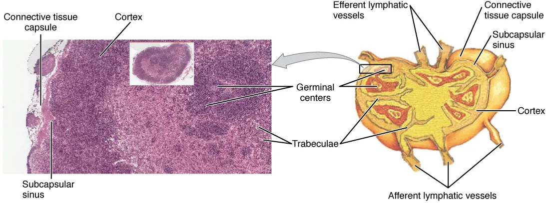
A bean-shaped organ, usually 2–10 mm, with a fibrous capsule that sends trabeculae inwards. Several afferent lymphatics pierce the capsule on the convex surface; at the hilum (concave side) an artery enters and a vein and one or two efferent lymphatics leave.
- 02
Lymph sinuses
Sinus nodi lymphoidei
Lymph flows from the subcapsular sinus through trabecular and medullary sinuses to the hilum. Reticular fibres and macrophages crossing the sinuses trap bacteria and tumour cells, which is why metastases first appear in the subcapsular sinus of the sentinel node.
- 03
Cortex
Cortex nodi lymphoidei
The outer layer, containing B-cell follicles; secondary follicles have germinal centres where B cells proliferate, undergo affinity maturation and class switching with help from T cells and follicular dendritic cells.
- 04
Paracortex
Paracortex
The zone between cortex and medulla, rich in T cells and antigen-presenting dendritic cells. It contains the high endothelial venules (HEVs) through which blood lymphocytes enter the node. It is depleted in congenital absence of the thymus.
- 05
Medulla
Medulla nodi lymphoidei
The inner part near the hilum, made of medullary cords containing plasma cells and macrophages, alternating with medullary sinuses. Plasma cells here secrete antibody straight into the outgoing lymph.
- 06
Head and neck nodes
Nodi lymphoidei capitis et colli
A superficial collar of nodes (submental, submandibular, parotid, mastoid, occipital) drains into the deep cervical chain along the internal jugular vein. The jugulodigastric node drains the palatine tonsil and is the one most often swollen in tonsillitis. An enlarged left supraclavicular (Virchow’s) node can signal abdominal cancer.
- 07
Axillary nodes
Nodi lymphoidei axillares
About 20–30 nodes in five groups: pectoral (anterior), humeral (lateral), subscapular (posterior), central and apical. They receive about 75% of the lymph from the breast and are therefore the first station of breast cancer spread; axillary clearance can injure the long thoracic nerve.
- 08
Abdominal and pelvic nodes
Nodi lymphoidei abdominis et pelvis
Visceral nodes follow the branches of the abdominal aorta (coeliac, superior and inferior mesenteric) and drain the gut; lumbar nodes around the aorta and inferior vena cava receive lymph from the kidneys, suprarenals, testes, ovaries and the iliac chains. All converge on the cisterna chyli and thoracic duct.
- 09
Inguinal nodes
Nodi lymphoidei inguinales
Superficial inguinal nodes lie below the inguinal ligament and drain the lower limb, buttock, abdominal wall below the umbilicus, perineum and anal canal below the pectinate line; deep inguinal nodes lie beside the femoral vein, the highest (Cloquet’s) in the femoral ring. Note that the testis drains not to the groin but to the lumbar nodes.
Position and relations
Lymph nodes form chains along blood vessels and groups where the limbs join the trunk: cervical (along the internal jugular vein), axillary and inguinal, with deep groups in the mediastinum, around the abdominal aorta, in the mesentery and in the pelvis. Each skin territory or organ drains to a given node group, the basis for clinical node examination and for sentinel-node mapping in cancer.
Form and structure
A node has a fibrous capsule, with afferent lymphatics entering its convex surface and the efferent vessel and blood vessels at the hilum. Lymph flows through the subcapsular, intermediate and medullary sinuses, where macrophages and dendritic cells filter it. The outer cortex holds B-cell follicles (with germinal centres when responding), and the paracortex is the T-cell zone with high endothelial venules, where lymphocytes leave the blood to enter the node.
Vessels and nerves
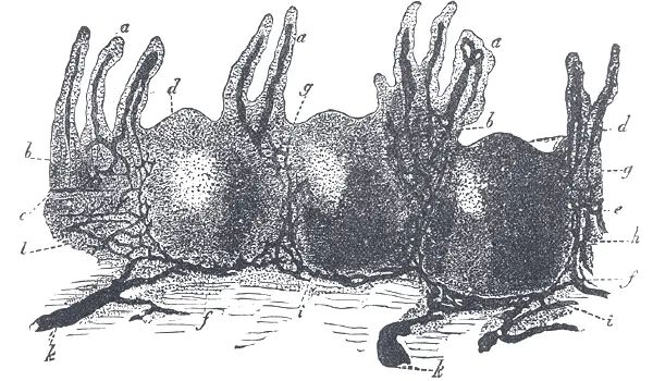
Lymph from the nodes gathers into lymphatic trunks that empty into two main ducts: the thoracic duct (beginning at the cisterna chyli at L1–L2 and ascending in the posterior mediastinum) drains most of the body into the left venous angle (the junction of the left internal jugular and subclavian veins); the right lymphatic duct drains the right half of the head and neck, the right upper limb and the right side of the thorax into the right venous angle.
Physiology
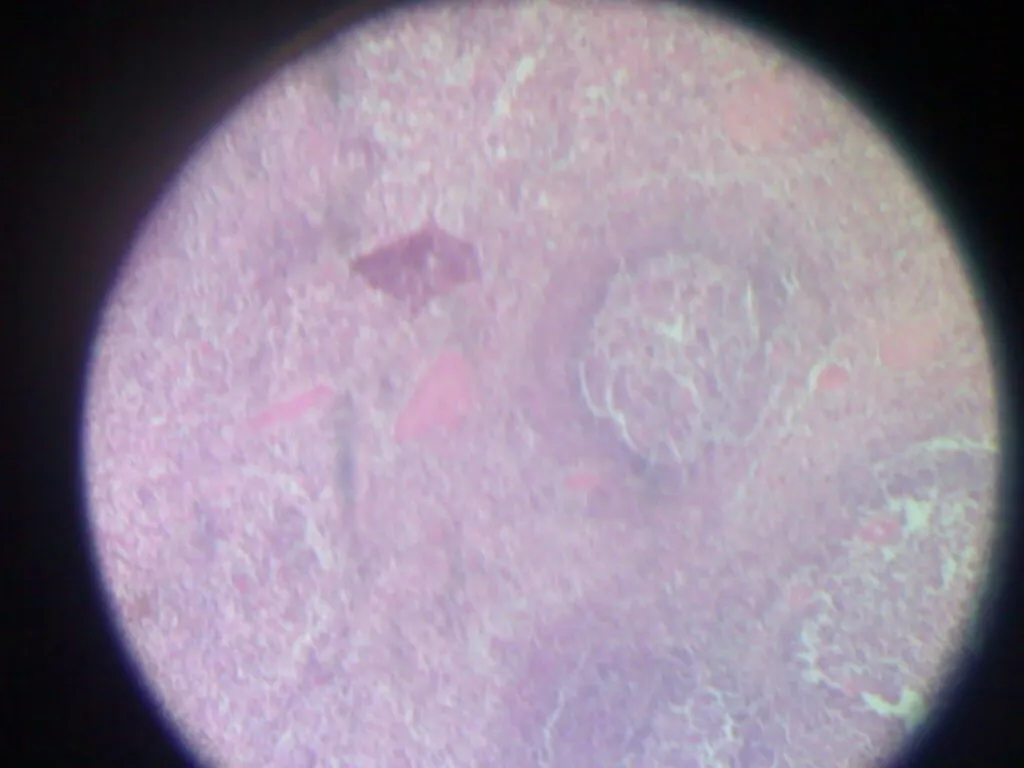
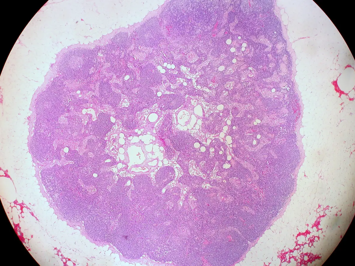
Lymph nodes have three compartments, cortex, paracortex and medulla; B and T cells home to separate areas, meet antigen-presenting cells and expand clonally. The basic anatomical and functional unit is the lymphoid lobule, built on a reticular meshwork. Within follicles, germinal centres are where B cells proliferate, undergo somatic hypermutation and are selected by affinity, producing high-affinity antibodies.[1][2]
Each node is constantly informed by lymph carrying metabolic products, proteins, pathogens and immune cells from the tissue it drains. Nodes differ in their capacity to generate immune responses, with consequences for vaccination and tumour control.[3]
Microanatomy, sinuses and lymphocyte traffic
By Starling’s law of the capillaries, fluid is filtered into the interstitium near arterioles and resorbed near venules; depending on the tissue, filtration exceeds resorption by about 10%, and this excess interstitial fluid is drained by the lymphatic system. Once it enters lymphatic capillaries the fluid is called lymph; it passes through collecting vessels, regional nodes and then collecting nodes (which receive lymph from several regional nodes), and returns to the blood at the jugulovenous angles only after passing through at least one node. The node is bean-shaped and 1–20 mm long depending on its site: afferent vessels enter the convex surface, while the efferent vessel leaves and blood vessels enter at the hilum on the concave side. A connective-tissue capsule surrounds it and sends trabeculae inwards. The supporting framework is made by reticular cells that synthesise type III collagen. The superficial cortex holds B cells with follicular dendritic cells arranged in primary follicles, which form germinal centres and become secondary follicles when the B cells are activated. The deep cortex is the T-cell zone with dendritic cells, organised into deep cortical units; activated T cells proliferate but do not form germinal centres. The medulla consists of medullary cords containing plasma cells derived from B cells of the superficial cortex.[4]
Sinuses are vessel-like spaces through which lymph flows, their lumen crossed by a meshwork of antigen-presenting-cell processes and type III collagen. Lymph enters the subcapsular sinus (between capsule and cortex), passes along trabecular sinuses to the medullary sinuses and leaves through efferent vessels. Naive B and T cells (mature but not yet exposed to their antigen) enter mainly through high endothelial venules by diapedesis aided by adhesion molecules, and chemokine signals sort them into the superficial and deep cortex; if they meet no antigen they leave through efferent vessels. Antigens and bacteria that have breached the first defences are trapped by the sinus meshwork and endocytosed, activating B and T cells; activated B cells become plasma cells, plasmablasts and memory B cells, and plasma cells move to the medullary cords to release antigen-specific antibody into lymph in the medullary sinuses. Cell debris, red cells or carbon particles are phagocytosed by sinus macrophages. Developmentally, nodes begin as mesenchymal condensations that bulge into lymph sacs; the T-cell region forms from week 13, the B-cell region from week 14 at the marginal sinus, and by week 20 primary follicles are present in the outer cortex. Recent descriptions add subcapsular proliferative foci, flat sheets of activated B cells that appear on re-exposure to particulate antigen and produce early antibody even before germinal centres, and intranodal lympho-venous shunts in mammals.[4]
Regional node groups and neck levels
In the lower limb, popliteal nodes in the popliteal fossa drain to the inguinal nodes, which comprise a vertical chain along the great saphenous vein and a horizontal group below the inguinal ligament and also receive lymph from the lower abdominal wall and lower back up to the umbilicus. Lymph then passes to the external iliac nodes (lateral, intermediate and medial groups around the external iliac vessels), the common iliac nodes (which also receive the internal iliac nodes of the pelvic viscera), and the left (beside the aorta), intermediate (between aorta and inferior vena cava) and right (beside the inferior vena cava) lumbar nodes. The two lumbar trunks join the intestinal trunk (from the mesentery and abdominal viscera) to form the cisterna chyli, a lymphatic sac of variable size and shape at L1–L2 that begins the thoracic duct. In the thorax, the lungs have two parallel drainage systems, peribronchial and subpleural, converging on the tracheobronchial nodes; these and the brachiocephalic nodes (draining thymus, thyroid and pericardium) empty into the bronchomediastinal trunks. In the upper limb, cubital nodes lie in the cubital fossa; axillary nodes receive the upper limb and the trunk wall down to the umbilicus and, in breast cancer, are divided into three levels by pectoralis minor, with spread proceeding from level I to III: level I lateral and inferior to the muscle (pectoral, subscapular and lateral axillary nodes), level II on or behind it (interpectoral and central nodes) and level III medial to it (apical nodes). The newer Clough classification replaces levels I–II with four zones, A–D, defined by the second intercostobrachial nerve and the lateral thoracic vein.[4]
Regional nodes of the head include the occipital nodes at the lateral border of trapezius, the mastoid nodes at the insertion of sternocleidomastoid on the mastoid process, superficial and deep parotid nodes, and facial nodes (malar, buccinator and mandibular), all draining down to the neck. Neck lymph is described both by chains and by six levels of dissection. The most important pathway is the deep lateral cervical chain occupying levels II, III and IV, with a common lateral border at the medial surface of sternocleidomastoid and a medial border at the internal carotid (level II) or common carotid artery (levels III and IV); its efferents drain into the jugular trunks and then one of the two great lymphatic ducts. The deep posterior cervical chain occupies level V. There are also superficial lateral jugular, superficial anterior jugular and deep anterior cervical chains, and the brain’s “glymphatic” system directs waste-laden cerebrospinal and interstitial fluid towards the cervical nodes. Node enlargement can be infectious, inflammatory or malignant; endoscopic ultrasound with fine-needle aspiration reaches a sensitivity of up to 85% and specificity of 100%, elastography acts as a “virtual biopsy” when nodes cannot be sampled, and PET/CT gives functional information at the cost of whole-body radiation. Current TNM staging no longer records simply “node-positive or negative” but the number of metastatic nodes, the nodal stations and the lymph-node ratio; in breast cancer pN1 is 1–3 axillary nodes, pN2 4–9 and pN3 ten or more or infraclavicular, supraclavicular or internal mammary nodes.[4]
Ia / Ib
- Boundaries
- Floor of the mouth (Ia in front of Ib)
- Main nodes
- Submental / submandibular
II
- Boundaries
- Skull base to hyoid; submandibular gland in front
- Main nodes
- Superior deep lateral cervical
III
- Boundaries
- Hyoid to cricoid cartilage
- Main nodes
- Middle deep lateral cervical
IV
- Boundaries
- Cricoid to clavicle
- Main nodes
- Inferior deep lateral cervical
V
- Boundaries
- Between posterior border of sternocleidomastoid and anterior border of trapezius; hyoid to transverse cervical vessels
- Main nodes
- Superior, middle and inferior deep posterior cervical
VI
- Boundaries
- Between anterior borders of both sternocleidomastoids; hyoid to manubrium
- Main nodes
- Anterior jugular, paratracheal and thyroid
| Level | Boundaries | Main nodes |
|---|---|---|
| Ia / Ib | Floor of the mouth (Ia in front of Ib) | Submental / submandibular |
| II | Skull base to hyoid; submandibular gland in front | Superior deep lateral cervical |
| III | Hyoid to cricoid cartilage | Middle deep lateral cervical |
| IV | Cricoid to clavicle | Inferior deep lateral cervical |
| V | Between posterior border of sternocleidomastoid and anterior border of trapezius; hyoid to transverse cervical vessels | Superior, middle and inferior deep posterior cervical |
| VI | Between anterior borders of both sternocleidomastoids; hyoid to manubrium | Anterior jugular, paratracheal and thyroid |
Full-text reading (StatPearls)
The StatPearls chapters on this topic, reproduced verbatim. Their CC BY-NC-ND 4.0 licence allows unmodified copies only, so they are not translated or shortened.
Anatomy, Lymph NodesWięcław M, Gupta V · StatPearls 2026 · NBK557717 · 21k characters
Loading…
Więcław M, Gupta V. Anatomy, Lymph Nodes. StatPearls [Internet]. Treasure Island (FL): StatPearls Publishing; 2026. NBK557717 · PMID 32491649 · CC BY-NC-ND 4.0. Reproduced verbatim, unmodified.
Anatomy, Head and Neck, Lymph NodesKoroulakis A, Jamal Z, Agarwal M · StatPearls 2022 · NBK513317 · 19k characters
Loading…
Koroulakis A, Jamal Z, Agarwal M. Anatomy, Head and Neck, Lymph Nodes. StatPearls [Internet]. Treasure Island (FL): StatPearls Publishing; 2022. NBK513317 · PMID 30020689 · CC BY-NC-ND 4.0. Reproduced verbatim, unmodified.
Anatomy, Head and Neck, Supraclavicular Lymph NodeBanjar FK, Wilson AM · StatPearls 2023 · NBK544300 · 11k characters
Loading…
Banjar FK, Wilson AM. Anatomy, Head and Neck, Supraclavicular Lymph Node. StatPearls [Internet]. Treasure Island (FL): StatPearls Publishing; 2023. NBK544300 · PMID 31335020 · CC BY-NC-ND 4.0. Reproduced verbatim, unmodified.
Anatomy, Abdomen and Pelvis: Inguinal Lymph NodeBui T, Bordoni B · StatPearls 2023 · NBK557639 · 10k characters
Loading…
Bui T, Bordoni B. Anatomy, Abdomen and Pelvis: Inguinal Lymph Node. StatPearls [Internet]. Treasure Island (FL): StatPearls Publishing; 2023. NBK557639 · PMID 32491571 · CC BY-NC-ND 4.0. Reproduced verbatim, unmodified.
Histology, Lymph NodesMercadante AA, Tadi P · StatPearls 2023 · NBK559053 · 7k characters
Loading…
Mercadante AA, Tadi P. Histology, Lymph Nodes. StatPearls [Internet]. Treasure Island (FL): StatPearls Publishing; 2023. NBK559053 · PMID 32644479 · CC BY-NC-ND 4.0. Reproduced verbatim, unmodified.
Organs in this system
References
Sources for the values and descriptions on this page, indexed in PubMed (US National Library of Medicine).
- [1]Normal structure, function, and histology of lymph nodesWillard-Mack CL, Toxicologic pathology 2006PMID 17067937
- [2]Germinal centersVictora GD et al., Annual review of immunology 2012PMID 22224772
- [3]Same yet different - how lymph node heterogeneity affects immune responsesCruz de Casas P et al., Nature reviews. Immunology 2024PMID 38097778
- [4]Anatomy, Lymph NodesWięcław M et al., StatPearls 2026PMID 32491649