Cardiovascular system
Model
Z-Anatomy anatomical model based on BodyParts3D, licensed CC BY-SA 4.0.
Overview
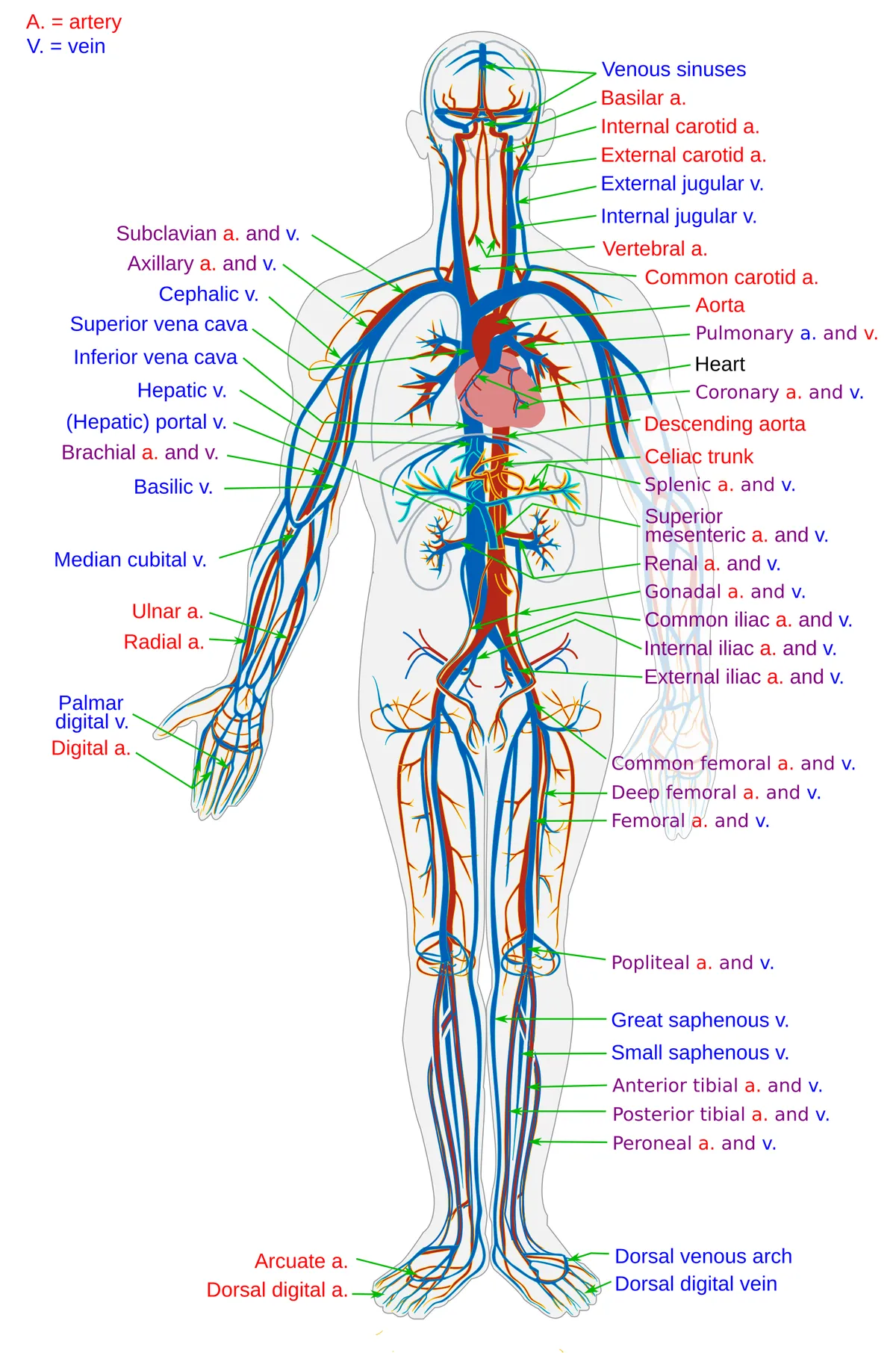
The cardiovascular system consists of the heart and the network of blood vessels (arteries, capillaries and veins). The heart drives blood through two circuits: the pulmonary circulation, for gas exchange, and the systemic circulation, which perfuses the tissues.
Functions
- 01Delivery of oxygen and nutrients to the tissues
- 02Removal of CO₂ and metabolic waste to the lungs and kidneys
- 03Contribution to thermoregulation
- 04Transport of hormones and immune cells
Heart wall and conduction system
The heart wall has three layers: endocardium, myocardium and epicardium. The epicardium is the visceral layer of the serous pericardium; in the embryo it arises from the proepicardial organ and provides progenitor cells for connective tissue and coronary vessels, as well as a signalling hub for myocardial development and regeneration.[1]
The impulse arises in the sinoatrial node and passes through the atrioventricular node and bundle of His to the subendocardial Purkinje network of the ventricles. Purkinje cells have a distinctive ultrastructure and electrophysiology that allow very rapid conduction, so that both ventricles contract almost simultaneously; for the same reason, Purkinje fibres are a frequent source and perpetuator of ventricular arrhythmias.[2]
Clinical correlations
The pericardium is a two-layered membrane enveloping the heart and the roots of the great vessels, yet it shows many anatomical variants, congenital anomalies and diseases (pericarditis, effusion, constriction). Echocardiography is usually the first modality; CT and MRI are increasingly used for diagnosis and follow-up.[3]
Two circuits through four chambers
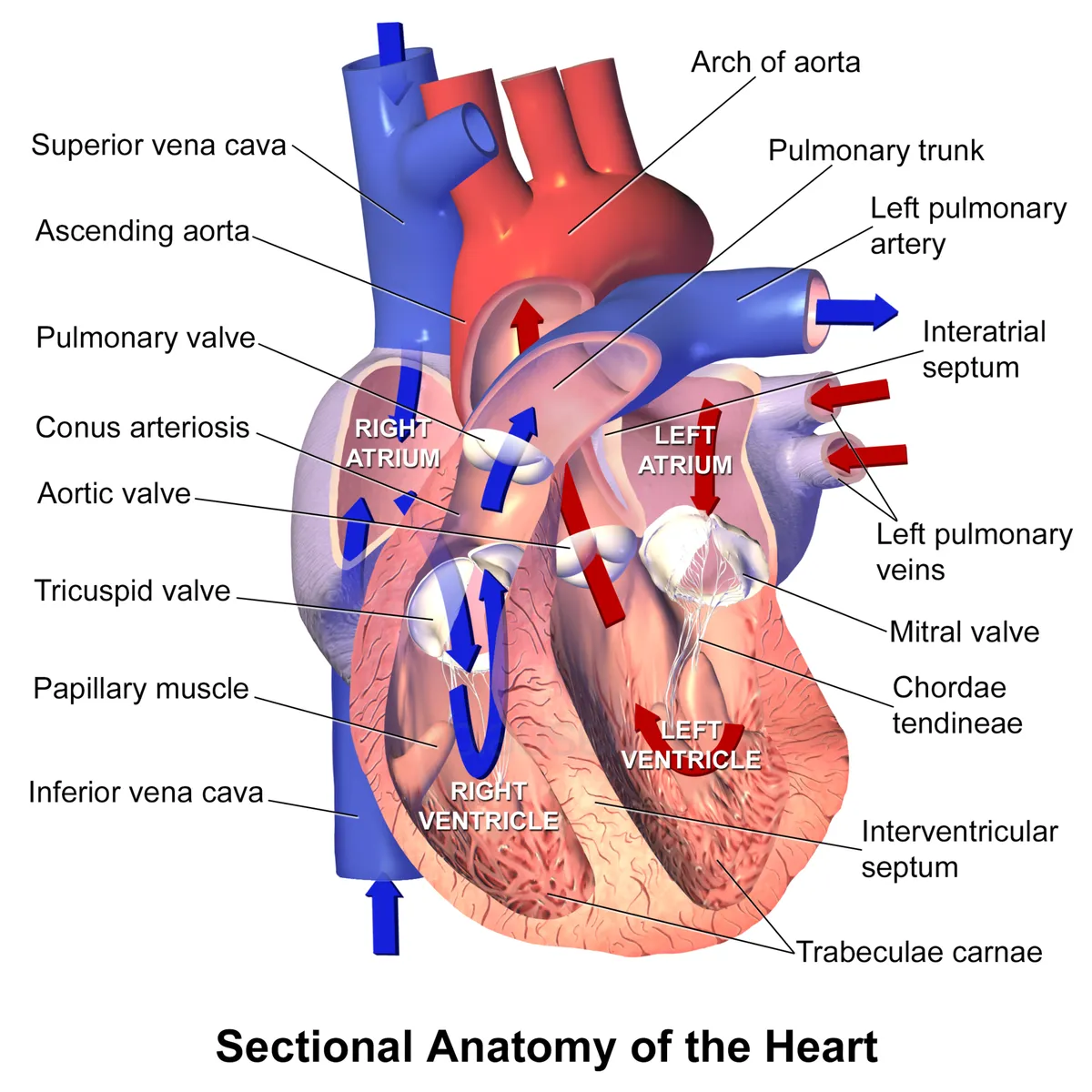
The heart has four chambers forming two pumps. The right atrium receives deoxygenated blood from the whole body except the lungs through the superior and inferior venae cavae, and blood from the heart muscle itself through the coronary sinus; blood crosses the tricuspid valve to the right ventricle, the main pumping chamber of the right heart, then passes the right ventricular outflow tract and pulmonary valve into the pulmonary trunk. In the pulmonary capillaries it takes up oxygen from the alveoli and returns through four pulmonary veins to the left atrium. The left atrium passes blood to the left ventricle across the mitral valve, both passively and by atrial contraction; the left ventricle ejects it through the aortic valve into the systemic circulation. All four valves have one purpose: to allow forward flow and prevent backflow.[4]
The sequence of contraction is driven by the conduction system: the sinus node, the main pacemaker, lies at the junction of the superior vena cava and right atrium; the impulse spreads through the atria to the atrioventricular node, bundle of His and Purkinje network, so that the atria contract first and the two ventricles almost simultaneously. In the fetus the interatrial septum is still open (foramen ovale) and the ductus arteriosus links the pulmonary artery to the aorta, diverting most blood away from the non-functioning lungs; both close quickly after birth, separating the pulmonary and systemic circulations as in the adult.[4]
The aorta: course and major branches
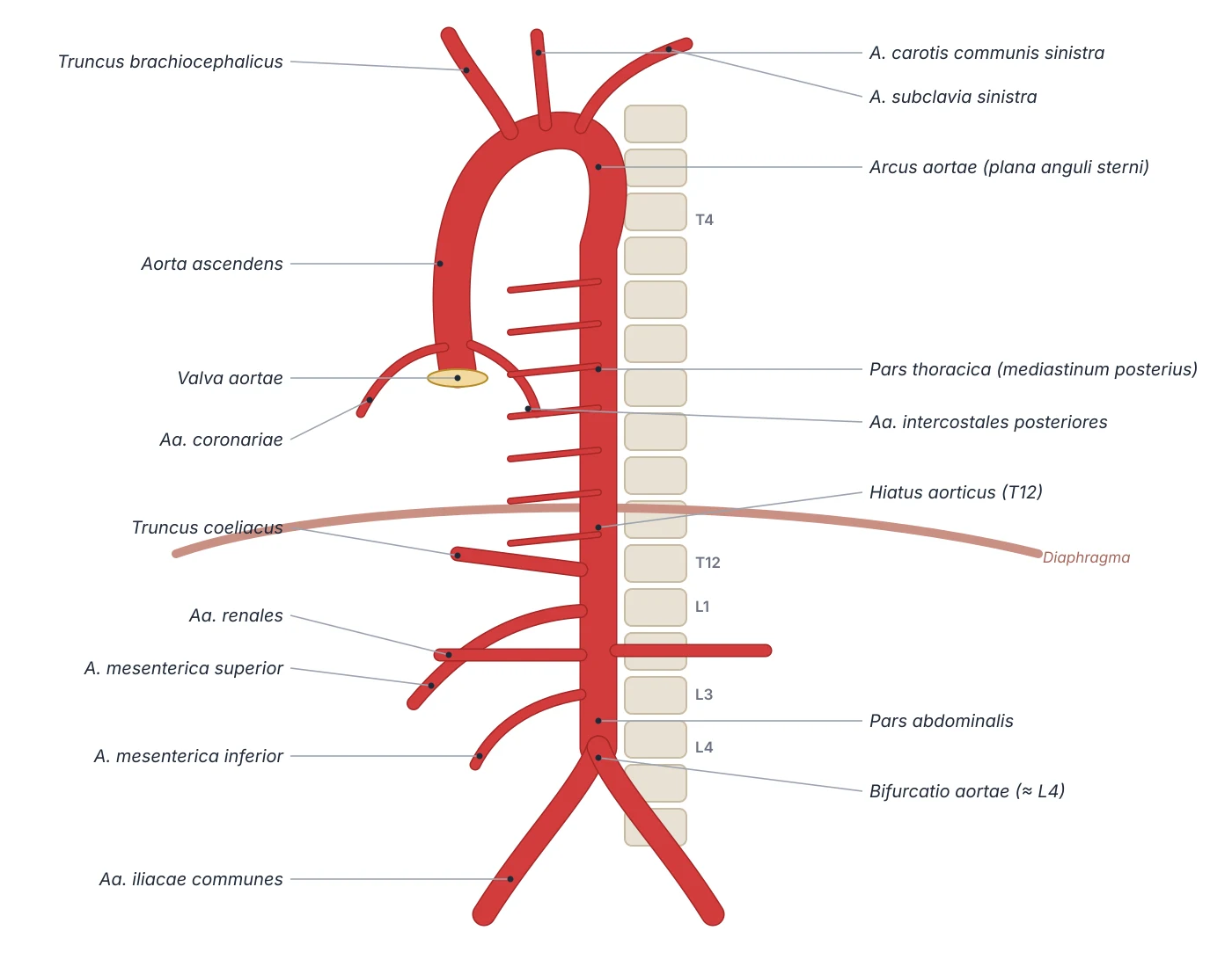
The aorta receives blood from the left ventricle through the three-cusped aortic valve. The ascending aorta is about 5 cm long and rises to the level of T4; the coronary arteries, arising from the valve sinuses, are its first branches. The arch curves backwards and to the left over the root of the left lung and in front of the trachea; its upper border lies near the transverse plane through the sternal angle (second sternocostal joint) and T4–T5, and it gives three branches: the brachiocephalic trunk, left common carotid and left subclavian arteries. The descending thoracic aorta lies in the posterior mediastinum to the left of the spine, drifting medially, gives visceral (pericardial, bronchial, oesophageal, mediastinal) and parietal (posterior intercostal, superior phrenic) branches, and passes the aortic hiatus of the diaphragm at T12 to become the abdominal aorta.[5]
Root and ascending aorta
- Extent
- Aortic valve → T4 (about 5 cm)
- Main branches
- Right and left coronary arteries
Arch
- Extent
- At the plane of the sternal angle
- Main branches
- Brachiocephalic trunk, left common carotid, left subclavian
Thoracic aorta
- Extent
- Posterior mediastinum → aortic hiatus at T12
- Main branches
- Bronchial, oesophageal, pericardial, posterior intercostal, superior phrenic
Abdominal aorta
- Extent
- T12 → bifurcation at about L4
- Main branches
- Coeliac trunk, superior mesenteric, renal, inferior mesenteric
| Segment | Extent | Main branches |
|---|---|---|
| Root and ascending aorta | Aortic valve → T4 (about 5 cm) | Right and left coronary arteries |
| Arch | At the plane of the sternal angle | Brachiocephalic trunk, left common carotid, left subclavian |
| Thoracic aorta | Posterior mediastinum → aortic hiatus at T12 | Bronchial, oesophageal, pericardial, posterior intercostal, superior phrenic |
| Abdominal aorta | T12 → bifurcation at about L4 | Coeliac trunk, superior mesenteric, renal, inferior mesenteric |
Fetal circulation and the changes at birth
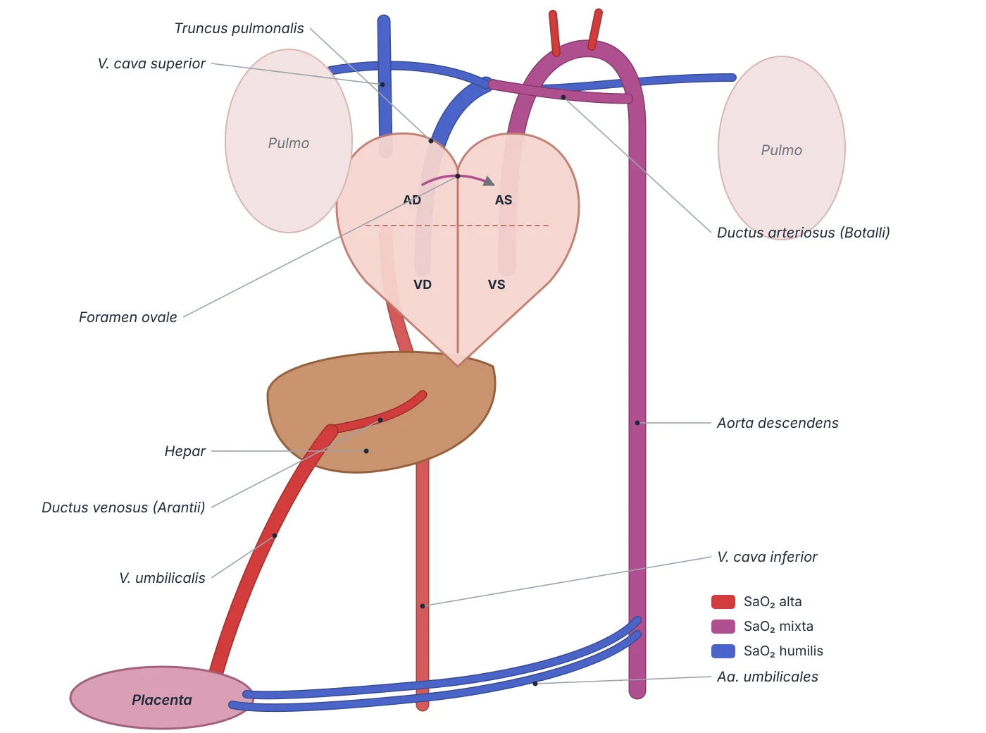
In the fetus blood is oxygenated in the placenta and returns via the inferior vena cava to the right atrium, while the lungs are collapsed, filled with amniotic fluid and their arterioles constricted, so pulmonary resistance is very high. Two shunts carry blood past the lungs. The foramen ovale between the atria, bounded by the septum primum and septum secundum, lets blood from the higher-pressure right atrium push the flap of the septum primum open into the left atrium, then to the left ventricle and aorta. The ductus arteriosus, derived from the sixth aortic arch, joins the pulmonary trunk to the aortic isthmus and diverts most right ventricular output into the systemic circulation; it is kept open by prostaglandin E (especially PGE2) made by the placenta and the duct itself.[6][7]
With the first breath the lungs expand, the pulmonary arterioles dilate and pulmonary blood flow rises steeply; at the same time the cord is clamped, removing the placenta from the circulation. Increased pulmonary venous return raises left atrial pressure above right, pressing the septum primum against the septum secundum and closing the foramen ovale, which later leaves the fossa ovalis. The ductus constricts as oxygen rises and prostaglandins fall. If it stays open (patent ductus arteriosus, associated with congenital rubella), blood flows from aorta to pulmonary artery down the pressure gradient, eventually causing pulmonary hypertension, shunt reversal and cyanosis (Eisenmenger syndrome) with risk of arrhythmia and sudden death. Prostaglandin E1 infusion can deliberately keep the duct open in duct-dependent congenital heart disease.[7][6]
Formation of the great vessels
The heart forms from two endocardial tubes that fuse, loop and are septated. The aorta and pulmonary trunk both come from the truncus arteriosus: right and left conotruncal ridges grow in a spiral and fuse into the aorticopulmonary septum in weeks 5–8, which is why the two great vessels twist around each other as they leave the heart; mesenchyme of the ridges also forms the aortic and pulmonary valves. Errors in this spiralling and septation underlie many conotruncal heart defects. The superior vena cava develops mainly from the right anterior and right common cardinal veins; the inferior vena cava is a composite of segments of the posterior, subcardinal and supracardinal veins with a hepatic segment, which explains variants such as a duplicated inferior vena cava or a persistent left superior vena cava. The pulmonary veins are incorporated into the wall of the left atrium, and the sinus venosus contributes to the crista terminalis of the right atrium.[8][4]
Principal organs
A hollow muscular organ with four chambers (two atria, two ventricles) lying in the middle mediastinum, with about two-thirds of its mass to the left of the midline. Resting stroke volume is about 70 mL.
AortaAortaThe largest artery, arising from the left ventricle; it comprises the ascending aorta, the aortic arch and the descending (thoracic and abdominal) aorta.
Pulmonary trunkTruncus pulmonalisA short arterial trunk (about 5 cm) leaving the conus arteriosus of the right ventricle, running upwards and backwards to divide below the aortic arch, at T5–T6, into the right and left pulmonary arteries. After birth it is the only artery carrying deoxygenated blood; in the fetus the ductus arteriosus links it to the aortic arch.
Venae cavaeVena cava superior et inferiorThe two largest veins, returning systemic blood to the right atrium. The superior vena cava (about 7 cm) forms behind the right first costal cartilage from the two brachiocephalic veins and receives the azygos vein. The inferior vena cava begins at L5 from the two common iliac veins, ascends to the right of the aorta, pierces the diaphragm at T8 and enters the right atrium.
Hepatic portal veinVena portae hepatisA vein about 8 cm long that brings blood from the gut, spleen and pancreas to the liver, providing about 75% of hepatic blood flow. Portal blood carries nutrients and absorbed substances for the liver to process before they reach the general circulation.
Reference values
- At rest the heart beats 60–100 times per minute, roughly 100,000 times a day.
- Resting cardiac output in an adult is about 5 L/min.
References
Sources for the values and descriptions on this page, indexed in PubMed (US National Library of Medicine).
- [1]Epicardium in Heart DevelopmentCao Y et al., Cold Spring Harbor perspectives in biology 2020PMID 31451510
- [2]Cardiac Purkinje cellsBoyden PA et al., Heart rhythm 2010PMID 19939742
- [3]Imaging of Pericardial DiseaseKligerman S, Radiologic clinics of North America 2019PMID 30454812
- [4]Anatomy, Thorax, HeartRehman I et al., StatPearls 2023PMID 29262022
- [5]Anatomy, Abdomen and Pelvis: AortaWhite HJ et al., StatPearls 2023PMID 30726004
- [6]Anatomy, Thorax, Heart Foramen OvaleYee K et al., StatPearls 2023PMID 31424787
- [7]Anatomy, Thorax, Heart Ductus ArteriosusHuff T et al., StatPearls 2023PMID 29262217
- [8]Anatomy, Thorax, Heart Great VesselsBamalan OA et al., StatPearls 2023PMID 31613460
- [9]Physiology, Cardiac OutputStatPearls (NCBI Bookshelf)PMID 29262215Supports: Heart rate, cardiac output
- [10]Physiology, CardiovascularStatPearls (NCBI Bookshelf)PMID 29630249