Interlobar sulci
Model
Anatomical regions
Sulci interlobares
Interlobar sulci
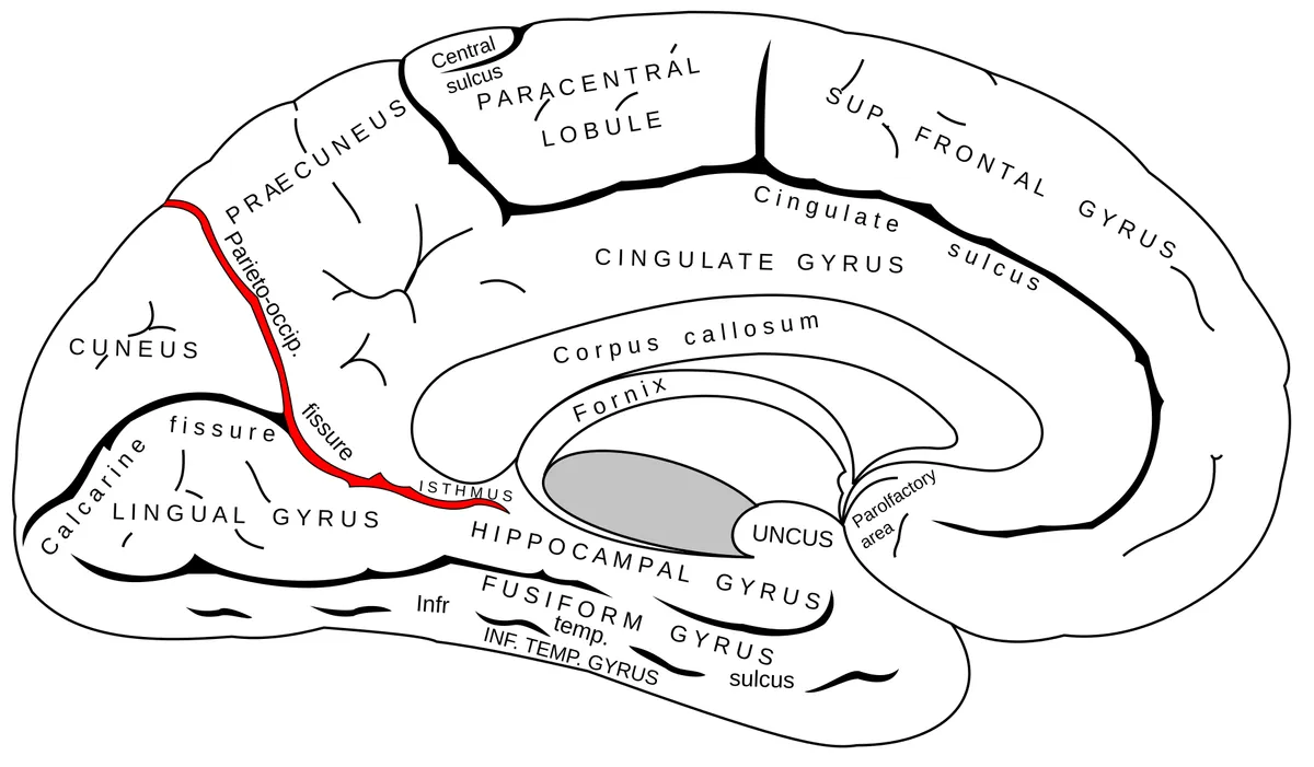
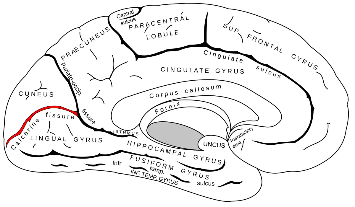
The sulci that mark the boundaries between lobes: the central sulcus (of Rolando), the lateral sulcus (of Sylvius), the parieto-occipital sulcus and the calcarine sulcus.
Functions
- Anatomical landmarks for cortical localisation
- Folding increases cortical surface area: about two-thirds of the cortex lies within sulci
Description
The sulci that mark the boundaries between lobes: the central sulcus (of Rolando), the lateral sulcus (of Sylvius), the parieto-occipital sulcus and the calcarine sulcus.
Key structures
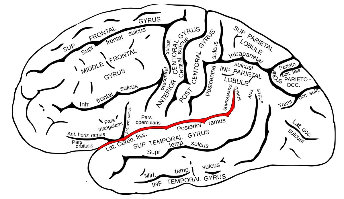
Sulcus centralis
- Name
- Central sulcus (Rolando)
- Role, notes
- Separates frontal from parietal lobe, motor from sensory cortex
Sulcus lateralis
- Name
- Lateral sulcus (Sylvius)
- Role, notes
- Separates temporal from frontal and parietal lobes; hides the insula
Sulcus parietooccipitalis
- Name
- Parieto-occipital sulcus
- Role, notes
- Parieto-occipital boundary on the medial surface
Sulcus cinguli
- Name
- Cingulate sulcus
- Role, notes
- Bounds the cingulate gyrus; its marginal ramus reaches the superior border
Fissura longitudinalis cerebri
- Name
- Longitudinal cerebral fissure
- Role, notes
- Separates the hemispheres, holds the falx cerebri
| Terminologia Anatomica | Name | Role, notes |
|---|---|---|
| Sulcus centralis | Central sulcus (Rolando) | Separates frontal from parietal lobe, motor from sensory cortex |
| Sulcus lateralis | Lateral sulcus (Sylvius) | Separates temporal from frontal and parietal lobes; hides the insula |
| Sulcus parietooccipitalis | Parieto-occipital sulcus | Parieto-occipital boundary on the medial surface |
| Sulcus cinguli | Cingulate sulcus | Bounds the cingulate gyrus; its marginal ramus reaches the superior border |
| Fissura longitudinalis cerebri | Longitudinal cerebral fissure | Separates the hemispheres, holds the falx cerebri |
Boundaries and structure
The main interlobar sulci are the central (Rolandic), lateral (Sylvian) and parieto-occipital sulci. Cortical folding varies considerably between individuals; buried transitional gyri (“plis de passage”) lie in the depths of the main sulci, such as the one in the middle of the central sulcus.[1]
Connections and function
Cortical folding develops during gestation as neural progenitors proliferate, so that cortical area grows faster than skull volume. Disturbances of this process cause malformations of cortical development (lissencephaly, polymicrogyria, etc.), which can now be detected prenatally by imaging.[2][3]
Functions
- 01Anatomical landmarks for cortical localisation
- 02Folding increases cortical surface area: about two-thirds of the cortex lies within sulci
Regions
References
Sources for the values and descriptions on this page, indexed in PubMed (US National Library of Medicine).
- [1]"Plis de passage" Deserve a Role in Models of the Cortical Folding ProcessMangin JF et al., Brain topography 2019PMID 31583493
- [2]Cortical progenitor biology: key features mediating proliferation versus differentiationUzquiano A et al., Journal of neurochemistry 2018PMID 29570795
- [3]Fetal malformations of cortical development: review and clinical guidanceRuss JB et al., Brain 2025PMID 40048696
- [4]Automatic parcellation of human cortical gyri and sulci using standard anatomical nomenclatureDestrieux C et al., 2010PMID 20547229Supports: Nomenclature of cortical gyri and sulci
- [5]Neuroanatomy, Cerebral HemisphereStatPearls (NCBI Bookshelf)PMID 31747196
- [6]Neuroanatomy, CerebellumStatPearls (NCBI Bookshelf)PMID 30844194
- [7]Current Controversies on Wernicke's Area and its Role in LanguagePMID 28656532Supports: Wernicke’s area
- [8]The human brain in numbers: a linearly scaled-up primate brainHerculano-Houzel S, 2009PMID 19915731