Frontal lobe
Model
Anatomical regions
Lobus frontalis
Frontal lobe
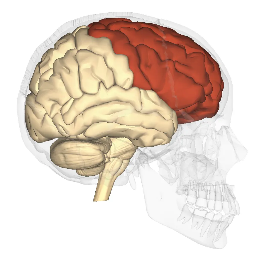
The largest lobe of the cerebral hemisphere, anterior to the central sulcus and superior to the lateral sulcus. The precentral gyrus contains the primary motor cortex (Brodmann area 4); the prefrontal cortex subserves executive functions.
Functions
- Voluntary movement (primary motor and premotor cortex)
- Executive functions: planning, response inhibition, working memory
- Expressive language (Broca’s area, Brodmann 44–45, usually in the dominant hemisphere)
- Social behaviour and personality
Description
The largest lobe of the cerebral hemisphere, anterior to the central sulcus and superior to the lateral sulcus. The precentral gyrus contains the primary motor cortex (Brodmann area 4); the prefrontal cortex subserves executive functions.
Key structures
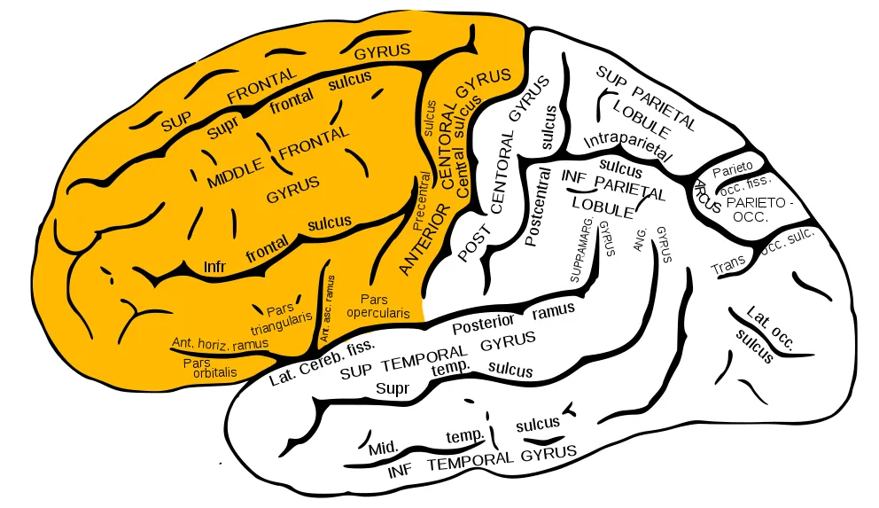
Gyrus precentralis
- Name
- Precentral gyrus
- Role, notes
- Primary motor cortex (Brodmann 4), inverted body map
Gyrus frontalis superior
- Name
- Superior frontal gyrus
- Role, notes
- Supplementary motor area on the medial surface
Gyrus frontalis medius
- Name
- Middle frontal gyrus
- Role, notes
- Dorsolateral prefrontal cortex, frontal eye field
Gyrus frontalis inferior
- Name
- Inferior frontal gyrus
- Role, notes
- Opercular and triangular parts: Broca’s area (Brodmann 44, 45)
Gyri orbitales, gyrus rectus
- Name
- Orbital gyri, gyrus rectus
- Role, notes
- Orbitofrontal cortex: reward valuation, social behaviour
Lobulus paracentralis (pars anterior)
- Name
- Paracentral lobule (anterior part)
- Role, notes
- Lower-limb motor area; bladder control
| Terminologia Anatomica | Name | Role, notes |
|---|---|---|
| Gyrus precentralis | Precentral gyrus | Primary motor cortex (Brodmann 4), inverted body map |
| Gyrus frontalis superior | Superior frontal gyrus | Supplementary motor area on the medial surface |
| Gyrus frontalis medius | Middle frontal gyrus | Dorsolateral prefrontal cortex, frontal eye field |
| Gyrus frontalis inferior | Inferior frontal gyrus | Opercular and triangular parts: Broca’s area (Brodmann 44, 45) |
| Gyri orbitales, gyrus rectus | Orbital gyri, gyrus rectus | Orbitofrontal cortex: reward valuation, social behaviour |
| Lobulus paracentralis (pars anterior) | Paracentral lobule (anterior part) | Lower-limb motor area; bladder control |
Boundaries and structure
The frontal lobe lies in front of the central sulcus and above the lateral sulcus. Its lateral surface comprises the precentral gyrus (primary motor cortex) and the superior, middle and inferior frontal gyri; the inferior frontal gyrus contains the triangular and opercular parts (Broca’s area in the dominant hemisphere). The prefrontal cortex is most developed in humans, making up about 30% of the total cortical area.[1][2]
Connections and function
Frontal function rests on a web of short and long association fibres that give direct access to sensory information and control over planning and motor regions. The prefrontal cortex exerts top-down cognitive control: maintaining goals, inhibiting inappropriate responses and switching strategies. For language, the classic Broca–Wernicke model has given way to a distributed network view that also includes subcortical structures.[2][3][4]
Functions
- 01Voluntary movement (primary motor and premotor cortex)
- 02Executive functions: planning, response inhibition, working memory
- 03Expressive language (Broca’s area, Brodmann 44–45, usually in the dominant hemisphere)
- 04Social behaviour and personality
Regions
References
Sources for the values and descriptions on this page, indexed in PubMed (US National Library of Medicine).
- [1]What constitutes the prefrontal cortex?Carlén M, Science 2017PMID 29074767
- [2]The anatomy of the human frontal lobeCatani M, Handbook of clinical neurology 2019PMID 31590750
- [3]Goal-direction and top-down controlBuschman TJ et al., Philosophical transactions of the Royal Society of London. Series B, Biological sciences 2014PMID 25267814
- [4]Broca and Wernicke are dead, or moving past the classic model of language neurobiologyTremblay P et al., Brain and language 2016PMID 27584714
- [5]Automatic parcellation of human cortical gyri and sulci using standard anatomical nomenclatureDestrieux C et al., 2010PMID 20547229Supports: Nomenclature of cortical gyri and sulci
- [6]Neuroanatomy, Cerebral HemisphereStatPearls (NCBI Bookshelf)PMID 31747196
- [7]Neuroanatomy, CerebellumStatPearls (NCBI Bookshelf)PMID 30844194
- [8]Current Controversies on Wernicke's Area and its Role in LanguagePMID 28656532Supports: Wernicke’s area
- [9]The human brain in numbers: a linearly scaled-up primate brainHerculano-Houzel S, 2009PMID 19915731