Cerebellum
Model
Anatomical regions
Cerebellum
Cerebellum
Lies in the posterior cranial fossa beneath the tentorium cerebelli and consists of the vermis and two cerebellar hemispheres. It occupies about 10% of brain volume yet contains more than half of all neurons.
Functions
- Balance and posture
- Motor coordination and precision
- Motor learning
Description
Lies in the posterior cranial fossa beneath the tentorium cerebelli and consists of the vermis and two cerebellar hemispheres. It occupies about 10% of brain volume yet contains more than half of all neurons.
Key structures
Vermis cerebelli
- Name
- Vermis
- Role, notes
- Axial balance, gait
Hemispherium cerebelli
- Name
- Cerebellar hemisphere
- Role, notes
- Coordination of the ipsilateral limbs
Lobus flocculonodularis
- Name
- Flocculonodular lobe
- Role, notes
- Vestibulocerebellum: eye movements, posture
Nucleus dentatus
- Name
- Dentate nucleus
- Role, notes
- Largest deep nucleus; exits via the superior peduncle to the thalamus
Pedunculi cerebellares
- Name
- Cerebellar peduncles
- Role, notes
- Superior (output), middle (input from pons), inferior (input from cord and vestibular nuclei)
| Terminologia Anatomica | Name | Role, notes |
|---|---|---|
| Vermis cerebelli | Vermis | Axial balance, gait |
| Hemispherium cerebelli | Cerebellar hemisphere | Coordination of the ipsilateral limbs |
| Lobus flocculonodularis | Flocculonodular lobe | Vestibulocerebellum: eye movements, posture |
| Nucleus dentatus | Dentate nucleus | Largest deep nucleus; exits via the superior peduncle to the thalamus |
| Pedunculi cerebellares | Cerebellar peduncles | Superior (output), middle (input from pons), inferior (input from cord and vestibular nuclei) |
Boundaries and structure
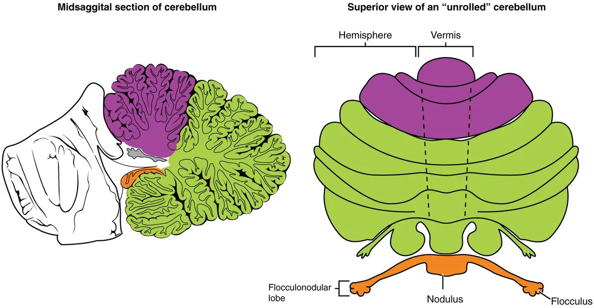
The cerebellum lies in the posterior cranial fossa, behind the brainstem and below the tentorium; it comprises the midline vermis and two hemispheres, connected to the brainstem by three pairs of peduncles. Its cortex has three layers (molecular, Purkinje cell, granular) with a highly regular repeating microcircuit that projects to the deep cerebellar nuclei.[1][2]
Connections and function
The cerebellum is essential for motor coordination and motor learning, and its role in cognition and emotion is increasingly recognised.[1]
Clinical correlations
Cerebellar infarction often presents with nausea, vomiting and dizziness, mimicking gastroenteritis or labyrinthitis. Large infarcts may swell, compress the brainstem and cause obstructive hydrocephalus, requiring emergency management.[3]
Cortex, deep nuclei and peduncles
The cerebellum accounts for only about 10% of brain weight yet holds up to 80% of its neurons, thanks to its tightly folded structure: a white matter core covered by grey matter. The cortex has three layers. The inner granular layer, the input layer, contains granule cell bodies, Golgi, Lugaro and unipolar brush cells and Purkinje axons. The middle layer, mostly Purkinje and Bergmann cells, is the output layer. The outer molecular layer holds stellate and basket cells, granule cell axons and Purkinje dendrites. Purkinje, Golgi, stellate and basket cells tend to be inhibitory, whereas granule and unipolar brush cells are excitatory.[2]
In sagittal section the branching white matter resembles a tree, which Winslow named the arbor vitae (“tree of life”) in 1740. Embedded in it are three pairs of deep nuclei: fastigial, interposed (globose and emboliform) and dentate, the largest. All cerebellar output leaves from these nuclei, and Purkinje cells are the sole output of the cerebellar cortex.[2]
Three pairs of peduncles join the cerebellum to the brainstem. The middle peduncle carries mostly pontocerebellar fibres from the contralateral pontine nuclei; the inferior peduncle, medial to it, carries both afferent and efferent fibres; the superior peduncle is mainly efferent, from the dentate, interposed and fastigial nuclei, and also receives the ventral spinocerebellar tract. Most input arrives as mossy and climbing fibres, both excitatory: mossy fibres from the pontine nuclei, spinal cord, reticular formation and vestibular nuclei; climbing fibres exclusively from the inferior olive.[2]
Lobes, lobules, functional divisions and the cerebellar homunculus
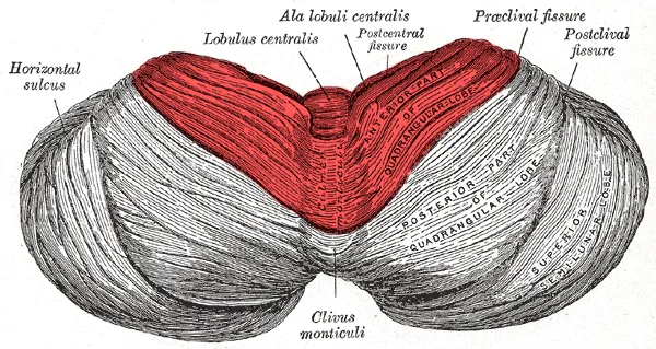
Two transverse fissures divide each hemisphere into three lobes: the primary fissure separates anterior from posterior lobe, and the posterolateral fissure separates posterior from flocculonodular lobe. In the Schmahmann classification, fissures further divide the cerebellum into 10 lobules numbered I to X radially in the sagittal plane, each with a central vermian part and two lateral hemispheric parts.[2]
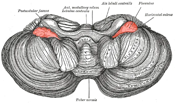
Functionally there are three divisions. The cerebrocerebellum, the largest, mainly the lateral hemispheres, receives cortical input via the pontine nuclei and projects to the thalamus and red nucleus. The spinocerebellum (anterior lobe, vermis, fastigial and interposed nuclei) receives proprioceptive input from the dorsal columns and sends back signals that fine-tune movement. The vestibulocerebellum (flocculonodular lobe and paravermis) receives vestibular input and projects to the vestibular nuclei and spinal cord to maintain trunk balance. The cerebellum thus compares intended with executed movement and corrects it through polysynaptic connections to the motor cortex.[2]
The cerebellum carries a somatotopic map, the cerebellar homunculus, like the motor and sensory cortices: clear in the anterior lobe, arranged front to back from foot to hand to head, and less consistent in the posterior lobe. Upper limb ataxia is linked to lobules IV–VI and lower limb ataxia to lobules III–IV; action tremor to ipsilateral anterior lobe lesions; hypermetria and decomposition of multi-joint movement to the dentate nucleus. The Guillain–Mollaret triangle (dentato-rubro-olivary pathway) takes part in generating several tremors.[2]
Beyond movement, the cerebellum contributes to language, working memory, executive and autonomic function and affect, though cognitive signs of cerebellar lesions are usually subtle and mostly seen acutely or in congenital disorders. Extensive vermal damage has been associated with anxiety, aggression and disinhibited behaviour, including in children after cerebellar tumour resection, consistent with links between the vermis and the limbic system. Eye movement disorders (nystagmus, saccadic dysmetria, saccadic pursuit) involve lobules VI–VII, the flocculus, paraflocculus, uvula and nodulus.[2]
Arteries, veins and infarct syndromes
Three main arteries supply the cerebellum. The superior cerebellar artery (SCA), the most constant, arises from the basilar artery or the first segment of the posterior cerebral artery and supplies the upper hemispheres and vermis, part of the white matter and the dentate nucleus. SCA infarcts commonly cause vertigo (60%), dysarthria (55%), nystagmus (50%), limb ataxia (48%), gait ataxia (45%) and dysmetria (43%); proximal occlusion adds brainstem dysfunction, distal occlusion gives purely cerebellar signs.[2]
The anterior inferior cerebellar artery (AICA) usually arises from the lower third of the basilar artery and supplies the anteroinferior cerebellum (including the flocculus), the middle peduncle, the lateral pons and the inner ear, so its infarcts can include hearing loss. The posterior inferior cerebellar artery (PICA) usually arises from the vertebral artery and supplies the lower hemispheres and vermis and the dorsolateral medulla; its ischaemia produces the lateral medullary (Wallenberg) syndrome: vertigo with nystagmus, dysphagia, dysphonia, hiccup, ipsilateral ataxia and Horner syndrome, and loss of pain and temperature on the ipsilateral face and contralateral body. PICA and AICA territories are reciprocal: a hypoplastic PICA means a larger AICA territory.[2]
Cerebellar veins form three groups: the anterior (petrosal) group, including the petrosal vein, drains into the superior and inferior petrosal sinuses; the posterior (tentorial) group, with the inferior vermian veins, drains towards the tentorium; and the superior (Galenic) group drains the upper hemispheres, vermis and midbrain into the vein of Galen and straight sinus.[2]
Functions
- 01Balance and posture
- 02Motor coordination and precision
- 03Motor learning
Regions
References
Sources for the values and descriptions on this page, indexed in PubMed (US National Library of Medicine).
- [1]The human cerebellum: a review of physiologic neuroanatomyRoostaei T et al., Neurologic clinics 2014PMID 25439284
- [2]A journey through the anatomy of the cerebellumTamanini JVG et al., Arquivos de neuro-psiquiatria 2025PMID 41429150
- [3]Cerebellar infarctionDatar S et al., Neurologic clinics 2014PMID 25439292
- [4]Automatic parcellation of human cortical gyri and sulci using standard anatomical nomenclatureDestrieux C et al., 2010PMID 20547229Supports: Nomenclature of cortical gyri and sulci
- [5]Neuroanatomy, Cerebral HemisphereStatPearls (NCBI Bookshelf)PMID 31747196
- [6]Neuroanatomy, CerebellumStatPearls (NCBI Bookshelf)PMID 30844194
- [7]Current Controversies on Wernicke's Area and its Role in LanguagePMID 28656532Supports: Wernicke’s area
- [8]The human brain in numbers: a linearly scaled-up primate brainHerculano-Houzel S, 2009PMID 19915731