Parietal lobe
Model
Anatomical regions
Lobus parietalis
Parietal lobe
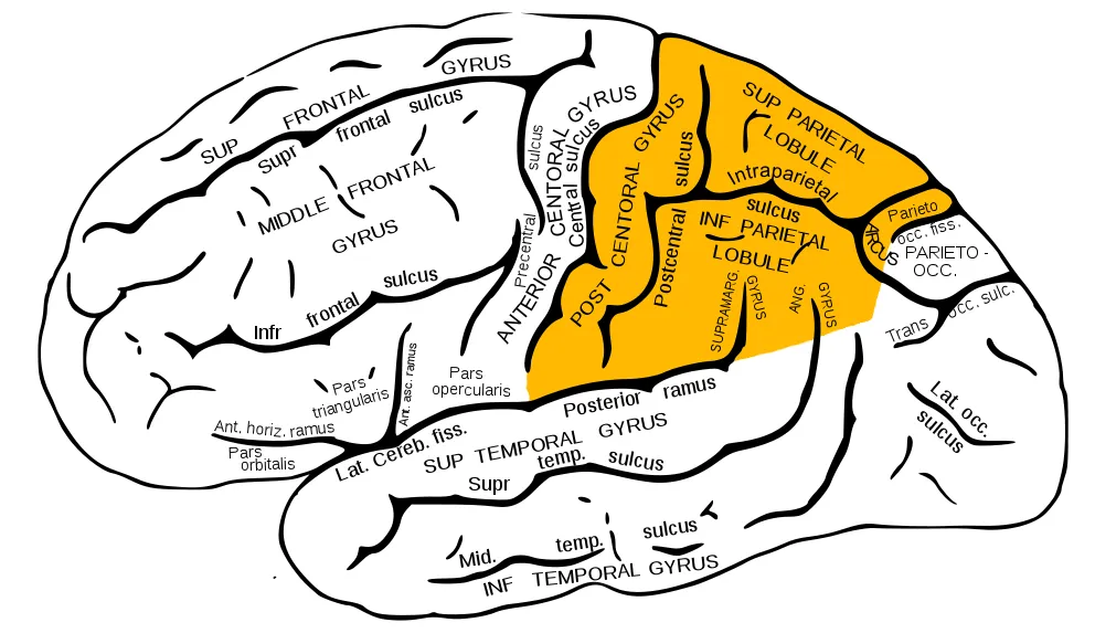
Lies posterior to the central sulcus, superior to the lateral sulcus and anterior to the parieto-occipital sulcus. The postcentral gyrus contains the primary somatosensory cortex (Brodmann areas 3, 1, 2).
Functions
- Somatic sensation: touch, proprioception, pain and temperature
- Spatial integration and attention
- Reading, writing and calculation (supramarginal and angular gyri)
Description
Lies posterior to the central sulcus, superior to the lateral sulcus and anterior to the parieto-occipital sulcus. The postcentral gyrus contains the primary somatosensory cortex (Brodmann areas 3, 1, 2).
Key structures
Gyrus postcentralis
- Name
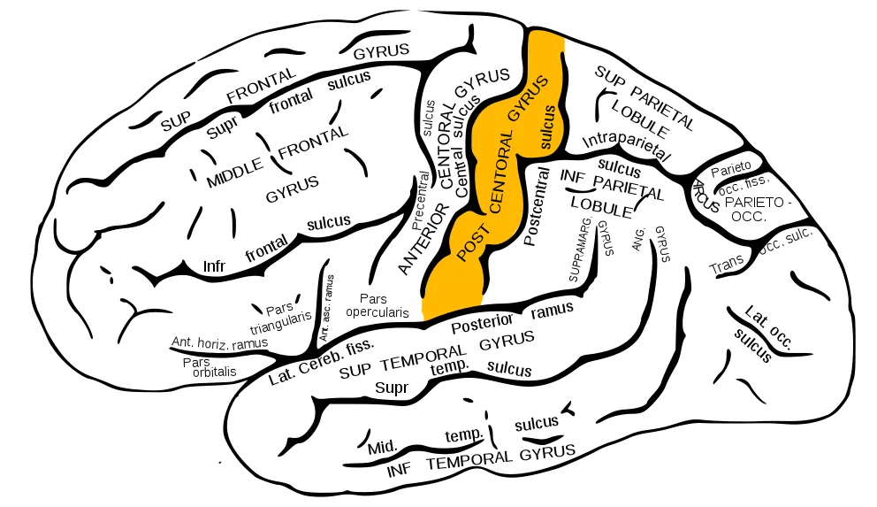
- Postcentral gyrus
- Role, notes
- Primary somatosensory cortex (Brodmann 3, 1, 2)
Lobulus parietalis superior
- Name
- Superior parietal lobule
- Role, notes
- Spatial integration, visually guided reaching
Gyrus supramarginalis
- Name
- Supramarginal gyrus
- Role, notes
- Praxis, phonological processing
Gyrus angularis
- Name
- Angular gyrus
- Role, notes
- Reading, writing, calculation; dominant lesions cause Gerstmann syndrome
Precuneus
- Name
- Precuneus
- Role, notes
- Default mode network, self-referential imagery
| Terminologia Anatomica | Name | Role, notes |
|---|---|---|
| Gyrus postcentralis | Postcentral gyrus | Primary somatosensory cortex (Brodmann 3, 1, 2) |
| Lobulus parietalis superior | Superior parietal lobule | Spatial integration, visually guided reaching |
| Gyrus supramarginalis | Supramarginal gyrus | Praxis, phonological processing |
| Gyrus angularis | Angular gyrus | Reading, writing, calculation; dominant lesions cause Gerstmann syndrome |
| Precuneus | Precuneus | Default mode network, self-referential imagery |
Boundaries and structure
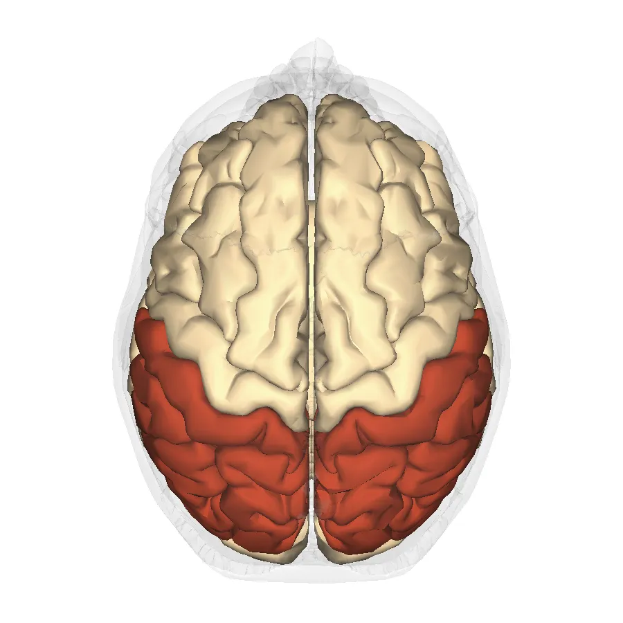
The parietal lobe lies behind the central sulcus, above the lateral sulcus and in front of the parieto-occipital sulcus. It comprises the postcentral gyrus (primary somatosensory cortex), the superior and inferior parietal lobules (supramarginal and angular gyri) and, medially, the precuneus.
Connections and function
The parietal cortex contains topographic maps of the body surface; these maps are not proportional to the body but magnify important regions such as the hand and face (the sensory homunculus). The precuneus is among the most active regions of the default-mode network during conscious rest, and it deactivates during sleep, anaesthesia and disorders of consciousness.[1][2]
Clinical correlations
Functions
- 01Somatic sensation: touch, proprioception, pain and temperature
- 02Spatial integration and attention
- 03Reading, writing and calculation (supramarginal and angular gyri)
Regions
References
Sources for the values and descriptions on this page, indexed in PubMed (US National Library of Medicine).
- [1]Somatosensory mapsHarding-Forrester S et al., Handbook of clinical neurology 2018PMID 29519481
- [2]The precuneus and consciousnessCavanna AE, CNS spectrums 2007PMID 17603406
- [3]Somatosensory deficitsKlingner CM et al., Handbook of clinical neurology 2018PMID 29519458
- [4]Constructional apraxiaGainotti G et al., Handbook of clinical neurology 2018PMID 29519467
- [5]Automatic parcellation of human cortical gyri and sulci using standard anatomical nomenclatureDestrieux C et al., 2010PMID 20547229Supports: Nomenclature of cortical gyri and sulci
- [6]Neuroanatomy, Cerebral HemisphereStatPearls (NCBI Bookshelf)PMID 31747196
- [7]Neuroanatomy, CerebellumStatPearls (NCBI Bookshelf)PMID 30844194
- [8]Current Controversies on Wernicke's Area and its Role in LanguagePMID 28656532Supports: Wernicke’s area
- [9]The human brain in numbers: a linearly scaled-up primate brainHerculano-Houzel S, 2009PMID 19915731