Brainstem
Model
Anatomical regions
Truncus encephali
Brainstem
Comprises the midbrain, pons and medulla oblongata, connecting the diencephalon to the spinal cord; it houses the nuclei of cranial nerves III–XII and the vital centres.
Functions
- Respiratory and cardiovascular control
- Conduction of ascending and descending tracts
- Swallowing, cough and vomiting reflexes
- Reticular activating system: maintenance of arousal
Description
Comprises the midbrain, pons and medulla oblongata, connecting the diencephalon to the spinal cord; it houses the nuclei of cranial nerves III–XII and the vital centres.
Key structures
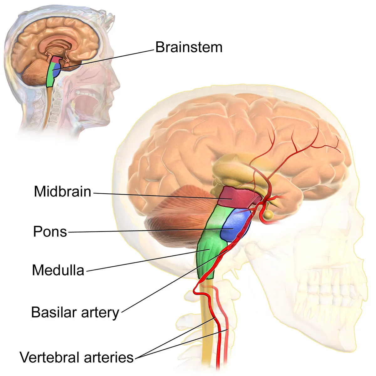
Mesencephalon
- Name
- Midbrain
- Role, notes
- Nuclei of III and IV; substantia nigra, red nucleus; colliculi
Pons
- Name
- Pons
- Role, notes
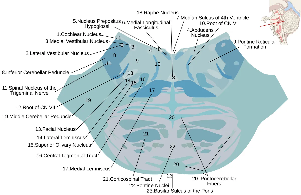
- Nuclei of V–VIII; pontine nuclei relay cortex to cerebellum
Medulla oblongata
- Name
- Medulla oblongata
- Role, notes
- Nuclei of IX–XII; respiratory and cardiovascular centres; pyramids and decussation
Formatio reticularis
- Name
- Reticular formation
- Role, notes
- Ascending reticular activating system: arousal
Lemniscus medialis
- Name
- Medial lemniscus
- Role, notes
- Carries proprioception and fine touch to the thalamus
| Terminologia Anatomica | Name | Role, notes |
|---|---|---|
| Mesencephalon | Midbrain | Nuclei of III and IV; substantia nigra, red nucleus; colliculi |
| Pons | Pons | Nuclei of V–VIII; pontine nuclei relay cortex to cerebellum |
| Medulla oblongata | Medulla oblongata | Nuclei of IX–XII; respiratory and cardiovascular centres; pyramids and decussation |
| Formatio reticularis | Reticular formation | Ascending reticular activating system: arousal |
| Lemniscus medialis | Medial lemniscus | Carries proprioception and fine touch to the thalamus |
Boundaries and structure
The brainstem comprises the midbrain, pons and medulla oblongata, linking the diencephalon to the spinal cord and housing the nuclei of cranial nerves III–XII. The dorsal midbrain bears the superior (visual) and inferior (auditory) colliculi; the ventral tegmental area and substantia nigra contain dopaminergic neurons.[1][2]
Connections and function
Functions
- 01Respiratory and cardiovascular control
- 02Conduction of ascending and descending tracts
- 03Swallowing, cough and vomiting reflexes
- 04Reticular activating system: maintenance of arousal
Regions
References
Sources for the values and descriptions on this page, indexed in PubMed (US National Library of Medicine).
- [1]Cranial Nerve AnatomyTraylor KS et al., Neuroimaging clinics of North America 2022PMID 35843663
- [2]The superior colliculusHoy JL et al., Current biology 2025PMID 40068606
- [3]Ventral tegmental area: cellular heterogeneity, connectivity and behaviourMorales M et al., Nature reviews. Neuroscience 2017PMID 28053327
- [4]The Parabrachial Nucleus: CGRP Neurons Function as a General AlarmPalmiter RD, Trends in neurosciences 2018PMID 29703377
- [5]Automatic parcellation of human cortical gyri and sulci using standard anatomical nomenclatureDestrieux C et al., 2010PMID 20547229Supports: Nomenclature of cortical gyri and sulci
- [6]Neuroanatomy, Cerebral HemisphereStatPearls (NCBI Bookshelf)PMID 31747196
- [7]Neuroanatomy, CerebellumStatPearls (NCBI Bookshelf)PMID 30844194
- [8]Current Controversies on Wernicke's Area and its Role in LanguagePMID 28656532Supports: Wernicke’s area
- [9]The human brain in numbers: a linearly scaled-up primate brainHerculano-Houzel S, 2009PMID 19915731