Occipital lobe
Model
Anatomical regions
Lobus occipitalis
Occipital lobe
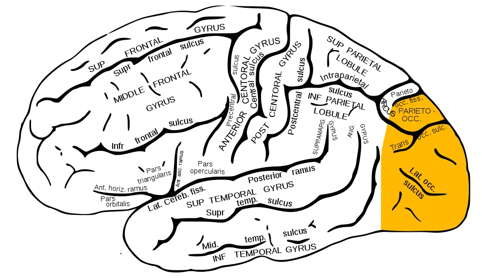
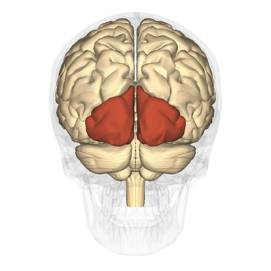
Lies posterior to the parieto-occipital sulcus. The primary visual cortex (Brodmann area 17, striate cortex) lines the banks of the calcarine sulcus on the medial surface.
Functions
- Primary visual processing
- Analysis of colour, form and motion (visual association cortex)
Description
Lies posterior to the parieto-occipital sulcus. The primary visual cortex (Brodmann area 17, striate cortex) lines the banks of the calcarine sulcus on the medial surface.
Key structures
Sulcus calcarinus
- Name
- Calcarine sulcus
- Role, notes
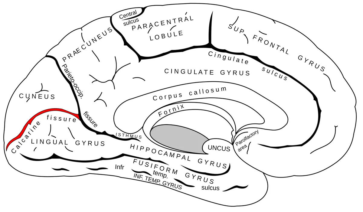
- Its banks hold primary visual cortex V1 (Brodmann 17, stria of Gennari)
Cuneus
- Name
- Cuneus
- Role, notes
- Above the calcarine sulcus: contralateral lower visual field
Gyrus lingualis
- Name
- Lingual gyrus
- Role, notes
- Below the calcarine sulcus: contralateral upper field; colour
Polus occipitalis
- Name
- Occipital pole
- Role, notes
- Represents the macula (central vision)
Gyri occipitales laterales
- Name
- Lateral occipital gyri
- Role, notes
- Visual association cortex (V2–V5), motion in MT/V5
| Terminologia Anatomica | Name | Role, notes |
|---|---|---|
| Sulcus calcarinus | Calcarine sulcus | Its banks hold primary visual cortex V1 (Brodmann 17, stria of Gennari) |
| Cuneus | Cuneus | Above the calcarine sulcus: contralateral lower visual field |
| Gyrus lingualis | Lingual gyrus | Below the calcarine sulcus: contralateral upper field; colour |
| Polus occipitalis | Occipital pole | Represents the macula (central vision) |
| Gyri occipitales laterales | Lateral occipital gyri | Visual association cortex (V2–V5), motion in MT/V5 |
Boundaries and structure
The occipital lobe is the most posterior part of the hemisphere, bounded medially by the parieto-occipital sulcus. The calcarine sulcus divides its medial surface into the cuneus above and the lingual gyrus below; the primary visual cortex (area 17, striate cortex) lies on either bank of the calcarine sulcus.
Connections and function
The visual cortex is organised into retinotopic maps in which neurons with similar receptive fields lie next to each other (ocular-dominance and orientation columns). Signals from both eyes converge here to create binocular vision and depth perception; from the occipital cortex, information flows along the ventral (recognition) and dorsal (location, motion) streams.[1][2]
Functions
- 01Primary visual processing
- 02Analysis of colour, form and motion (visual association cortex)
Regions
References
Sources for the values and descriptions on this page, indexed in PubMed (US National Library of Medicine).
- [1]Cortical MapsBednar JA et al., The Neuroscientist 2016PMID 26290447
- [2]Binocular visionBlake R et al., Vision research 2011PMID 20951722
- [3]Automatic parcellation of human cortical gyri and sulci using standard anatomical nomenclatureDestrieux C et al., 2010PMID 20547229Supports: Nomenclature of cortical gyri and sulci
- [4]Neuroanatomy, Cerebral HemisphereStatPearls (NCBI Bookshelf)PMID 31747196
- [5]Neuroanatomy, CerebellumStatPearls (NCBI Bookshelf)PMID 30844194
- [6]Current Controversies on Wernicke's Area and its Role in LanguagePMID 28656532Supports: Wernicke’s area
- [7]The human brain in numbers: a linearly scaled-up primate brainHerculano-Houzel S, 2009PMID 19915731