Basal nuclei and ventricles
Model
Anatomical regions
Nuclei basales et ventriculi
Basal nuclei and ventricles
The deep grey-matter masses of the hemisphere (caudate nucleus, putamen, globus pallidus, amygdala) and the ventricular system that contains cerebrospinal fluid.
Functions
- Modulation of movement initiation and amplitude
- Procedural learning and habit formation
- Production (choroid plexus) and circulation of cerebrospinal fluid
Description
The deep grey-matter masses of the hemisphere (caudate nucleus, putamen, globus pallidus, amygdala) and the ventricular system that contains cerebrospinal fluid.
Key structures
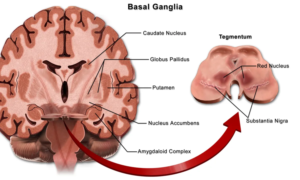
Nucleus caudatus
- Name
- Caudate nucleus
- Role, notes
- Striatum; atrophies early in Huntington disease
Putamen
- Name
- Putamen
- Role, notes
- Striatum, receiving motor cortical input
Globus pallidus
- Name
- Globus pallidus
- Role, notes
- Main basal ganglia output to the thalamus
Capsula interna
- Name
- Internal capsule
- Role, notes
- Corticospinal fibres in the posterior limb; a small infarct causes pure hemiparesis
Ventriculus lateralis
- Name
- Lateral ventricle
- Role, notes
- Contains choroid plexus that secretes cerebrospinal fluid
| Terminologia Anatomica | Name | Role, notes |
|---|---|---|
| Nucleus caudatus | Caudate nucleus | Striatum; atrophies early in Huntington disease |
| Putamen | Putamen | Striatum, receiving motor cortical input |
| Globus pallidus | Globus pallidus | Main basal ganglia output to the thalamus |
| Capsula interna | Internal capsule | Corticospinal fibres in the posterior limb; a small infarct causes pure hemiparesis |
| Ventriculus lateralis | Lateral ventricle | Contains choroid plexus that secretes cerebrospinal fluid |
Boundaries and structure
The basal ganglia are a group of interconnected subcortical nuclei: the striatum (caudate nucleus and putamen), globus pallidus, subthalamic nucleus and substantia nigra. Deep within the hemisphere, they are closely related to the internal capsule, thalamus and lateral ventricles.[1]
Connections and function
Cortical information loops through the basal ganglia and back to the cortex via the thalamus, serving motor planning and initiation, executive functions, emotion and habit formation. Dopamine from the substantia nigra keeps this network in balance; when dopamine falls, as in Parkinson’s disease, basal ganglia dysfunction causes movement disorders.[1][2]
Functions
- 01Modulation of movement initiation and amplitude
- 02Procedural learning and habit formation
- 03Production (choroid plexus) and circulation of cerebrospinal fluid
Regions
References
Sources for the values and descriptions on this page, indexed in PubMed (US National Library of Medicine).
- [1]Functional Neuroanatomy of the Normal and Pathological Basal GangliaLanciego JL et al., Cold Spring Harbor perspectives in medicine 2026PMID 39694693
- [2]The role of the basal ganglia in habit formationYin HH et al., Nature reviews. Neuroscience 2006PMID 16715055
- [3]Automatic parcellation of human cortical gyri and sulci using standard anatomical nomenclatureDestrieux C et al., 2010PMID 20547229Supports: Nomenclature of cortical gyri and sulci
- [4]Neuroanatomy, Cerebral HemisphereStatPearls (NCBI Bookshelf)PMID 31747196
- [5]Neuroanatomy, CerebellumStatPearls (NCBI Bookshelf)PMID 30844194
- [6]Current Controversies on Wernicke's Area and its Role in LanguagePMID 28656532Supports: Wernicke’s area
- [7]The human brain in numbers: a linearly scaled-up primate brainHerculano-Houzel S, 2009PMID 19915731