Limbic lobe
Model
Anatomical regions
Lobus limbicus
Limbic lobe
Formed by the structures on the medial surface of the hemisphere that encircle the corpus callosum and diencephalon: the cingulate gyrus, parahippocampal gyrus and hippocampus. It is the core of the limbic system.
Functions
- Emotion
- Memory consolidation (hippocampus)
- Motivation and behaviour
Description
Formed by the structures on the medial surface of the hemisphere that encircle the corpus callosum and diencephalon: the cingulate gyrus, parahippocampal gyrus and hippocampus. It is the core of the limbic system.
Key structures
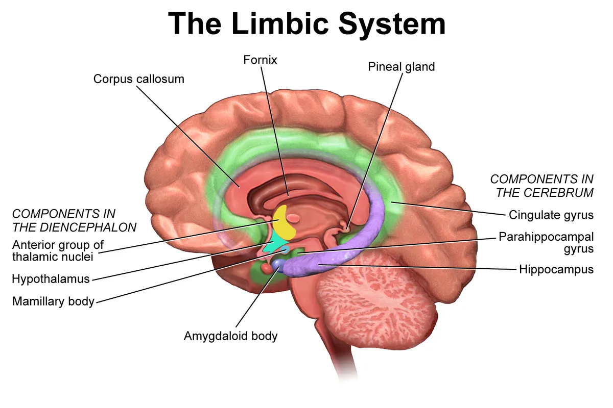
Gyrus cinguli
- Name
- Cingulate gyrus
- Role, notes
- Anterior: emotion, conflict monitoring; posterior: spatial memory
Hippocampus
- Name
- Hippocampus
- Role, notes
- Encoding of declarative memory, spatial navigation
Gyrus parahippocampalis
- Name
- Parahippocampal gyrus
- Role, notes
- Entorhinal cortex: gateway to the hippocampus
Corpus amygdaloideum
- Name
- Amygdala
- Role, notes
- Fear, emotional salience
Fornix
- Name
- Fornix
- Role, notes
- Main hippocampal output to the mammillary bodies (Papez circuit)
| Terminologia Anatomica | Name | Role, notes |
|---|---|---|
| Gyrus cinguli | Cingulate gyrus | Anterior: emotion, conflict monitoring; posterior: spatial memory |
| Hippocampus | Hippocampus | Encoding of declarative memory, spatial navigation |
| Gyrus parahippocampalis | Parahippocampal gyrus | Entorhinal cortex: gateway to the hippocampus |
| Corpus amygdaloideum | Amygdala | Fear, emotional salience |
| Fornix | Fornix | Main hippocampal output to the mammillary bodies (Papez circuit) |
Boundaries and structure
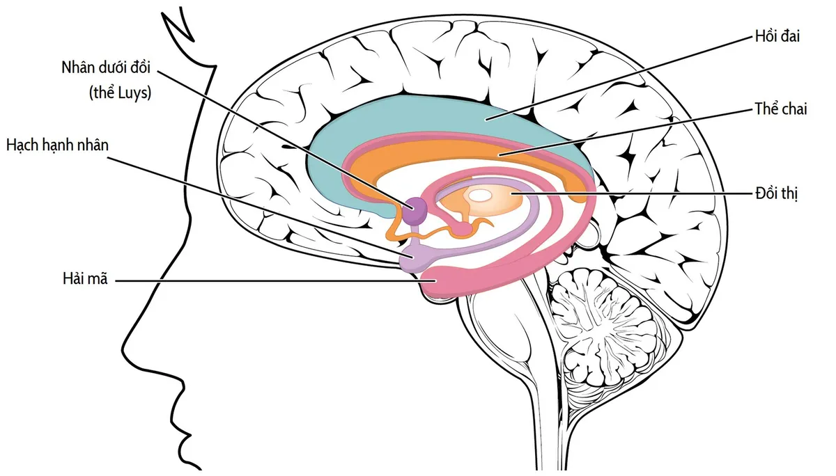
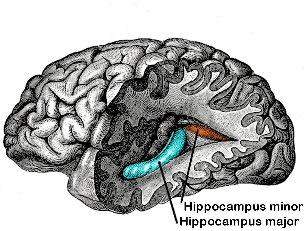
The hippocampal formation is a C-shaped structure bulging into the floor of the temporal horn of the lateral ventricle. The hippocampus proper has three subfields (CA1–CA3); together with the dentate gyrus, the subicular complex and the entorhinal cortex it forms the hippocampal formation. The amygdala lies just in front of the hippocampal head, in the medial temporal lobe.[1]
Connections and function
Processed multimodal sensory information is funnelled into the hippocampus by the entorhinal cortex, where episodic and spatial memories are formed. The dentate gyrus continues to generate new neurons in adulthood, contributing to learning. The amygdala processes both fearful and rewarding stimuli through distinct circuits.[1][2][3]
Clinical correlations
Functions
- 01Emotion
- 02Memory consolidation (hippocampus)
- 03Motivation and behaviour
Regions
References
Sources for the values and descriptions on this page, indexed in PubMed (US National Library of Medicine).
- [1]Anatomy of the hippocampal formationSchultz C et al., Frontiers of neurology and neuroscience 2014PMID 24777126
- [2]Adult Neurogenesis in the Hippocampus: From Stem Cells to BehaviorGonçalves JT et al., Cell 2016PMID 27814520
- [3]From circuits to behaviour in the amygdalaJanak PH et al., Nature 2015PMID 25592533
- [4]Stress Effects on Neuronal Structure: Hippocampus, Amygdala, and Prefrontal CortexMcEwen BS et al., Neuropsychopharmacology 2016PMID 26076834
- [5]The relationship between adult hippocampal neurogenesis and cognitive impairment in Alzheimer's diseaseGeigenmüller JN et al., Alzheimer's & dementia 2024PMID 39166771
- [6]Automatic parcellation of human cortical gyri and sulci using standard anatomical nomenclatureDestrieux C et al., 2010PMID 20547229Supports: Nomenclature of cortical gyri and sulci
- [7]Neuroanatomy, Cerebral HemisphereStatPearls (NCBI Bookshelf)PMID 31747196
- [8]Neuroanatomy, CerebellumStatPearls (NCBI Bookshelf)PMID 30844194
- [9]Current Controversies on Wernicke's Area and its Role in LanguagePMID 28656532Supports: Wernicke’s area
- [10]The human brain in numbers: a linearly scaled-up primate brainHerculano-Houzel S, 2009PMID 19915731